9KPL
 
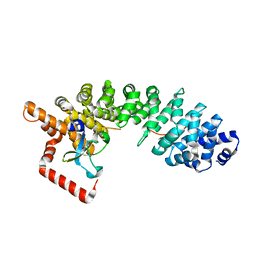
 | | Crystal structure of T. rubripes Mincle with glucose | | 分子名称: | C-type lectin domain-containing protein, CALCIUM ION, alpha-D-glucopyranose, ... | | 著者 | Ito, T, Nagae, M, Yamasaki, S. | | 登録日 | 2024-11-23 | | 公開日 | 2025-04-02 | | 実験手法 | X-RAY DIFFRACTION (1.8 Å) | | 主引用文献 | Phylogenetic and structural insights into the origin of C-type lectin Mincle in vertebrates.
Immunogenetics, 77, 2025
|
|
8PE0
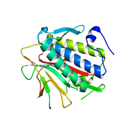
 | | X-ray structure of the Thermus thermophilus K167L mutant of the PilF-GSPIIB domain in the c-di-GMP bound state | | 分子名称: | 9,9'-[(2R,3R,3aS,5S,7aR,9R,10R,10aS,12S,14aR)-3,5,10,12-tetrahydroxy-5,12-dioxidooctahydro-2H,7H-difuro[3,2-d:3',2'-j][1,3,7,9,2,8]tetraoxadiphosphacyclododecine-2,9-diyl]bis(2-amino-1,9-dihydro-6H-purin-6-one), ACETATE ION, SULFATE ION, ... | | 著者 | Neissner, K, Woehnert, J. | | 登録日 | 2023-06-13 | | 公開日 | 2024-06-26 | | 最終更新日 | 2025-01-08 | | 実験手法 | X-RAY DIFFRACTION (1.9 Å) | | 主引用文献 | The structural basis for high-affinity c-di-GMP binding to the GSPII-B domain of the traffic ATPase PilF from Thermus thermophilus.
J.Biol.Chem., 301, 2024
|
|
8PDK
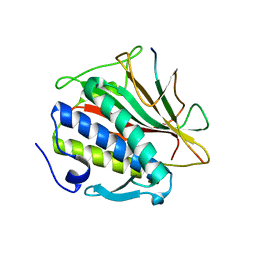
 | | X-ray structure of the Thermus thermophilus PilF-GSPIIB domain in the c-di-GMP bound state | | 分子名称: | 9,9'-[(2R,3R,3aS,5S,7aR,9R,10R,10aS,12S,14aR)-3,5,10,12-tetrahydroxy-5,12-dioxidooctahydro-2H,7H-difuro[3,2-d:3',2'-j][1,3,7,9,2,8]tetraoxadiphosphacyclododecine-2,9-diyl]bis(2-amino-1,9-dihydro-6H-purin-6-one), ACETATE ION, SULFATE ION, ... | | 著者 | Neissner, K, Woehnert, J. | | 登録日 | 2023-06-12 | | 公開日 | 2024-06-26 | | 最終更新日 | 2025-01-08 | | 実験手法 | X-RAY DIFFRACTION (2 Å) | | 主引用文献 | The structural basis for high-affinity c-di-GMP binding to the GSPII-B domain of the traffic ATPase PilF from Thermus thermophilus.
J.Biol.Chem., 301, 2024
|
|
8PFA
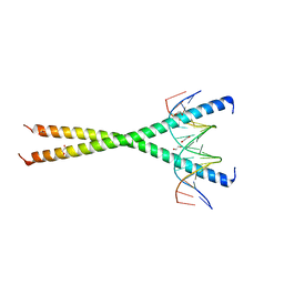
 | | X-ray structure of the Thermus thermophilus K167R mutant of the PilF-GSPIIB domain in the c-di-GMP bound state | | 分子名称: | 9,9'-[(2R,3R,3aS,5S,7aR,9R,10R,10aS,12S,14aR)-3,5,10,12-tetrahydroxy-5,12-dioxidooctahydro-2H,7H-difuro[3,2-d:3',2'-j][1,3,7,9,2,8]tetraoxadiphosphacyclododecine-2,9-diyl]bis(2-amino-1,9-dihydro-6H-purin-6-one), ACETATE ION, SULFATE ION, ... | | 著者 | Neissner, K, Woehnert, J. | | 登録日 | 2023-06-15 | | 公開日 | 2024-06-26 | | 最終更新日 | 2025-01-08 | | 実験手法 | X-RAY DIFFRACTION (1.8 Å) | | 主引用文献 | The structural basis for high-affinity c-di-GMP binding to the GSPII-B domain of the traffic ATPase PilF from Thermus thermophilus.
J.Biol.Chem., 301, 2024
|
|
7KWM
 | | CtBP1 (28-375) L182F/V185T - AMP | | 分子名称: | ADENOSINE MONOPHOSPHATE, C-terminal-binding protein 1, CALCIUM ION | | 著者 | Royer, W.E, Del Campo, M. | | 登録日 | 2020-12-01 | | 公開日 | 2021-02-03 | | 最終更新日 | 2023-10-18 | | 実験手法 | X-RAY DIFFRACTION (2.3 Å) | | 主引用文献 | NAD(H) phosphates mediate tetramer assembly of human C-terminal binding protein (CtBP).
J.Biol.Chem., 296, 2021
|
|
2LSK
 | | C-terminal domain of human REV1 in complex with DNA-polymerase H (eta) | | 分子名称: | DNA polymerase eta, DNA repair protein REV1 | | 著者 | Pozhidaeva, A, Pustovalova, Y, Bezsonova, I, Korzhnev, D. | | 登録日 | 2012-05-01 | | 公開日 | 2012-06-27 | | 最終更新日 | 2024-05-15 | | 実験手法 | SOLUTION NMR | | 主引用文献 | NMR structure and dynamics of the C-terminal domain from human Rev1 and its complex with Rev1 interacting region of DNA polymerase eta.
Biochemistry, 51, 2012
|
|
3EFK
 | | Structure of c-Met with pyrimidone inhibitor 50 | | 分子名称: | 5-{4-[(6,7-dimethoxyquinolin-4-yl)oxy]-3-fluorophenyl}-2-[(4-fluorophenyl)amino]-3-methylpyrimidin-4(3H)-one, Hepatocyte growth factor receptor | | 著者 | Bellon, S.F, D'Angelo, N, Whittington, D, Dussault, I. | | 登録日 | 2008-09-09 | | 公開日 | 2008-10-07 | | 最終更新日 | 2023-08-30 | | 実験手法 | X-RAY DIFFRACTION (2.2 Å) | | 主引用文献 | Design, synthesis, and biological evaluation of potent c-Met inhibitors.
J.Med.Chem., 51, 2008
|
|
3EFJ
 | | Structure of c-Met with pyrimidone inhibitor 7 | | 分子名称: | 2-benzyl-5-{4-[(6,7-dimethoxyquinolin-4-yl)oxy]-3-fluorophenyl}-3-methylpyrimidin-4(3H)-one, Hepatocyte growth factor receptor | | 著者 | D'Angelo, N, Bellon, S, Whittington, D. | | 登録日 | 2008-09-09 | | 公開日 | 2008-10-07 | | 最終更新日 | 2023-08-30 | | 実験手法 | X-RAY DIFFRACTION (2.6 Å) | | 主引用文献 | Design, synthesis, and biological evaluation of potent c-Met inhibitors.
J.Med.Chem., 51, 2008
|
|
4IZY
 | |
4TRK
 | |
4TZJ
 | |
5UTS
 | |
2XR4
 | | C-terminal domain of BC2L-C Lectin from Burkholderia cenocepacia | | 分子名称: | LECTIN, SULFATE ION | | 著者 | Sulak, O, Cioci, G, Lameignere, E, Delia, M, Wimmerova, M, Imberty, A. | | 登録日 | 2010-09-09 | | 公開日 | 2011-08-03 | | 最終更新日 | 2023-12-20 | | 実験手法 | X-RAY DIFFRACTION (1.9 Å) | | 主引用文献 | Burkholderia Cenocepacia Bc2L-C is a Super Lectin with Dual Specificity and Proinflammatory Activity.
Plos Pathog., 7, 2011
|
|
2JDQ
 | | C-terminal domain of influenza A virus polymerase PB2 subunit in complex with human importin alpha5 | | 分子名称: | IMPORTIN ALPHA-1 SUBUNIT, POLYMERASE BASIC PROTEIN 2 | | 著者 | Tarendeau, F, Guilligay, D, Mas, P, Boulo, S, Baudin, F, Ruigrok, R.W.H, Hart, D.J, Cusack, S. | | 登録日 | 2007-01-11 | | 公開日 | 2007-02-27 | | 最終更新日 | 2023-12-13 | | 実験手法 | X-RAY DIFFRACTION (2.2 Å) | | 主引用文献 | Structure and Nuclear Import Function of the C- Terminal Domain of Influenza Virus Polymerase Pb2 Subunit
Nat.Struct.Mol.Biol., 14, 2007
|
|
4TZL
 | |
4TZO
 | |
7L4V
 | |
6B6X
 | | Crystal structure of Desulfovibrio vulgaris carbon monoxide dehydrogenase, dithionite-reduced (protein batch 2), canonical C-cluster | | 分子名称: | CHLORIDE ION, Carbon monoxide dehydrogenase, FE(4)-NI(1)-S(4) CLUSTER, ... | | 著者 | Wittenborn, E.C, Drennan, C.L. | | 登録日 | 2017-10-03 | | 公開日 | 2018-10-03 | | 最終更新日 | 2023-10-04 | | 実験手法 | X-RAY DIFFRACTION (1.84 Å) | | 主引用文献 | Redox-dependent rearrangements of the NiFeS cluster of carbon monoxide dehydrogenase.
Elife, 7, 2018
|
|
6B6Y
 | | Crystal structure of Desulfovibrio vulgaris carbon monoxide dehydrogenase, dithionite-reduced then oxygen-exposed (protein batch 2), oxidized C-cluster | | 分子名称: | Carbon monoxide dehydrogenase, FE2/S2 (INORGANIC) CLUSTER, Fe(4)-Ni(1)-S(4) cluster, ... | | 著者 | Wittenborn, E.C, Drennan, C.L. | | 登録日 | 2017-10-03 | | 公開日 | 2018-10-03 | | 最終更新日 | 2023-10-04 | | 実験手法 | X-RAY DIFFRACTION (2.6 Å) | | 主引用文献 | Redox-dependent rearrangements of the NiFeS cluster of carbon monoxide dehydrogenase.
Elife, 7, 2018
|
|
6B6V
 | | Crystal structure of Desulfovibrio vulgaris carbon monoxide dehydrogenase, as-isolated (protein batch 1), canonical C-cluster | | 分子名称: | Carbon monoxide dehydrogenase, FE(4)-NI(1)-S(4) CLUSTER, FE2/S2 (INORGANIC) CLUSTER, ... | | 著者 | Wittenborn, E.C, Drennan, C.L. | | 登録日 | 2017-10-03 | | 公開日 | 2018-10-03 | | 最終更新日 | 2023-10-04 | | 実験手法 | X-RAY DIFFRACTION (2.5 Å) | | 主引用文献 | Redox-dependent rearrangements of the NiFeS cluster of carbon monoxide dehydrogenase.
Elife, 7, 2018
|
|
3PYY
 | | Discovery and Characterization of a Cell-Permeable, Small-molecule c-Abl Kinase Activator that Binds to the Myristoyl Binding Site | | 分子名称: | (5R)-5-[3-(4-fluorophenyl)-1-phenyl-1H-pyrazol-4-yl]imidazolidine-2,4-dione, 4-(4-METHYL-PIPERAZIN-1-YLMETHYL)-N-[4-METHYL-3-(4-PYRIDIN-3-YL-PYRIMIDIN-2-YLAMINO)-PHENYL]-BENZAMIDE, GLYCEROL, ... | | 著者 | Yang, J, Campobasso, N, Biju, M.P, Fisher, K, Pan, X.Q, Cottom, J, Galbraith, S, Ho, T, Zhang, H, Hong, X, Ward, P, Hofmann, G, Siegfried, B. | | 登録日 | 2010-12-13 | | 公開日 | 2011-03-09 | | 最終更新日 | 2025-05-07 | | 実験手法 | X-RAY DIFFRACTION (1.85 Å) | | 主引用文献 | Discovery and Characterization of a Cell-Permeable, Small-Molecule c-Abl Kinase Activator that Binds to the Myristoyl Binding Site.
Chem.Biol., 18, 2011
|
|
1RMJ
 | | C-terminal domain of insulin-like growth factor (IGF) binding protein-6: structure and interaction with IGF-II | | 分子名称: | Insulin-like growth factor binding protein 6 | | 著者 | Headey, S.J, Keizer, D.W, Yao, S, Brasier, G, Kantharidis, P, Bach, L.A, Norton, R.S. | | 登録日 | 2003-11-28 | | 公開日 | 2004-09-14 | | 最終更新日 | 2024-11-20 | | 実験手法 | SOLUTION NMR | | 主引用文献 | C-terminal domain of insulin-like growth factor (IGF) binding protein-6: structure and interaction with IGF-II.
Mol.Endocrinol., 18, 2004
|
|
6B6W
 | | Crystal structure of Desulfovibrio vulgaris carbon monoxide dehydrogenase, as-isolated (protein batch 2), oxidized C-cluster | | 分子名称: | CHLORIDE ION, Carbon monoxide dehydrogenase, FE(4)-NI(1)-S(4) CLUSTER, ... | | 著者 | Wittenborn, E.C, Drennan, C.L. | | 登録日 | 2017-10-03 | | 公開日 | 2018-10-03 | | 最終更新日 | 2023-10-04 | | 実験手法 | X-RAY DIFFRACTION (1.72 Å) | | 主引用文献 | Redox-dependent rearrangements of the NiFeS cluster of carbon monoxide dehydrogenase.
Elife, 7, 2018
|
|
4TZS
 | |
1GUA
 | | HUMAN RAP1A, RESIDUES 1-167, DOUBLE MUTANT (E30D,K31E) COMPLEXED WITH GPPNHP AND THE RAS-BINDING-DOMAIN OF HUMAN C-RAF1, RESIDUES 51-131 | | 分子名称: | C-RAF1, CALCIUM ION, MAGNESIUM ION, ... | | 著者 | Nassar, N, Wittinghofer, A. | | 登録日 | 1996-06-18 | | 公開日 | 1997-01-11 | | 最終更新日 | 2024-02-07 | | 実験手法 | X-RAY DIFFRACTION (2 Å) | | 主引用文献 | Ras/Rap effector specificity determined by charge reversal.
Nat.Struct.Biol., 3, 1996
|
|


