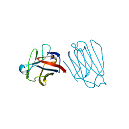
6GQH
 
 | |
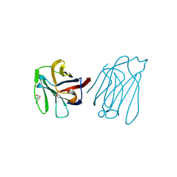
6GQG
 | |
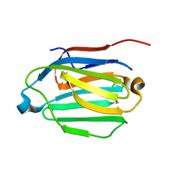
6MGH
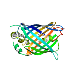
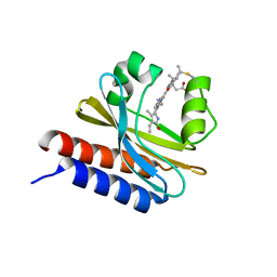 | | X-ray structure of monomeric near-infrared fluorescent protein miRFP670nano | | 分子名称: | 3-[2-[(~{Z})-[5-[(~{Z})-[(3~{S},4~{R})-3-ethenyl-4-methyl-5-oxidanylidene-pyrrolidin-2-ylidene]methyl]-3-(3-hydroxy-3-oxopropyl)-4-methyl-pyrrol-2-ylidene]methyl]-5-[(~{Z})-(4-ethenyl-3-methyl-5-oxidanylidene-pyrrol-2-ylidene)methyl]-4-methyl-1~{H}-pyrrol-3-yl]propanoic acid, GLYCEROL, ISOPROPYL ALCOHOL, ... | | 著者 | Pletnev, S. | | 登録日 | 2018-09-13 | | 公開日 | 2018-12-19 | | 最終更新日 | 2019-01-30 | | 実験手法 | X-RAY DIFFRACTION (1.95 Å) | | 主引用文献 | Smallest near-infrared fluorescent protein evolved from cyanobacteriochrome as versatile tag for spectral multiplexing.
Nat Commun, 10, 2019
|
|
6GO9
 | | Structure of GFPmut2 crystallized at pH 6 and transferred to pH 7 | | 分子名称: | (4R)-2-METHYLPENTANE-2,4-DIOL, (4S)-2-METHYL-2,4-PENTANEDIOL, Green fluorescent protein | | 著者 | Lolli, G, Raboni, S, Pasqualetto, E, Campanini, B, Mozzarelli, A, Bettati, S, Battistutta, R. | | 登録日 | 2018-06-01 | | 公開日 | 2018-12-19 | | 最終更新日 | 2024-01-17 | | 実験手法 | X-RAY DIFFRACTION (1.672 Å) | | 主引用文献 | Insight into GFPmut2 pH Dependence by Single Crystal Microspectrophotometry and X-ray Crystallography.
J.Phys.Chem.B, 122, 2018
|
|
6GO8
 | | Structure of GFPmut2 crystallized at pH 6 | | 分子名称: | (4R)-2-METHYLPENTANE-2,4-DIOL, (4S)-2-METHYL-2,4-PENTANEDIOL, Green fluorescent protein | | 著者 | Lolli, G, Raboni, S, Pasqualetto, E, Campanini, B, Mozzarelli, A, Bettati, S, Battistutta, R. | | 登録日 | 2018-06-01 | | 公開日 | 2018-12-19 | | 最終更新日 | 2024-01-17 | | 実験手法 | X-RAY DIFFRACTION (1.648 Å) | | 主引用文献 | Insight into GFPmut2 pH Dependence by Single Crystal Microspectrophotometry and X-ray Crystallography.
J.Phys.Chem.B, 122, 2018
|
|
6GRM
 | | Structure of GFPmut2 crystallized at pH 6 and transferred to pH 9 | | 分子名称: | Green fluorescent protein | | 著者 | Lolli, G, Raboni, S, Pasqualetto, E, Campanini, B, Mozzarelli, A, Bettati, S, Battistutta, R. | | 登録日 | 2018-06-11 | | 公開日 | 2018-12-19 | | 最終更新日 | 2024-01-17 | | 実験手法 | X-RAY DIFFRACTION (2.3 Å) | | 主引用文献 | Insight into GFPmut2 pH Dependence by Single Crystal Microspectrophotometry and X-ray Crystallography.
J.Phys.Chem.B, 122, 2018
|
|
6A62
 | | Placental protein 13/galectin-13 variant R53HH57RD33G with Lactose | | 分子名称: | Galactoside-binding soluble lectin 13, beta-D-galactopyranose-(1-4)-beta-D-glucopyranose | | 著者 | Su, J. | | 登録日 | 2018-06-26 | | 公開日 | 2018-12-26 | | 最終更新日 | 2020-07-29 | | 実験手法 | X-RAY DIFFRACTION (2.03 Å) | | 主引用文献 | Resetting the ligand binding site of placental protein 13/galectin-13 recovers its ability to bind lactose
Biosci. Rep., 38, 2018
|
|
6A1T
 | | Charcot-Leyden crystal protein/Galectin-10 variant E33A with lactose | | 分子名称: | Galectin-10, beta-D-galactopyranose-(1-4)-beta-D-glucopyranose | | 著者 | Su, J. | | 登録日 | 2018-06-08 | | 公開日 | 2018-12-26 | | 最終更新日 | 2024-03-27 | | 実験手法 | X-RAY DIFFRACTION (1.97 Å) | | 主引用文献 | Identification of key amino acid residues determining ligand binding specificity, homodimerization and cellular distribution of human galectin-10
Glycobiology, 29, 2019
|
|
6A1U
 | | Charcot-Leyden crystal protein/Galectin-10 variant E33D | | 分子名称: | Galectin-10 | | 著者 | Su, J. | | 登録日 | 2018-06-08 | | 公開日 | 2018-12-26 | | 最終更新日 | 2024-03-27 | | 実験手法 | X-RAY DIFFRACTION (1.62 Å) | | 主引用文献 | Identification of key amino acid residues determining ligand binding specificity, homodimerization and cellular distribution of human galectin-10
Glycobiology, 29, 2019
|
|
6A64
 | |
6A1Y
 | | Charcot-Leyden crystal protein/Galectin-10 variant Y35A | | 分子名称: | Galectin-10 | | 著者 | Su, J. | | 登録日 | 2018-06-08 | | 公開日 | 2018-12-26 | | 最終更新日 | 2024-03-27 | | 実験手法 | X-RAY DIFFRACTION (1.63 Å) | | 主引用文献 | Identification of key amino acid residues determining ligand binding specificity, homodimerization and cellular distribution of human galectin-10
Glycobiology, 29, 2019
|
|
6A65
 | | Placental protein 13/galectin-13 variant R53HR55N with Tris | | 分子名称: | 2-AMINO-2-HYDROXYMETHYL-PROPANE-1,3-DIOL, Galactoside-binding soluble lectin 13 | | 著者 | Su, J. | | 登録日 | 2018-06-26 | | 公開日 | 2018-12-26 | | 実験手法 | X-RAY DIFFRACTION (1.771 Å) | | 主引用文献 | Resetting the ligand binding site of placental protein 13/galectin-13 recovers its ability to bind lactose
Biosci. Rep., 38, 2018
|
|
6A1V
 | | Charcot-Leyden crystal protein/Galectin-10 variant E33Q | | 分子名称: | Galectin-10 | | 著者 | Su, J. | | 登録日 | 2018-06-08 | | 公開日 | 2018-12-26 | | 最終更新日 | 2024-03-27 | | 実験手法 | X-RAY DIFFRACTION (1.984 Å) | | 主引用文献 | Identification of key amino acid residues determining ligand binding specificity, homodimerization and cellular distribution of human galectin-10
Glycobiology, 29, 2019
|
|
6A1S
 | | Charcot-Leyden crystal protein/Galectin-10 variant E33A | | 分子名称: | Galectin-10 | | 著者 | Su, J. | | 登録日 | 2018-06-08 | | 公開日 | 2018-12-26 | | 最終更新日 | 2024-03-27 | | 実験手法 | X-RAY DIFFRACTION (1.63 Å) | | 主引用文献 | Identification of key amino acid residues determining ligand binding specificity, homodimerization and cellular distribution of human galectin-10
Glycobiology, 29, 2019
|
|
6A63
 | | Placental protein 13/galectin-13 variant R53HH57R with Lactose | | 分子名称: | Galactoside-binding soluble lectin 13, beta-D-galactopyranose-(1-4)-beta-D-glucopyranose | | 著者 | Su, J. | | 登録日 | 2018-06-26 | | 公開日 | 2018-12-26 | | 最終更新日 | 2020-07-29 | | 実験手法 | X-RAY DIFFRACTION (1.63 Å) | | 主引用文献 | Resetting the ligand binding site of placental protein 13/galectin-13 recovers its ability to bind lactose
Biosci. Rep., 38, 2018
|
|
6A66
 | | Placental protein 13/galectin-13 variant R53H with Tris | | 分子名称: | 2-AMINO-2-HYDROXYMETHYL-PROPANE-1,3-DIOL, Galactoside-binding soluble lectin 13 | | 著者 | Su, J.Y. | | 登録日 | 2018-06-26 | | 公開日 | 2018-12-26 | | 実験手法 | X-RAY DIFFRACTION (1.4 Å) | | 主引用文献 | Resetting the ligand binding site of placental protein 13/galectin-13 recovers its ability to bind lactose
Biosci. Rep., 38, 2018
|
|
6A1X
 | | Charcot-Leyden crystal protein/Galectin-10 variant W127A | | 分子名称: | Galectin-10 | | 著者 | Su, J. | | 登録日 | 2018-06-08 | | 公開日 | 2018-12-26 | | 最終更新日 | 2024-03-27 | | 実験手法 | X-RAY DIFFRACTION (1.99 Å) | | 主引用文献 | Identification of key amino acid residues determining ligand binding specificity, homodimerization and cellular distribution of human galectin-10
Glycobiology, 29, 2019
|
|
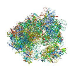
6I7O
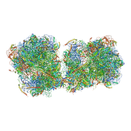 | | The structure of a di-ribosome (disome) as a unit for RQC and NGD quality control pathways recognition. | | 分子名称: | 18S ribosomal RNA, 25S ribosomal RNA, 40S ribosomal protein S0-A, ... | | 著者 | Tesina, P, Cheng, J, Becker, T, Beckmann, R. | | 登録日 | 2018-11-16 | | 公開日 | 2019-01-16 | | 最終更新日 | 2019-03-13 | | 実験手法 | ELECTRON MICROSCOPY (5.3 Å) | | 主引用文献 | Collided ribosomes form a unique structural interface to induce Hel2-driven quality control pathways.
EMBO J., 38, 2019
|
|
6EFR
 | | Crystal Structure of iNicSnFR 1.0 | | 分子名称: | iNicSnFR 1.0, a genetically encoded nicotine biosensor,Green fluorescent protein | | 著者 | Shivange, A.V, Borden, P.M. | | 登録日 | 2018-08-17 | | 公開日 | 2019-01-23 | | 最終更新日 | 2023-11-15 | | 実験手法 | X-RAY DIFFRACTION (2.4 Å) | | 主引用文献 | Nicotinic Drugs in the Endoplasmic Reticulum: Beginning the Inside-out Pathway of Addiction and Therapy
J.Gen.Physiol., 2019
|
|
6MDR
 | | Cryo-EM structure of the Ceru+32/GFP-17 protomer | | 分子名称: | Ceru+32, GFP-17 | | 著者 | Simon, A.J, Zhou, Y, Ramasubramani, V, Glaser, J, Pothukuchy, A, Golihar, J, Gerberich, J.C, Leggere, J.C, Morrow, B.R, Jung, C, Glotzer, S.C, Taylor, D.W, Ellington, A.D. | | 登録日 | 2018-09-05 | | 公開日 | 2019-01-23 | | 最終更新日 | 2024-03-13 | | 実験手法 | ELECTRON MICROSCOPY (3.47 Å) | | 主引用文献 | Supercharging enables organized assembly of synthetic biomolecules.
Nat Chem, 11, 2019
|
|
5YR2
 | |
6FLL
 | |
6HHQ
 | | Crystal structure of compound C45 bound to the yeast 80S ribosome | | 分子名称: | (3~{R})-3-[(1~{S})-2-[(1~{S},4~{a}~{R},6~{S},7~{S},8~{a}~{R})-6,7-bis(chloranyl)-5,5,8~{a}-trimethyl-2-methylidene-3,4,4~{a},6,7,8-hexahydro-1~{H}-naphthalen-1-yl]-1-oxidanyl-ethyl]pyrrolidine-2,5-dione, 18S ribosomal RNA, 25S ribosomal RNA, ... | | 著者 | Pellegrino, S, Vanderwal, C.D, Yusupov, M. | | 登録日 | 2018-08-28 | | 公開日 | 2019-02-20 | | 最終更新日 | 2024-05-15 | | 実験手法 | X-RAY DIFFRACTION (3.10000038 Å) | | 主引用文献 | Understanding the role of intermolecular interactions between lissoclimides and the eukaryotic ribosome.
Nucleic Acids Res., 47, 2019
|
|
6HOG
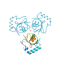 | | Structure of VPS34 LIR motif bound to GABARAP | | 分子名称: | 1,2-ETHANEDIOL, GLYCEROL, Phosphatidylinositol 3-kinase catalytic subunit type 3,Gamma-aminobutyric acid receptor-associated protein, ... | | 著者 | Mouilleron, S, Birgisdottir, A.B, Bhujbal, Z, Wirth, M, Sjottem, E, Evjen, G, Zhang, W, Lee, R, O'Reilly, N, Tooze, S, Lamark, T, Johansen, T. | | 登録日 | 2018-09-17 | | 公開日 | 2019-02-27 | | 最終更新日 | 2024-01-24 | | 実験手法 | X-RAY DIFFRACTION (1.26 Å) | | 主引用文献 | Members of the autophagy class III phosphatidylinositol 3-kinase complex I interact with GABARAP and GABARAPL1 via LIR motifs.
Autophagy, 15, 2019
|
|
6HOK
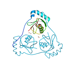 | | Structure of Beclin1 LIR (S96E) motif bound to GABARAP | | 分子名称: | 1,2-ETHANEDIOL, Beclin-1,Gamma-aminobutyric acid receptor-associated protein | | 著者 | Mouilleron, S, Birgisdottir, A.B, Bhujbal, Z, Wirth, M, Sjottem, E, Evjen, G, Zhang, W, Lee, R, O'Reilly, N, Tooze, S, Lamark, T, Johansen, T. | | 登録日 | 2018-09-17 | | 公開日 | 2019-02-27 | | 最終更新日 | 2024-01-24 | | 実験手法 | X-RAY DIFFRACTION (1.61 Å) | | 主引用文献 | Members of the autophagy class III phosphatidylinositol 3-kinase complex I interact with GABARAP and GABARAPL1 via LIR motifs.
Autophagy, 15, 2019
|
|


