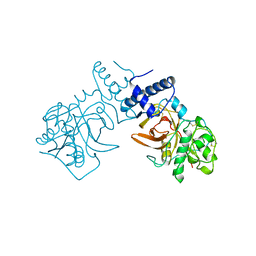
8KE6
 
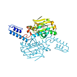 | | PylRS C-terminus domain mutant bound with L-3-chlorophenylalanine and AMPNP | | 分子名称: | 3-CHLORO-L-PHENYLALANINE, MAGNESIUM ION, PHOSPHOAMINOPHOSPHONIC ACID-ADENYLATE ESTER, ... | | 著者 | Weng, J.H, Tsai, M.D, Wang, Y.S. | | 登録日 | 2023-08-11 | | 公開日 | 2023-11-01 | | 最終更新日 | 2023-11-15 | | 実験手法 | X-RAY DIFFRACTION (1.89570856 Å) | | 主引用文献 | Rational design of the genetic code expansion toolkit for in vivo encoding of D-amino acids.
Front Genet, 14, 2023
|
|
8KE1
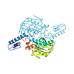 | | PylRS C-terminus domain mutant bound with L-3-bromophenylalanine and AMPNP | | 分子名称: | 3-bromo-L-phenylalanine, MAGNESIUM ION, PHOSPHOAMINOPHOSPHONIC ACID-ADENYLATE ESTER, ... | | 著者 | Weng, J.H, Tsai, M.D, Wang, Y.S. | | 登録日 | 2023-08-11 | | 公開日 | 2023-11-01 | | 最終更新日 | 2023-11-15 | | 実験手法 | X-RAY DIFFRACTION (2.50081539 Å) | | 主引用文献 | Rational design of the genetic code expansion toolkit for in vivo encoding of D-amino acids.
Front Genet, 14, 2023
|
|
8KE4
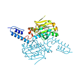 | | PylRS C-terminus domain mutant bound with D-3-bromophenylalanine and AMPNP | | 分子名称: | (2R)-2-azanyl-3-(3-bromophenyl)propanoic acid, MAGNESIUM ION, PHOSPHOAMINOPHOSPHONIC ACID-ADENYLATE ESTER, ... | | 著者 | Weng, J.H, Tsai, M.D, Wang, Y.S. | | 登録日 | 2023-08-11 | | 公開日 | 2023-11-01 | | 最終更新日 | 2023-11-15 | | 実験手法 | X-RAY DIFFRACTION (1.75050962 Å) | | 主引用文献 | Rational design of the genetic code expansion toolkit for in vivo encoding of D-amino acids.
Front Genet, 14, 2023
|
|
8JWD
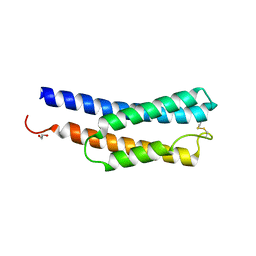 | | Histidine kinase QseE sensor domain of Escherichia coli O157:H7 | | 分子名称: | 1,2-ETHANEDIOL, histidine kinase | | 著者 | Matsumoto, K, Fukuda, Y, Inoue, T. | | 登録日 | 2023-06-28 | | 公開日 | 2023-11-15 | | 実験手法 | X-RAY DIFFRACTION (1.33 Å) | | 主引用文献 | Crystal structures of QseE and QseG: elements of a three-component system from Escherichia coli.
Acta Crystallogr.,Sect.F, 79, 2023
|
|
7PBC
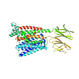
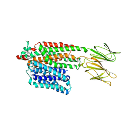
 | | Crystal structure of engineered TCR (796) complexed to HLA-A*02:01 presenting MAGE-A10 9-mer peptide | | 分子名称: | Beta-2-microglobulin, CHLORIDE ION, GLYCEROL, ... | | 著者 | Simister, P.C, Border, E.C, Vieira, J.F, Pumphrey, N.J. | | 登録日 | 2021-08-02 | | 公開日 | 2022-08-03 | | 最終更新日 | 2024-10-09 | | 実験手法 | X-RAY DIFFRACTION (2.04 Å) | | 主引用文献 | Structural insights into engineering a T-cell receptor targeting MAGE-A10 with higher affinity and specificity for cancer immunotherapy.
J Immunother Cancer, 10, 2022
|
|
4OE4
 | | Crystal Structure of Yeast ALDH4A1 Complexed with NAD+ | | 分子名称: | Delta-1-pyrroline-5-carboxylate dehydrogenase, mitochondrial, NICOTINAMIDE-ADENINE-DINUCLEOTIDE | | 著者 | Tanner, J.J. | | 登録日 | 2014-01-11 | | 公開日 | 2014-02-19 | | 最終更新日 | 2024-04-03 | | 実験手法 | X-RAY DIFFRACTION (2.168 Å) | | 主引用文献 | Structural Studies of Yeast Delta (1)-Pyrroline-5-carboxylate Dehydrogenase (ALDH4A1): Active Site Flexibility and Oligomeric State.
Biochemistry, 53, 2014
|
|
8JMR
 | | Crystal structure of hinokiresinol synthase in complex with 1,7-bis(4-hydroxyphenyl)hepta-1,6-dien-3-one | | 分子名称: | 1,7-bis(4-hydroxyphenyl)hepta-1,6-dien-3-one, Hinokiresinol synthase alpha subunit, Hinokiresinol synthase beta subunit, ... | | 著者 | Ding, Y, Ushimaru, R, Mori, T, Abe, I. | | 登録日 | 2023-06-05 | | 公開日 | 2023-12-20 | | 最終更新日 | 2024-10-16 | | 実験手法 | X-RAY DIFFRACTION (2.2 Å) | | 主引用文献 | Structural and Mechanistic Insights into the C-C Bond-Forming Rearrangement Reaction Catalyzed by Heterodimeric Hinokiresinol Synthase.
J.Am.Chem.Soc., 145, 2023
|
|
8JMQ
 | | Crystal structure of hinokiresinol synthase | | 分子名称: | Hinokiresinol synthase alpha subunit, Hinokiresinol synthase beta subunit | | 著者 | Ding, Y, Ushimaru, R, Mori, T, Abe, I. | | 登録日 | 2023-06-05 | | 公開日 | 2023-12-20 | | 最終更新日 | 2024-10-16 | | 実験手法 | X-RAY DIFFRACTION (2 Å) | | 主引用文献 | Structural and Mechanistic Insights into the C-C Bond-Forming Rearrangement Reaction Catalyzed by Heterodimeric Hinokiresinol Synthase.
J.Am.Chem.Soc., 145, 2023
|
|
7PCK
 | |
2IIM
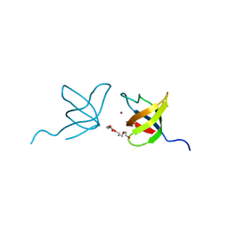 | | SH3 Domain of Human Lck | | 分子名称: | CALCIUM ION, Proto-oncogene tyrosine-protein kinase LCK, TETRAETHYLENE GLYCOL, ... | | 著者 | Romir, J, Egerer-Sieber, C, Muller, Y.A. | | 登録日 | 2006-09-28 | | 公開日 | 2006-11-07 | | 最終更新日 | 2023-08-30 | | 実験手法 | X-RAY DIFFRACTION (1 Å) | | 主引用文献 | Crystal structure analysis and solution studies of human Lck-SH3; zinc-induced homodimerization competes with the binding of proline-rich motifs.
J.Mol.Biol., 365, 2007
|
|
8OE6
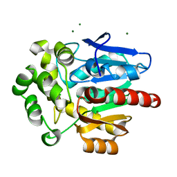 | | Structure of hyperstable haloalkane dehalogenase variant DhaA231 | | 分子名称: | CHLORIDE ION, MAGNESIUM ION, Structure of hyperstable haloalkane dehalogenase variant DhaA231 | | 著者 | Marek, M. | | 登録日 | 2023-03-10 | | 公開日 | 2024-01-17 | | 実験手法 | X-RAY DIFFRACTION (1.31 Å) | | 主引用文献 | Advancing Enzyme's Stability and Catalytic Efficiency through Synergy of Force-Field Calculations, Evolutionary Analysis, and Machine Learning.
Acs Catalysis, 13, 2023
|
|
2ITA
 | |
2JYL
 | |
8OE2
 | | Structure of hyperstable haloalkane dehalogenase variant DhaA223 | | 分子名称: | 2-AMINO-2-HYDROXYMETHYL-PROPANE-1,3-DIOL, CHLORIDE ION, GLYCEROL, ... | | 著者 | Marek, M. | | 登録日 | 2023-03-10 | | 公開日 | 2024-01-17 | | 実験手法 | X-RAY DIFFRACTION (1.51 Å) | | 主引用文献 | Advancing Enzyme's Stability and Catalytic Efficiency through Synergy of Force-Field Calculations, Evolutionary Analysis, and Machine Learning.
Acs Catalysis, 13, 2023
|
|
7PMU
 | | Crystal structure of native Iripin-8 | | 分子名称: | DI(HYDROXYETHYL)ETHER, HEXAETHYLENE GLYCOL, Serpin-8, ... | | 著者 | Polderdijk, S, Kotal, J, Chmelar, J, Huntington, J.A. | | 登録日 | 2021-09-02 | | 公開日 | 2021-10-13 | | 最終更新日 | 2024-01-31 | | 実験手法 | X-RAY DIFFRACTION (1.89 Å) | | 主引用文献 | Ixodes ricinus Salivary Serpin Iripin-8 Inhibits the Intrinsic Pathway of Coagulation and Complement.
Int J Mol Sci, 22, 2021
|
|
7PMW
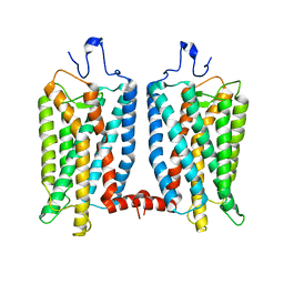
 | | HsPepT1 bound to Ala-Phe in the outward facing occluded conformation | | 分子名称: | ALA-PHE, Solute carrier family 15 member 1 | | 著者 | Killer, M, Wald, J, Pieprzyk, J, Marlovits, T.C, Loew, C. | | 登録日 | 2021-09-03 | | 公開日 | 2021-10-20 | | 最終更新日 | 2024-07-17 | | 実験手法 | ELECTRON MICROSCOPY (4.1 Å) | | 主引用文献 | Structural snapshots of human PepT1 and PepT2 reveal mechanistic insights into substrate and drug transport across epithelial membranes.
Sci Adv, 7, 2021
|
|
7PMX
 | | HsPepT1 bound to Ala-Phe in the outward facing open conformation | | 分子名称: | ALA-PHE, Solute carrier family 15 member 1 | | 著者 | Killer, M, Wald, J, Pieprzyk, J, Marlovits, T.C, Loew, C. | | 登録日 | 2021-09-03 | | 公開日 | 2021-10-20 | | 最終更新日 | 2024-07-17 | | 実験手法 | ELECTRON MICROSCOPY (3.5 Å) | | 主引用文献 | Structural snapshots of human PepT1 and PepT2 reveal mechanistic insights into substrate and drug transport across epithelial membranes.
Sci Adv, 7, 2021
|
|
6OFJ
 | |
7PMY
 | | HsPepT2 bound to Ala-Phe in the inward facing partially occluded conformation | | 分子名称: | ALA-PHE, Solute carrier family 15 member 2 | | 著者 | Killer, M, Wald, J, Pieprzyk, J, Marlovits, T.C, Loew, C. | | 登録日 | 2021-09-04 | | 公開日 | 2021-10-20 | | 最終更新日 | 2024-07-17 | | 実験手法 | ELECTRON MICROSCOPY (3.8 Å) | | 主引用文献 | Structural snapshots of human PepT1 and PepT2 reveal mechanistic insights into substrate and drug transport across epithelial membranes.
Sci Adv, 7, 2021
|
|
7PN1
 | | Apo HsPepT1 in the outward facing open conformation | | 分子名称: | Solute carrier family 15 member 1 | | 著者 | Killer, M, Wald, J, Pieprzyk, J, Marlovits, T.C, Loew, C. | | 登録日 | 2021-09-04 | | 公開日 | 2021-10-20 | | 最終更新日 | 2024-07-17 | | 実験手法 | ELECTRON MICROSCOPY (3.9 Å) | | 主引用文献 | Structural snapshots of human PepT1 and PepT2 reveal mechanistic insights into substrate and drug transport across epithelial membranes.
Sci Adv, 7, 2021
|
|
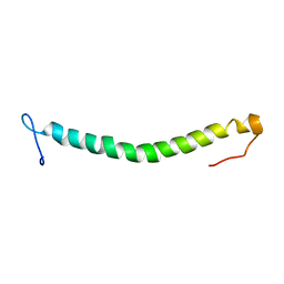
7Q16
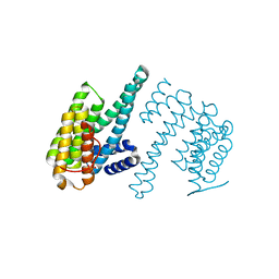 | | Human 14-3-3 zeta fused to the BAD peptide including phosphoserine-74 | | 分子名称: | 14-3-3 protein zeta/delta,Bcl2-associated agonist of cell death | | 著者 | Sluchanko, N.N, Tugaeva, K.V, Gushchin, I, Remeeva, A, Kovalev, K, Cooley, R.B. | | 登録日 | 2021-10-18 | | 公開日 | 2021-11-17 | | 最終更新日 | 2024-01-31 | | 実験手法 | X-RAY DIFFRACTION (2.356 Å) | | 主引用文献 | Crystal structure of human 14-3-3 zeta complexed with the noncanonical phosphopeptide from proapoptotic BAD.
Biochem.Biophys.Res.Commun., 583, 2021
|
|
2JX2
 | | Solution conformation of RNA-bound NELF-E RRM | | 分子名称: | Negative elongation factor E | | 著者 | Jampani, N, Schweimer, K, Wenzel, S, Woehrl, B.M, Roesch, P. | | 登録日 | 2007-11-02 | | 公開日 | 2008-04-08 | | 最終更新日 | 2024-05-08 | | 実験手法 | SOLUTION NMR | | 主引用文献 | NELF-E RRM Undergoes Major Structural Changes in Flexible Protein Regions on Target RNA Binding
Biochemistry, 47, 2008
|
|
8KDB
 | | Cryo-EM structure of the human parainfluenza virus hPIV3 L-P polymerase in dimeric form | | 分子名称: | MAGNESIUM ION, Phosphoprotein, RNA-directed RNA polymerase L, ... | | 著者 | Xie, J, Wang, L, Zhai, G, Wu, D, Lin, Z, Wang, M, Yan, X, Gao, L, Huang, X, Fearns, R, Chen, S. | | 登録日 | 2023-08-09 | | 公開日 | 2024-04-24 | | 実験手法 | ELECTRON MICROSCOPY (2.7 Å) | | 主引用文献 | Structural basis for dimerization of a paramyxovirus polymerase complex.
Nat Commun, 15, 2024
|
|
3KXE
 | |
8JOR
 | | Structure of an acyltransferase involved in mannosylerythritol lipid formation from Pseudozyma tsukubaensis in type A crystal | | 分子名称: | Acyltransferase, PENTAETHYLENE GLYCOL | | 著者 | Nakamichi, Y, Saika, A, Watanabe, M, Fujii, T, Morita, T. | | 登録日 | 2023-06-08 | | 公開日 | 2024-04-17 | | 実験手法 | X-RAY DIFFRACTION (1.45 Å) | | 主引用文献 | Structural identification of catalytic His158 of PtMAC2p from Pseudozyma tsukubaensis , an acyltransferase involved in mannosylerythritol lipids formation.
Front Bioeng Biotechnol, 11, 2023
|
|


