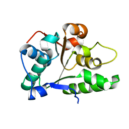
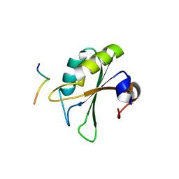
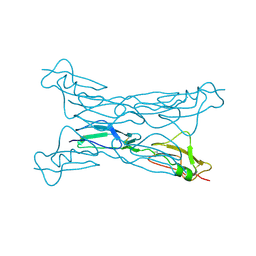
2OX9
 
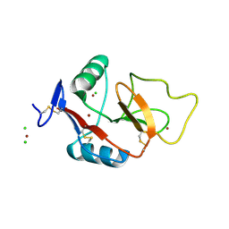
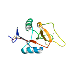 | | Mouse Scavenger Receptor C-type Lectin carbohydrate-recognition domain. | | 分子名称: | CALCIUM ION, Collectin placenta 1, alpha-L-fucopyranose-(1-3)-[beta-D-galactopyranose-(1-4)]2-acetamido-2-deoxy-beta-D-glucopyranose | | 著者 | Weis, W.I, Feinberg, H, Drickamer, K, Taylor, M.E. | | 登録日 | 2007-02-20 | | 公開日 | 2007-04-03 | | 最終更新日 | 2020-07-29 | | 実験手法 | X-RAY DIFFRACTION (1.95 Å) | | 主引用文献 | Scavenger receptor C-type lectin binds to the leukocyte cell surface glycan Lewis(x) by a novel mechanism.
J.Biol.Chem., 282, 2007
|
|
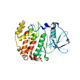
2OXX
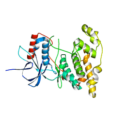
 | | Protein kinase CK2 in complex with tetrabromobenzoimidazole derivatives K17, K22 and K32 | | 分子名称: | 4,5,6,7-TETRABROMO-1H,3H-BENZIMIDAZOL-2-THIONE, Casein kinase II subunit alpha | | 著者 | Battistutta, R, Zanotti, G, Cendron, L. | | 登録日 | 2007-02-21 | | 公開日 | 2007-09-25 | | 最終更新日 | 2024-02-21 | | 実験手法 | X-RAY DIFFRACTION (2.3 Å) | | 主引用文献 | The ATP-Binding Site of Protein Kinase CK2 Holds a Positive Electrostatic Area and Conserved Water Molecules.
Chembiochem, 8, 2007
|
|
2G0S
 | | Unphotolyzed CO-bound L29F Myoglobin, crystal 2 | | 分子名称: | CARBON MONOXIDE, Myoglobin, PROTOPORPHYRIN IX CONTAINING FE, ... | | 著者 | Aranda, R, Levin, E.J, Schotte, F, Anfinrud, P.A, Phillips Jr, G.N. | | 登録日 | 2006-02-13 | | 公開日 | 2006-07-04 | | 最終更新日 | 2023-08-30 | | 実験手法 | X-RAY DIFFRACTION (1.9 Å) | | 主引用文献 | Time-dependent atomic coordinates for the dissociation of carbon monoxide from myoglobin.
Acta Crystallogr.,Sect.D, 62, 2006
|
|
2G0Z
 | | Photolyzed CO L29F Myoglobin: 1ns | | 分子名称: | CARBON MONOXIDE, Myoglobin, PROTOPORPHYRIN IX CONTAINING FE, ... | | 著者 | Aranda, R, Levin, E.J, Schotte, F, Anfinrud, P.A, Phillips Jr, G.N. | | 登録日 | 2006-02-13 | | 公開日 | 2006-07-04 | | 最終更新日 | 2023-08-30 | | 実験手法 | X-RAY DIFFRACTION (1.95 Å) | | 主引用文献 | Time-dependent atomic coordinates for the dissociation of carbon monoxide from myoglobin.
Acta Crystallogr.,Sect.D, 62, 2006
|
|
2P65
 | | Crystal Structure of the first nucleotide binding domain of chaperone ClpB1, putative, (Pv089580) from Plasmodium Vivax | | 分子名称: | Hypothetical protein PF08_0063 | | 著者 | Wernimont, A.K, Lew, J, Kozieradzki, I, Lin, Y.H, Hassanali, A, Zhao, Y, Arrowsmith, C.H, Edwards, A.M, Weigelt, J, Sundstrom, M, Bochkarev, A, Hui, R, Artz, J.D, Structural Genomics Consortium (SGC) | | 登録日 | 2007-03-16 | | 公開日 | 2007-04-03 | | 最終更新日 | 2023-08-30 | | 実験手法 | X-RAY DIFFRACTION (1.7 Å) | | 主引用文献 | Crystal Structure of the first nucleotide binding domain of chaperone ClpB1, putative, (Pv089580) from Plasmodium Vivax
To be Published
|
|
2G4M
 | | Insulin collected at 2.0 A wavelength | | 分子名称: | Insulin A chain, Insulin B chain | | 著者 | Mueller-Dieckmann, C, Weiss, M.S. | | 登録日 | 2006-02-22 | | 公開日 | 2007-02-20 | | 最終更新日 | 2011-07-13 | | 実験手法 | X-RAY DIFFRACTION (1.8 Å) | | 主引用文献 | On the routine use of soft X-rays in macromolecular crystallography. Part IV. Efficient determination of anomalous substructures in biomacromolecules using longer X-ray wavelengths.
Acta Crystallogr.,Sect.D, 63, 2007
|
|
2GFC
 | | cAMP-dependent protein kinase PKA catalytic subunit with PKI-5-24 | | 分子名称: | N-OCTANE, cAMP-dependent protein kinase inhibitor alpha, cAMP-dependent protein kinase, ... | | 著者 | Bonn, S, Herrero, S, Breitenlechner, C.B, Engh, R.A, Gassel, M, Bossemeyer, D. | | 登録日 | 2006-03-21 | | 公開日 | 2006-05-23 | | 最終更新日 | 2024-04-03 | | 実験手法 | X-RAY DIFFRACTION (1.87 Å) | | 主引用文献 | Structural Analysis of Protein Kinase A Mutants with Rho-kinase Inhibitor Specificity.
J.Biol.Chem., 281, 2006
|
|
2OPU
 | |
4LL5
 | | Crystal Structure of Pim-1 in complex with the fluorescent compound SKF86002 | | 分子名称: | 6-(4-fluorophenyl)-5-(pyridin-4-yl)-2,3-dihydroimidazo[2,1-b][1,3]thiazole, CALCIUM ION, GLYCEROL, ... | | 著者 | Parker, L.J, Tanaka, A, Handa, N, Honda, K, Tomabechi, Y, Shirouzu, M, Yokoyama, S. | | 登録日 | 2013-07-09 | | 公開日 | 2014-02-12 | | 最終更新日 | 2023-11-08 | | 実験手法 | X-RAY DIFFRACTION (2 Å) | | 主引用文献 | Kinase crystal identification and ATP-competitive inhibitor screening using the fluorescent ligand SKF86002.
Acta Crystallogr.,Sect.D, 70, 2014
|
|
2GHI
 | | Crystal Structure of Plasmodium yoelii Multidrug Resistance Protein 2 | | 分子名称: | SULFATE ION, transport protein | | 著者 | Dong, A, Gao, M, Choe, J, Zhao, Y, Lew, J, Wasney, G, Alam, Z, Melone, M, Kozieradzki, I, Vedadi, M, Edwards, A.M, Arrowsmith, C.H, Weigelt, J, Sundstrom, M, Bochkarev, A, Hui, R, Artz, J.D, Structural Genomics Consortium (SGC) | | 登録日 | 2006-03-27 | | 公開日 | 2006-04-25 | | 最終更新日 | 2023-08-30 | | 実験手法 | X-RAY DIFFRACTION (2.2 Å) | | 主引用文献 | Genome-scale protein expression and structural biology of Plasmodium falciparum and related Apicomplexan organisms.
Mol.Biochem.Parasitol., 151, 2007
|
|
2PEH
 | | Crystal structure of the UHM domain of human SPF45 in complex with SF3b155-ULM5 | | 分子名称: | Splicing factor 3B subunit 1, Splicing factor 45 | | 著者 | Corsini, L, Basquin, J, Hothorn, M, Sattler, M. | | 登録日 | 2007-04-03 | | 公開日 | 2007-06-26 | | 最終更新日 | 2023-08-30 | | 実験手法 | X-RAY DIFFRACTION (2.11 Å) | | 主引用文献 | U2AF-homology motif interactions are required for alternative splicing regulation by SPF45.
Nat.Struct.Mol.Biol., 14, 2007
|
|
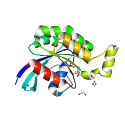
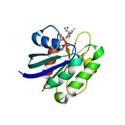
2OXY
 | | Protein kinase CK2 in complex with tetrabromobenzoimidazole derivatives K17, K22 and K32 | | 分子名称: | 4,5,6,7-TETRABROMO-BENZIMIDAZOLE, Casein kinase II subunit alpha | | 著者 | Battistutta, R, Zanotti, G, Cendron, L. | | 登録日 | 2007-02-21 | | 公開日 | 2007-09-25 | | 最終更新日 | 2024-02-21 | | 実験手法 | X-RAY DIFFRACTION (1.812 Å) | | 主引用文献 | The ATP-Binding Site of Protein Kinase CK2 Holds a Positive Electrostatic Area and Conserved Water Molecules.
Chembiochem, 8, 2007
|
|
4LFI
 | | Crystal structure of scCK2 alpha in complex with GMPPNP | | 分子名称: | Casein kinase II subunit alpha, GLYCEROL, MANGANESE (II) ION, ... | | 著者 | Liu, H. | | 登録日 | 2013-06-27 | | 公開日 | 2014-03-05 | | 最終更新日 | 2024-03-20 | | 実験手法 | X-RAY DIFFRACTION (1.85 Å) | | 主引用文献 | The multiple nucleotide-divalent cation binding modes of Saccharomyces cerevisiae CK2 alpha indicate a possible co-substrate hydrolysis product (ADP/GDP) release pathway.
Acta Crystallogr.,Sect.D, 70, 2014
|
|
2GCN
 | |
2G78
 | |
2G7I
 | |
2P3G
 | | Crystal structure of a pyrrolopyridine inhibitor bound to MAPKAP Kinase-2 | | 分子名称: | 2-[2-(2-FLUOROPHENYL)PYRIDIN-4-YL]-1,5,6,7-TETRAHYDRO-4H-PYRROLO[3,2-C]PYRIDIN-4-ONE, MAP kinase-activated protein kinase 2 | | 著者 | Kurumbail, R.G, Caspers, N. | | 登録日 | 2007-03-08 | | 公開日 | 2007-06-12 | | 最終更新日 | 2024-04-03 | | 実験手法 | X-RAY DIFFRACTION (3.8 Å) | | 主引用文献 | Pyrrolopyridine Inhibitors of Mitogen-Activated Protein Kinase-Activated Protein Kinase 2 (MK-2).
J.Med.Chem., 50, 2007
|
|
2P42
 | | Complex of a camelid single-domain vhh antibody fragment with RNASE A at 1.8A resolution: SE3-mono-2 crystal form with three se-met sites (M34, M51, M83) in vhh scaffold | | 分子名称: | ANTIBODY CAB-RN05, MAGNESIUM ION, Ribonuclease pancreatic | | 著者 | Tereshko, V, Uysal, S, Koide, A, Margalef, K, Koide, S, Kossiakoff, A.A. | | 登録日 | 2007-03-11 | | 公開日 | 2008-03-11 | | 最終更新日 | 2011-07-13 | | 実験手法 | X-RAY DIFFRACTION (1.8 Å) | | 主引用文献 | Toward chaperone-assisted crystallography: protein engineering enhancement of crystal packing and X-ray phasing capabilities of a camelid single-domain antibody (VHH) scaffold
Protein Sci., 17, 2008
|
|
2J9K
 | | Atomic-resolution Crystal Structure of Chemically-Synthesized HIV-1 Protease Complexed with Inhibitor MVT-101 | | 分子名称: | ACETATE ION, GLYCEROL, N-{(2S)-2-[(N-acetyl-L-threonyl-L-isoleucyl)amino]hexyl}-L-norleucyl-L-glutaminyl-N~5~-[amino(iminio)methyl]-L-ornithinamide, ... | | 著者 | Malito, E, Shen, Y, Johnson, E.C.B, Tang, W.J. | | 登録日 | 2006-11-11 | | 公開日 | 2007-08-28 | | 最終更新日 | 2023-12-13 | | 実験手法 | X-RAY DIFFRACTION (1.2 Å) | | 主引用文献 | Insights from Atomic-Resolution X-Ray Structures of Chemically Synthesized HIV-1 Protease in Complex with Inhibitors.
J.Mol.Biol., 373, 2007
|
|
2J9M
 | | Crystal Structure of CDK2 in complex with Macrocyclic Aminopyrimidine | | 分子名称: | 6-BROMO-13-THIA-2,4,8,12,19-PENTAAZATRICYCLO[12.3.1.1~3,7~]NONADECA-1(18),3(19),4,6,14,16-HEXAENE 13,13-DIOXIDE, CELL DIVISION PROTEIN KINASE 2 | | 著者 | Schaefer, M, Luecking, U, Siemeister, G, Briem, H, Krueger, M, Lienau, P, Jautelat, R. | | 登録日 | 2006-11-13 | | 公開日 | 2007-11-06 | | 最終更新日 | 2023-12-13 | | 実験手法 | X-RAY DIFFRACTION (2.5 Å) | | 主引用文献 | Macrocyclic Aminopyrimidines as Multitarget Cdk and Vegf-R Inhibitors with Potent Antiproliferative Activities.
Chemmedchem, 2, 2007
|
|
2J9N
 | | Robotically harvested Trypsin complexed with Benzamidine containing polypeptide mediated crystal contacts | | 分子名称: | BENZAMIDINE, CALCIUM ION, CATIONIC TRYPSIN, ... | | 著者 | Viola, R, Carman, P, Walsh, J, Miller, E, Benning, M, Frankel, D, McPherson, A, Cudney, R, Rupp, B. | | 登録日 | 2006-11-14 | | 公開日 | 2007-04-03 | | 最終更新日 | 2023-12-13 | | 実験手法 | X-RAY DIFFRACTION (1.5 Å) | | 主引用文献 | Operator Assisted Harvesting of Protein Crystals Using a Universal Micromanipulation Robot.
J.Appl.Crystallogr., 40, 2007
|
|
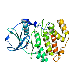
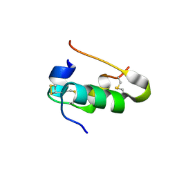
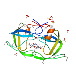
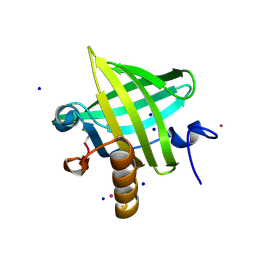
2OX8
 | | Human Scavenger Receptor C-type Lectin carbohydrate-recognition domain. | | 分子名称: | CALCIUM ION, CHLORIDE ION, Scavenger receptor with C-type lectin type I, ... | | 著者 | Weis, W.I, Feinberg, H, Drickamer, K, Taylor, M.E. | | 登録日 | 2007-02-20 | | 公開日 | 2007-04-03 | | 最終更新日 | 2011-07-13 | | 実験手法 | X-RAY DIFFRACTION (2.5 Å) | | 主引用文献 | Scavenger receptor C-type lectin binds to the leukocyte cell surface glycan Lewis(x) by a novel mechanism.
J.Biol.Chem., 282, 2007
|
|
4KKG
 | |
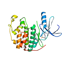
2OZQ
 | | Crystal Structure of apo-MUP | | 分子名称: | CADMIUM ION, Novel member of the major urinary protein (Mup) gene family, SODIUM ION | | 著者 | Dennis, C.A, Homans, S.W, Phillips, S.E.V, Syme, N.R. | | 登録日 | 2007-02-27 | | 公開日 | 2008-01-15 | | 最終更新日 | 2023-08-30 | | 実験手法 | X-RAY DIFFRACTION (1.8 Å) | | 主引用文献 | Origin of heat capacity changes in a "nonclassical" hydrophobic interaction.
Chembiochem, 8, 2007
|
|
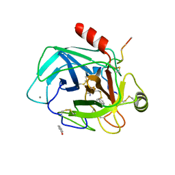
2J5X
 | | STRUCTURE OF THE SMALL G PROTEIN ARF6 IN COMPLEX WITH GTPGAMMAS | | 分子名称: | 5'-GUANOSINE-DIPHOSPHATE-MONOTHIOPHOSPHATE, ADP-RIBOSYLATION FACTOR 6, MAGNESIUM ION | | 著者 | Pasqualato, S, Menetrey, J, Franco, M, Cherfils, J. | | 登録日 | 2006-09-20 | | 公開日 | 2006-09-21 | | 最終更新日 | 2024-05-01 | | 実験手法 | X-RAY DIFFRACTION (2.8 Å) | | 主引用文献 | The Structural Gdp-GTP Cycle of Human Arf6.
Embo Rep., 2, 2001
|
|


