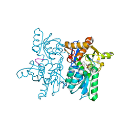
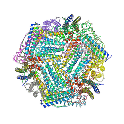
4NU8
 
 | |
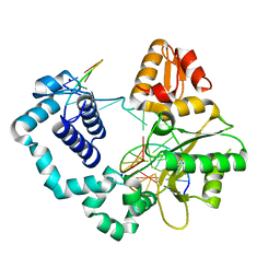
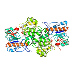
4O5C
 | |
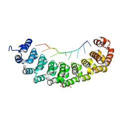
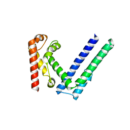
3BX2
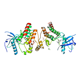
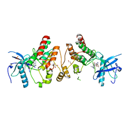
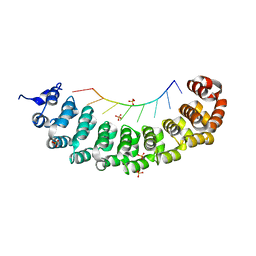
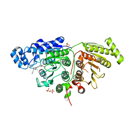
 | | Puf4 RNA binding domain bound to HO endonuclease RNA 3' UTR recognition sequence | | 分子名称: | HO endonuclease 3' UTR binding sequence, Protein PUF4, SODIUM ION, ... | | 著者 | Miller, M.T, Higgin, J.J, Hall, T.M.T. | | 登録日 | 2008-01-11 | | 公開日 | 2008-03-11 | | 最終更新日 | 2024-02-21 | | 実験手法 | X-RAY DIFFRACTION (2.84 Å) | | 主引用文献 | Basis of altered RNA-binding specificity by PUF proteins revealed by crystal structures of yeast Puf4p
Nat.Struct.Mol.Biol., 15, 2008
|
|
4F63
 | | Crystal structure of Human Fibroblast Growth Factor Receptor 1 Kinase domain in complex with compound 1 | | 分子名称: | 1,2-ETHANEDIOL, 5-bromo-N~4~-(3-methyl-1H-pyrazol-5-yl)-N~2~-[2-(pyridin-3-yl)ethyl]pyrimidine-2,4-diamine, Fibroblast growth factor receptor 1 | | 著者 | Norman, R.A, Breed, J, Ogg, D. | | 登録日 | 2012-05-14 | | 公開日 | 2012-06-06 | | 最終更新日 | 2023-09-13 | | 実験手法 | X-RAY DIFFRACTION (2.55 Å) | | 主引用文献 | Protein-Ligand Crystal Structures Can Guide the Design of Selective Inhibitors of the FGFR Tyrosine Kinase.
J.Med.Chem., 55, 2012
|
|
4F64
 | | Crystal structure of Human Fibroblast Growth Factor Receptor 1 Kinase domain in complex with compound 6 | | 分子名称: | 1,2-ETHANEDIOL, 5-bromo-N~4~-[3-(3-methoxypropyl)-1H-pyrazol-5-yl]-N~2~-[(3-methyl-1,2-oxazol-5-yl)methyl]pyrimidine-2,4-diamine, Fibroblast growth factor receptor 1, ... | | 著者 | Norman, R.A, Breed, J, Ogg, D. | | 登録日 | 2012-05-14 | | 公開日 | 2012-06-06 | | 最終更新日 | 2023-09-13 | | 実験手法 | X-RAY DIFFRACTION (2.05 Å) | | 主引用文献 | Protein-Ligand Crystal Structures Can Guide the Design of Selective Inhibitors of the FGFR Tyrosine Kinase.
J.Med.Chem., 55, 2012
|
|
4NXB
 | | Crystal structure of iLOV-I486(2LT) at pH 7.0 | | 分子名称: | FLAVIN MONONUCLEOTIDE, Phototropin-2 | | 著者 | Wang, J, Li, J, Liu, X. | | 登録日 | 2013-12-09 | | 公開日 | 2014-09-24 | | 最終更新日 | 2023-11-08 | | 実験手法 | X-RAY DIFFRACTION (2.561 Å) | | 主引用文献 | Significant expansion of fluorescent protein sensing ability through the genetic incorporation of superior photo-induced electron-transfer quenchers.
J.Am.Chem.Soc., 136, 2014
|
|
4NXL
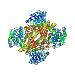 | | Dibenzothiophene monooxygenase (DszC) from Rhodococcus erythropolis | | 分子名称: | DszC | | 著者 | Zhang, L, Duan, X, Li, X, Rao, Z. | | 登録日 | 2013-12-09 | | 公開日 | 2014-07-23 | | 最終更新日 | 2024-02-28 | | 実験手法 | X-RAY DIFFRACTION (2.3 Å) | | 主引用文献 | Structural insights into the stabilization of active, tetrameric DszC by its C-terminus.
Proteins, 82, 2014
|
|
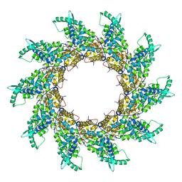
4FAZ
 | |
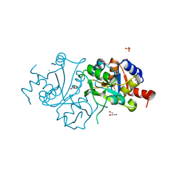
4O21
 | | Product complex of metal-free PKAc, ATP-gamma-S and SP20. | | 分子名称: | ADENOSINE-5'-DIPHOSPHATE, Thio-phosphorylated peptide pSP20, cAMP-dependent protein kinase catalytic subunit alpha | | 著者 | Das, A, Kovalevsky, A.Y, Gerlits, O, Langan, P, Heller, W.T, Keshwani, M, Taylor, S. | | 登録日 | 2013-12-16 | | 公開日 | 2014-05-28 | | 実験手法 | X-RAY DIFFRACTION (1.95 Å) | | 主引用文献 | Metal-Free cAMP-Dependent Protein Kinase Can Catalyze Phosphoryl Transfer.
Biochemistry, 53, 2014
|
|
3BX3
 | |
3BZK
 | |
3GV6
 | | Crystal Structure of human chromobox homolog 6 (CBX6) with H3K9 peptide | | 分子名称: | Chromobox protein homolog 6, Histone H3K9me3 peptide | | 著者 | Dong, A, Amaya, M.F, Li, Z, Loppnau, P, Kozieradzki, I, Edwards, A.M, Arrowsmith, C.H, Weigelt, J, Bountra, C, Bochkarev, A, Min, J, Ouyang, H, Structural Genomics Consortium (SGC) | | 登録日 | 2009-03-30 | | 公開日 | 2009-04-21 | | 最終更新日 | 2023-09-06 | | 実験手法 | X-RAY DIFFRACTION (1.76 Å) | | 主引用文献 | Recognition and specificity determinants of the human cbx chromodomains.
J.Biol.Chem., 286, 2011
|
|
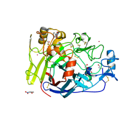
4O72
 | | Crystal structure of the first bromodomain of human BRD4 in complex with NU7441 | | 分子名称: | 1,2-ETHANEDIOL, 8-(dibenzo[b,d]thiophen-4-yl)-2-(morpholin-4-yl)-4H-chromen-4-one, Bromodomain-containing protein 4, ... | | 著者 | Zhu, J.-Y, Ember, S.W, Watts, C, Schonbrunn, E. | | 登録日 | 2013-12-24 | | 公開日 | 2014-03-05 | | 最終更新日 | 2023-09-20 | | 実験手法 | X-RAY DIFFRACTION (1.4 Å) | | 主引用文献 | Acetyl-lysine Binding Site of Bromodomain-Containing Protein 4 (BRD4) Interacts with Diverse Kinase Inhibitors.
Acs Chem.Biol., 9, 2014
|
|
4FFJ
 | |
4F0K
 | |
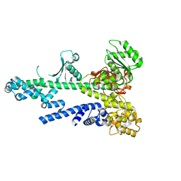
4OI6
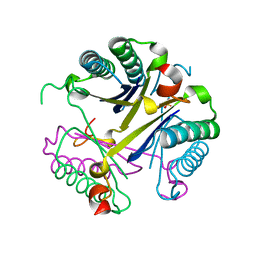
 | | Crystal structure analysis of nickel-bound form SCO4226 from Streptomyces coelicolor A3(2) | | 分子名称: | CITRIC ACID, NICKEL (II) ION, Nickel responsive protein | | 著者 | Lu, M, Jiang, Y.L, Wang, S, Cheng, W, Zhang, R.G, Virolle, M.J, Chen, Y, Zhou, C.Z. | | 登録日 | 2014-01-18 | | 公開日 | 2014-09-10 | | 最終更新日 | 2018-01-24 | | 実験手法 | X-RAY DIFFRACTION (2.04 Å) | | 主引用文献 | Streptomyces coelicolor SCO4226 Is a Nickel Binding Protein.
Plos One, 9, 2014
|
|
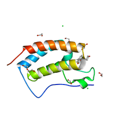
4FQO
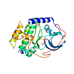
 | | Crystal Structure of Calcium-Loaded S100B Bound to SBi4211 | | 分子名称: | 4,4'-[heptane-1,7-diylbis(oxy)]dibenzenecarboximidamide, CALCIUM ION, Protein S100-B | | 著者 | McKnight, L.E, Raman, E.P, Bezawada, P, Kudrimoti, S, Wilder, P.T, Hartman, K.G, Toth, E.A, Coop, A, MacKerrell, A.D, Weber, D.J. | | 登録日 | 2012-06-25 | | 公開日 | 2012-10-17 | | 最終更新日 | 2024-03-13 | | 実験手法 | X-RAY DIFFRACTION (1.65 Å) | | 主引用文献 | Structure-Based Discovery of a Novel Pentamidine-Related Inhibitor of the Calcium-Binding Protein S100B.
ACS Med Chem Lett, 3, 2012
|
|
4V0Z
 | |
4V1W
 | |
7Y74
 | |
7YA3
 | |
6X1H
 | |
4V5V
 | | Structure of respiratory syncytial virus nucleocapsid protein, P1 crystal form | | 分子名称: | RESPIRATORY SYNCYTIAL VIRUS NUCLEOCAPSID PROTEIN, RNA | | 著者 | El Omari, K, Dhaliwal, B, Ren, J, Abrescia, N.G.A, Lockyer, M, Powell, K.L, Hawkins, A.R, Stammers, D.K. | | 登録日 | 2011-05-04 | | 公開日 | 2014-07-09 | | 最終更新日 | 2024-01-10 | | 実験手法 | X-RAY DIFFRACTION (3.6 Å) | | 主引用文献 | Structures of Respiratory Syncytial Virus Nucleocapsid Protein from Two Crystal Forms: Details of Potential Packing Interactions in the Native Helical Form.
Acta Crystallogr.,Sect.F, 67, 2011
|
|
6X2D
 | | Crystal Structure of DNase I Domain of Ribonuclease E from Vibrio cholerae | | 分子名称: | IODIDE ION, Ribonuclease E | | 著者 | Minasov, G, Shuvalova, L, Kiryukhina, O, Dubrovska, I, Wiersum, G, Satchell, K.J.F, Center for Structural Genomics of Infectious Diseases (CSGID) | | 登録日 | 2020-05-20 | | 公開日 | 2020-06-10 | | 最終更新日 | 2023-11-15 | | 実験手法 | X-RAY DIFFRACTION (1.85 Å) | | 主引用文献 | Crystal Structure of DNase I Domain of Ribonuclease E from Vibrio cholerae.
To Be Published
|
|
6WDW
 | |


