6DAR
 
 | | Discovery of Potent 2-Aryl-6,7-Dihydro-5HPyrrolo[ 1,2-a]imidazoles as WDR5 WIN-site Inhibitors Using Fragment-Based Methods and Structure-Based Design | | 分子名称: | DIMETHYL SULFOXIDE, N-(cyclopropylmethyl)-N-{[3-(6,7-dihydro-5H-pyrrolo[1,2-a]imidazol-2-yl)phenyl]methyl}-3-methoxybenzamide, SULFATE ION, ... | | 著者 | Phan, J, Fesik, S.W. | | 登録日 | 2018-05-01 | | 公開日 | 2018-09-05 | | 最終更新日 | 2023-10-04 | | 実験手法 | X-RAY DIFFRACTION (1.88 Å) | | 主引用文献 | Discovery of Potent 2-Aryl-6,7-dihydro-5 H-pyrrolo[1,2- a]imidazoles as WDR5-WIN-Site Inhibitors Using Fragment-Based Methods and Structure-Based Design.
J. Med. Chem., 61, 2018
|
|
3O71
 | | Crystal structure of ERK2/DCC peptide complex | | 分子名称: | Mitogen-activated protein kinase 1, Peptide of Deleted in Colorectal Cancer, THIOCYANATE ION | | 著者 | Ma, W.F, Shang, Y, Wei, Z.Y, Wen, W.Y, Wang, W.N, Zhang, M.J. | | 登録日 | 2010-07-30 | | 公開日 | 2011-06-15 | | 最終更新日 | 2024-03-20 | | 実験手法 | X-RAY DIFFRACTION (1.95 Å) | | 主引用文献 | Phosphorylation of DCC by ERK2 is facilitated by direct docking of the receptor P1 domain to the kinase
Structure, 18, 2010
|
|
2IFQ
 | |
6DAS
 | | Discovery of Potent 2-Aryl-6,7-Dihydro-5HPyrrolo[ 1,2-a]imidazoles as WDR5 WIN-site Inhibitors Using Fragment-Based Methods and Structure-Based Design | | 分子名称: | N-[(1R)-6-(6,7-dihydro-5H-pyrrolo[1,2-a]imidazol-2-yl)-2,3-dihydro-1H-inden-1-yl]-3-methoxy-4-methylbenzamide, WD repeat-containing protein 5 | | 著者 | Phan, J, Fesik, S.W. | | 登録日 | 2018-05-01 | | 公開日 | 2018-07-18 | | 最終更新日 | 2023-10-04 | | 実験手法 | X-RAY DIFFRACTION (1.8 Å) | | 主引用文献 | Discovery of Potent 2-Aryl-6,7-dihydro-5 H-pyrrolo[1,2- a]imidazoles as WDR5-WIN-Site Inhibitors Using Fragment-Based Methods and Structure-Based Design.
J. Med. Chem., 61, 2018
|
|
1Y0J
 | | Zinc fingers as protein recognition motifs: structural basis for the GATA-1/Friend of GATA interaction | | 分子名称: | Erythroid transcription factor, ZINC ION, Zinc-finger protein ush | | 著者 | Liew, C.K, Simpson, R.J.Y, Kwan, A.H.Y, Crofts, L.A, Loughlin, F.E, Matthews, J.M, Crossley, M, Mackay, J.P. | | 登録日 | 2004-11-15 | | 公開日 | 2005-01-25 | | 最終更新日 | 2024-05-29 | | 実験手法 | SOLUTION NMR | | 主引用文献 | Zinc fingers as protein recognition motifs: Structural basis for the GATA-1/Friend of GATA interaction
Proc.Natl.Acad.Sci.Usa, 102, 2005
|
|
6AE3
 | | Crystal structure of GSK3beta complexed with Morin | | 分子名称: | 2-[2,4-bis(oxidanyl)phenyl]-3,5,7-tris(oxidanyl)chromen-4-one, GLYCEROL, Glycogen synthase kinase-3 beta | | 著者 | Kim, K.L, Cha, J.S, Kim, J.S, Ahn, J.S, Ha, N.C, Cho, H.S. | | 登録日 | 2018-08-03 | | 公開日 | 2018-09-19 | | 最終更新日 | 2018-10-03 | | 実験手法 | X-RAY DIFFRACTION (2.14 Å) | | 主引用文献 | Crystal structure of GSK3 beta in complex with the flavonoid, morin
Biochem. Biophys. Res. Commun., 504, 2018
|
|
4BTE
 | | DJ-1 Cu(I) complex | | 分子名称: | COPPER (I) ION, PROTEIN DJ-1 | | 著者 | Puno, M.R.A, Odell, M, Moody, P.C.E. | | 登録日 | 2013-06-14 | | 公開日 | 2013-11-06 | | 最終更新日 | 2023-12-20 | | 実験手法 | X-RAY DIFFRACTION (1.38 Å) | | 主引用文献 | Structure of Cu(I)-Bound Dj-1 Reveals a Biscysteinate Metal Binding Site at the Homodimer Interface: Insights Into Mutational Inactivation of Dj-1 in Parkinsonism.
J.Am.Chem.Soc., 135, 2013
|
|
6KX3
 | | Crystal structure of RhoA protein with covalent inhibitor DC-Rhoin | | 分子名称: | GUANOSINE-5'-DIPHOSPHATE, Transforming protein RhoA, prop-2-enyl (3R)-1,1-bis(oxidanylidene)-2,3-dihydro-1-benzothiophene-3-carboxylate | | 著者 | Zhang, H, Luo, C. | | 登録日 | 2019-09-09 | | 公開日 | 2020-08-19 | | 最終更新日 | 2023-11-22 | | 実験手法 | X-RAY DIFFRACTION (1.981 Å) | | 主引用文献 | Covalent Inhibitors Allosterically Block the Activation of Rho Family Proteins and Suppress Cancer Cell Invasion.
Adv Sci, 7, 2020
|
|
6KX2
 | | Crystal structure of GDP bound RhoA protein | | 分子名称: | GUANOSINE-5'-DIPHOSPHATE, Transforming protein RhoA | | 著者 | Zhang, H, Luo, C. | | 登録日 | 2019-09-09 | | 公開日 | 2020-08-19 | | 最終更新日 | 2023-11-22 | | 実験手法 | X-RAY DIFFRACTION (1.454 Å) | | 主引用文献 | Covalent Inhibitors Allosterically Block the Activation of Rho Family Proteins and Suppress Cancer Cell Invasion.
Adv Sci, 7, 2020
|
|
1V9U
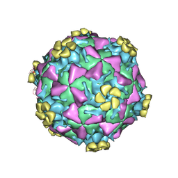 | | Human Rhinovirus 2 bound to a fragment of its cellular receptor protein | | 分子名称: | CALCIUM ION, Coat protein VP1, Coat protein VP2, ... | | 著者 | Verdaguer, N, Fita, I, Reithmayer, M, Moser, R, Blaas, D. | | 登録日 | 2004-02-03 | | 公開日 | 2004-05-04 | | 最終更新日 | 2023-10-25 | | 実験手法 | X-RAY DIFFRACTION (3.6 Å) | | 主引用文献 | X-ray structure of a minor group human rhinovirus bound to a fragment of its cellular receptor protein
NAT.STRUCT.MOL.BIOL., 11, 2004
|
|
4RM8
 | | Crystal structure of human ezrin in space group P21 | | 分子名称: | Ezrin | | 著者 | Phang, J.M, Harrop, S.J, Davies, R, Duff, A.P, Wilk, K.E, Curmi, P.M.G. | | 登録日 | 2014-10-20 | | 公開日 | 2015-12-09 | | 最終更新日 | 2023-09-20 | | 実験手法 | X-RAY DIFFRACTION (1.9 Å) | | 主引用文献 | Structural characterization suggests models for monomeric and dimeric forms of full-length ezrin.
Biochem. J., 473, 2016
|
|
4RM9
 | | Crystal structure of human ezrin in space group C2221 | | 分子名称: | Ezrin | | 著者 | Phang, J.M, Harrop, S.J, Davies, R, Duff, A.P, Wilk, K.E, Curmi, P.M.G. | | 登録日 | 2014-10-21 | | 公開日 | 2015-12-09 | | 最終更新日 | 2023-09-20 | | 実験手法 | X-RAY DIFFRACTION (2 Å) | | 主引用文献 | Structural characterization suggests models for monomeric and dimeric forms of full-length ezrin.
Biochem. J., 473, 2016
|
|
2HSH
 | |
2YPA
 | | Structure of the SCL:E47:LMO2:LDB1 complex bound to DNA | | 分子名称: | EBOX FORWARD, EBOX REVERSE, LIM DOMAIN-BINDING PROTEIN 1, ... | | 著者 | El Omari, K, Hoosdally, S.J, Tuladhar, K, Karia, D, Ponsele, E, Platonova, O, Vyas, P, Patient, R, Porcher, C, Mancini, E.J. | | 登録日 | 2012-10-30 | | 公開日 | 2013-07-31 | | 最終更新日 | 2024-05-08 | | 実験手法 | X-RAY DIFFRACTION (2.8 Å) | | 主引用文献 | Structural Basis for Lmo2-Driven Recruitment of the Scl:E47bHLH Heterodimer to Hematopoietic-Specific Transcriptional Targets.
Cell Rep., 4, 2013
|
|
1BH8
 | | HTAFII18/HTAFII28 HETERODIMER CRYSTAL STRUCTURE | | 分子名称: | TAFII18, TAFII28 | | 著者 | Birck, C, Poch, O, Romier, C, Ruff, M, Mengus, G, Lavigne, A.-C, Davidson, I, Moras, D. | | 登録日 | 1998-06-16 | | 公開日 | 1999-06-22 | | 最終更新日 | 2024-02-07 | | 実験手法 | X-RAY DIFFRACTION (3 Å) | | 主引用文献 | Human TAF(II)28 and TAF(II)18 interact through a histone fold encoded by atypical evolutionary conserved motifs also found in the SPT3 family.
Cell(Cambridge,Mass.), 94, 1998
|
|
2YMN
 | | Organization of the Influenza Virus Replication Machinery | | 分子名称: | NUCLEOPROTEIN | | 著者 | Moeller, A, Kirchdoerfer, R.N, Potter, C.S, Carragher, B, Wilson, I.A. | | 登録日 | 2012-10-09 | | 公開日 | 2012-12-05 | | 最終更新日 | 2024-05-08 | | 実験手法 | ELECTRON MICROSCOPY (20 Å) | | 主引用文献 | Organization of the Influenza Virus Replication Machinery.
Science, 338, 2012
|
|
1BH9
 | | HTAFII18/HTAFII28 HETERODIMER CRYSTAL STRUCTURE WITH BOUND PCMBS | | 分子名称: | PARA-MERCURY-BENZENESULFONIC ACID, TAFII18, TAFII28 | | 著者 | Birck, C, Poch, O, Romier, C, Ruff, M, Mengus, G, Lavigne, A.-C, Davidson, I, Moras, D. | | 登録日 | 1998-06-16 | | 公開日 | 1999-06-22 | | 最終更新日 | 2024-02-07 | | 実験手法 | X-RAY DIFFRACTION (2.6 Å) | | 主引用文献 | Human TAF(II)28 and TAF(II)18 interact through a histone fold encoded by atypical evolutionary conserved motifs also found in the SPT3 family.
Cell(Cambridge,Mass.), 94, 1998
|
|
3D23
 | | Main protease of HCoV-HKU1 | | 分子名称: | 3C-like proteinase, N-[(5-METHYLISOXAZOL-3-YL)CARBONYL]ALANYL-L-VALYL-N~1~-((1R,2Z)-4-(BENZYLOXY)-4-OXO-1-{[(3R)-2-OXOPYRROLIDIN-3-YL]METHYL}BUT-2-ENYL)-L-LEUCINAMIDE | | 著者 | Zhao, Q, Chen, C, Li, S, Zou, Y. | | 登録日 | 2008-05-07 | | 公開日 | 2008-09-16 | | 最終更新日 | 2023-11-15 | | 実験手法 | X-RAY DIFFRACTION (2.5 Å) | | 主引用文献 | Structure of the main protease from a global infectious human coronavirus, HCoV-HKU1.
J.Virol., 82, 2008
|
|
6RT0
 | | cryo-em structure of alpha-synuclein fibril polymorph 2A | | 分子名称: | Alpha-synuclein | | 著者 | Guerrero-Ferreira, R, Taylor, N.M.I, Arteni, A.A, Melki, R, Meier, B.H, Bockmann, A, Bousset, L, Stahlberg, H. | | 登録日 | 2019-05-22 | | 公開日 | 2019-06-26 | | 最終更新日 | 2024-05-22 | | 実験手法 | ELECTRON MICROSCOPY (3.1 Å) | | 主引用文献 | Two new polymorphic structures of human full-length alpha-synuclein fibrils solved by cryo-electron microscopy.
Elife, 8, 2019
|
|
6UPL
 | | Structure of FACT_subnucleosome complex 2 | | 分子名称: | DNA (79-mer), FACT complex subunit SPT16, FACT complex subunit SSRP1, ... | | 著者 | Zhou, K, Tan, Y.Z, Wei, H, Liu, Y, Carragher, B, Potter, C, Luger, K. | | 登録日 | 2019-10-17 | | 公開日 | 2019-12-11 | | 最終更新日 | 2024-03-20 | | 実験手法 | ELECTRON MICROSCOPY (7.4 Å) | | 主引用文献 | FACT caught in the act of manipulating the nucleosome.
Nature, 577, 2020
|
|
6UPK
 | | Structure of FACT_subnucleosome complex 1 | | 分子名称: | DNA (79-mer), FACT complex subunit SPT16, FACT complex subunit SSRP1, ... | | 著者 | Zhou, K, Tan, Y.Z, Wei, H, Liu, Y, Carragher, B, Potter, C, Luger, K. | | 登録日 | 2019-10-17 | | 公開日 | 2019-12-11 | | 最終更新日 | 2024-03-20 | | 実験手法 | ELECTRON MICROSCOPY (4.9 Å) | | 主引用文献 | FACT caught in the act of manipulating the nucleosome.
Nature, 577, 2020
|
|
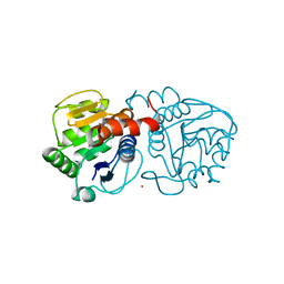
2HRV
 | | 2A CYSTEINE PROTEINASE FROM HUMAN RHINOVIRUS 2 | | 分子名称: | 2A CYSTEINE PROTEINASE, ZINC ION | | 著者 | Petersen, J.F.W, Cherney, M.M, Liebig, H.-D, Skern, T, Kuechler, E, James, M.N.G. | | 登録日 | 1999-04-29 | | 公開日 | 2000-05-03 | | 最終更新日 | 2023-12-27 | | 実験手法 | X-RAY DIFFRACTION (1.95 Å) | | 主引用文献 | The structure of the 2A proteinase from a common cold virus: a proteinase responsible for the shut-off of host-cell protein synthesis.
EMBO J., 18, 1999
|
|
3C6D
 | |
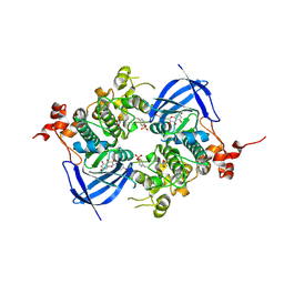
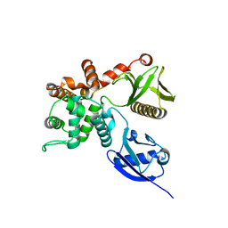
3VW9
 | | Human Glyoxalase I with an N-hydroxypyridone inhibitor | | 分子名称: | 1-hydroxy-6-[1-(3-methoxypropyl)-1H-pyrrolo[2,3-b]pyridin-5-yl]-4-phenylpyridin-2(1H)-one, 4-(2-HYDROXYETHYL)-1-PIPERAZINE ETHANESULFONIC ACID, Lactoylglutathione lyase, ... | | 著者 | Fukami, T.A, Irie, M, Matsuura, T. | | 登録日 | 2012-08-10 | | 公開日 | 2012-12-12 | | 最終更新日 | 2023-11-08 | | 実験手法 | X-RAY DIFFRACTION (1.47 Å) | | 主引用文献 | Design and evaluation of azaindole-substituted N-hydroxypyridones as glyoxalase I inhibitors
Bioorg.Med.Chem.Lett., 22, 2012
|
|
1LB1
 | | Crystal Structure of the Dbl and Pleckstrin homology domains of Dbs in complex with RhoA | | 分子名称: | Guanine nucleotide exchange factor DBS, Transforming protein RhoA | | 著者 | Snyder, J.T, Worthylake, D.K, Rossman, K.L, Betts, L, Pruitt, W.M, Siderovski, D.P, Der, C.J, Sondek, J. | | 登録日 | 2002-04-01 | | 公開日 | 2002-05-29 | | 最終更新日 | 2023-08-16 | | 実験手法 | X-RAY DIFFRACTION (2.81 Å) | | 主引用文献 | Structural basis for the selective activation of Rho GTPases by Dbl exchange factors.
Nat.Struct.Biol., 9, 2002
|
|


