3ZUJ
 
 | |
7V80
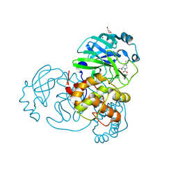
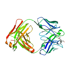
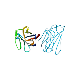
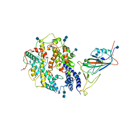 | | Local refinement of SARS-CoV-2 S-Beta variant (B.1.351) RBD and Angiotensin-converting enzyme 2 (ACE2) ectodomain | | 分子名称: | 2-acetamido-2-deoxy-beta-D-glucopyranose, 2-acetamido-2-deoxy-beta-D-glucopyranose-(1-4)-2-acetamido-2-deoxy-beta-D-glucopyranose, Angiotensin-converting enzyme 2,Green fluorescent protein, ... | | 著者 | Yang, T.J, Yu, P.Y, Chang, Y.C, Hsu, S.T.D. | | 登録日 | 2021-08-22 | | 公開日 | 2021-10-06 | | 実験手法 | ELECTRON MICROSCOPY (3.9 Å) | | 主引用文献 | Local refinement of SARS-CoV-2 S-Beta variant (B.1.351) RBD and Angiotensin-converting enzyme 2 (ACE2) ectodomain
To Be Published
|
|
6DGV
 | |
6F2W
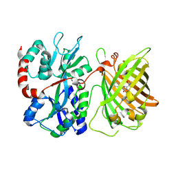
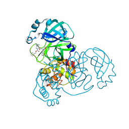
 | | Bacterial asc transporter crystal structure in open to in conformation | | 分子名称: | ALPHA-AMINOISOBUTYRIC ACID, Nanobody 74, Putative amino acid/polyamine transport protein, ... | | 著者 | Fort, J, Errasti-Murugarren, E, Carpena, X, Palacin, M, Fita, I. | | 登録日 | 2017-11-27 | | 公開日 | 2019-04-24 | | 最終更新日 | 2024-01-17 | | 実験手法 | X-RAY DIFFRACTION (3.4 Å) | | 主引用文献 | L amino acid transporter structure and molecular bases for the asymmetry of substrate interaction.
Nat Commun, 10, 2019
|
|
1DV0
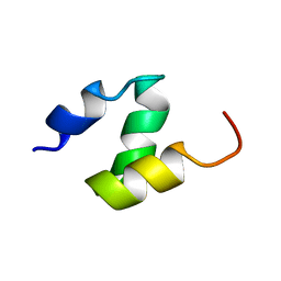 | |
1F4I
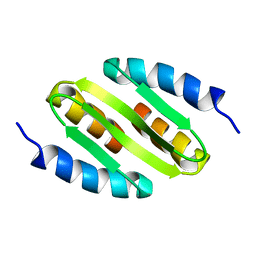
 | | SOLUTION STRUCTURE OF THE HHR23A UBA(2) MUTANT P333E, DEFICIENT IN BINDING THE HIV-1 ACCESSORY PROTEIN VPR | | 分子名称: | UV EXCISION REPAIR PROTEIN PROTEIN RAD23 HOMOLOG A | | 著者 | Withers-Ward, E.S, Mueller, T.D, Chen, I.S, Feigon, J. | | 登録日 | 2000-06-07 | | 公開日 | 2000-12-20 | | 最終更新日 | 2024-05-22 | | 実験手法 | SOLUTION NMR | | 主引用文献 | Biochemical and structural analysis of the interaction between the UBA(2) domain of the DNA repair protein HHR23A and HIV-1 Vpr.
Biochemistry, 39, 2000
|
|
2IOV
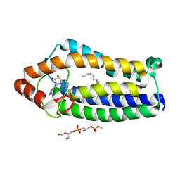
 | | Bright-state structure of the reversibly switchable fluorescent protein Dronpa | | 分子名称: | Fluorescent protein Dronpa | | 著者 | Stiel, A.C, Trowitzsch, S, Weber, G, Andresen, M, Eggeling, C, Hell, S.W, Jakobs, S, Wahl, M.C. | | 登録日 | 2006-10-11 | | 公開日 | 2006-12-05 | | 最終更新日 | 2023-11-15 | | 実験手法 | X-RAY DIFFRACTION (1.8 Å) | | 主引用文献 | 1.8 A bright-state structure of the reversibly switchable fluorescent protein Dronpa guides the generation of fast switching variants
Biochem.J., 402, 2007
|
|
7LYI
 | | Crystal structure of the SARS-CoV-2 (COVID-19) main protease in complex with inhibitor UAWJ9-36-3 | | 分子名称: | 3C-like proteinase, GLYCEROL, SODIUM ION, ... | | 著者 | Sacco, M, Wang, J, Chen, Y. | | 登録日 | 2021-03-07 | | 公開日 | 2021-03-17 | | 最終更新日 | 2023-10-18 | | 実験手法 | X-RAY DIFFRACTION (1.9 Å) | | 主引用文献 | Rational Design of Hybrid SARS-CoV-2 Main Protease Inhibitors Guided by the Superimposed Cocrystal Structures with the Peptidomimetic Inhibitors GC-376, Telaprevir, and Boceprevir.
Acs Pharmacol Transl Sci, 4, 2021
|
|
7LYH
 | | Crystal structure of the SARS-CoV-2 (COVID-19) main protease in complex with inhibitor UAWJ9-36-1 | | 分子名称: | 3C-like proteinase, GLYCEROL, benzyl (1S,3aR,6aS)-1-({(2S)-1-hydroxy-3-[(3S)-2-oxopyrrolidin-3-yl]propan-2-yl}carbamoyl)hexahydrocyclopenta[c]pyrrole-2(1H)-carboxylate | | 著者 | Sacco, M, Wang, J, Chen, Y. | | 登録日 | 2021-03-07 | | 公開日 | 2021-03-17 | | 最終更新日 | 2023-10-18 | | 実験手法 | X-RAY DIFFRACTION (1.9 Å) | | 主引用文献 | Rational Design of Hybrid SARS-CoV-2 Main Protease Inhibitors Guided by the Superimposed Cocrystal Structures with the Peptidomimetic Inhibitors GC-376, Telaprevir, and Boceprevir.
Acs Pharmacol Transl Sci, 4, 2021
|
|
3MMI
 | |
4A2N
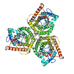
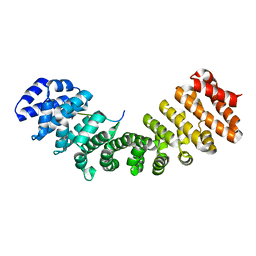
 | | Crystal Structure of Ma-ICMT | | 分子名称: | CARDIOLIPIN, ISOPRENYLCYSTEINE CARBOXYL METHYLTRANSFERASE, PALMITIC ACID, ... | | 著者 | Yang, J, Kulkarni, K, Manolaridis, I, Zhang, Z, Dodd, R.B, Mas-Droux, C, Barford, D. | | 登録日 | 2011-09-27 | | 公開日 | 2012-01-11 | | 最終更新日 | 2024-05-08 | | 実験手法 | X-RAY DIFFRACTION (3.4 Å) | | 主引用文献 | Mechanism of Isoprenylcysteine Carboxyl Methylation from the Crystal Structure of the Integral Membrane Methyltransferase Icmt.
Mol.Cell, 44, 2011
|
|
4AUR
 | |
3RA7
 | |
3RWA
 | |
8GW7
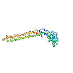
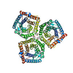 | | AtSLAC1 6D mutant in open state | | 分子名称: | CHLORIDE ION, CHOLESTEROL HEMISUCCINATE, Guard cell S-type anion channel SLAC1,Green fluorescent protein (Fragment) | | 著者 | Lee, Y, Lee, S. | | 登録日 | 2022-09-16 | | 公開日 | 2023-11-15 | | 最終更新日 | 2023-11-29 | | 実験手法 | ELECTRON MICROSCOPY (3.3 Å) | | 主引用文献 | Cryo-EM structures of the plant anion channel SLAC1 from Arabidopsis thaliana suggest a combined activation model.
Nat Commun, 14, 2023
|
|
8GW6
 | | AtSLAC1 6D mutant in closed state | | 分子名称: | CHLORIDE ION, CHOLESTEROL HEMISUCCINATE, Guard cell S-type anion channel SLAC1,Green fluorescent protein (Fragment) | | 著者 | Lee, Y, Lee, S. | | 登録日 | 2022-09-16 | | 公開日 | 2023-11-15 | | 最終更新日 | 2023-11-29 | | 実験手法 | ELECTRON MICROSCOPY (3.3 Å) | | 主引用文献 | Cryo-EM structures of the plant anion channel SLAC1 from Arabidopsis thaliana suggest a combined activation model.
Nat Commun, 14, 2023
|
|
5XG8
 | |
5XG7
 | | Galectin-13/Placental Protein 13 crystal structure | | 分子名称: | Galactoside-binding soluble lectin 13 | | 著者 | Su, J.Y, Wang, Y. | | 登録日 | 2017-04-12 | | 公開日 | 2018-01-31 | | 最終更新日 | 2024-04-17 | | 実験手法 | X-RAY DIFFRACTION (1.55 Å) | | 主引用文献 | Galectin-13, a different prototype galectin, does not bind beta-galacto-sides and forms dimers via intermolecular disulfide bridges between Cys-136 and Cys-138
Sci Rep, 8, 2018
|
|
5WTS
 | |
5Y03
 | |
5NOC
 | |
6MLT
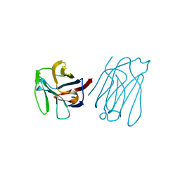
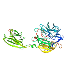 | | Crystal structure of the V. cholerae biofilm matrix protein Bap1 | | 分子名称: | CALCIUM ION, CITRATE ANION, GLYCEROL, ... | | 著者 | Kaus, K, Biester, A, Chupp, E, Lu, K, Vidsudharomn, C, Olson, R. | | 登録日 | 2018-09-28 | | 公開日 | 2019-08-28 | | 最終更新日 | 2024-03-13 | | 実験手法 | X-RAY DIFFRACTION (1.9 Å) | | 主引用文献 | The 1.9 angstrom crystal structure of the extracellular matrix protein Bap1 fromVibrio choleraeprovides insights into bacterial biofilm adhesion.
J.Biol.Chem., 294, 2019
|
|
8AAB
 | | S148F mutant of blue-to-red fluorescent timer mRubyFT | | 分子名称: | mRubyFT S148F mutant of blue-to-red fluorescent timer | | 著者 | Boyko, K.M, Nikolaeva, A.Y, Vlaskina, A.V, Dorovatovskii, P.V, Subach, O.M, Popov, V.O, Subach, F.V. | | 登録日 | 2022-06-30 | | 公開日 | 2022-08-17 | | 最終更新日 | 2024-02-07 | | 実験手法 | X-RAY DIFFRACTION (1.6 Å) | | 主引用文献 | mRubyFT/S147I, a mutant of blue-to-red fluorescent timer
Crystallography Reports, 2022
|
|
6OAM
 | | Crystal Structure of ChlaDUB2 DUB domain | | 分子名称: | Deubiquitinase and deneddylase Dub2, Ubiquitin | | 著者 | Hausman, J.M, Das, C. | | 登録日 | 2019-03-17 | | 公開日 | 2020-04-22 | | 最終更新日 | 2023-10-11 | | 実験手法 | X-RAY DIFFRACTION (2.503 Å) | | 主引用文献 | The Two Deubiquitinating Enzymes fromChlamydia trachomatisHave Distinct Ubiquitin Recognition Properties.
Biochemistry, 59, 2020
|
|
5HHG
 | |


