1ZQQ
 
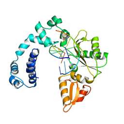 | |
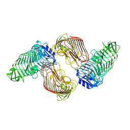
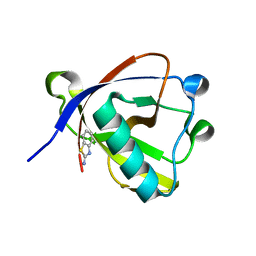
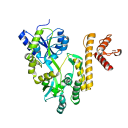
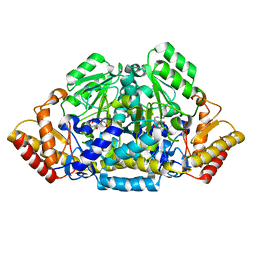
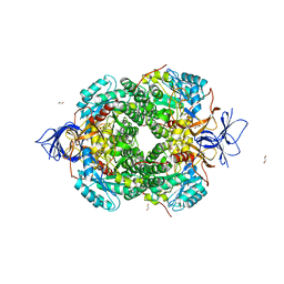
3FQ8
 | | M248I mutant of GSAM | | 分子名称: | 4'-DEOXY-4'-AMINOPYRIDOXAL-5'-PHOSPHATE, Glutamate-1-semialdehyde 2,1-aminomutase | | 著者 | Stetefeld, J. | | 登録日 | 2009-01-07 | | 公開日 | 2009-11-24 | | 最終更新日 | 2023-09-06 | | 実験手法 | X-RAY DIFFRACTION (2 Å) | | 主引用文献 | Absence of a catalytic water confers resistance to the neurotoxin gabaculine.
Faseb J., 24, 2010
|
|
8RF5
 | | Crystal structure of N-terminal SARS-CoV-2 nsp1 in complex with fragment hit 11A7 refined against the anomalous diffraction data | | 分子名称: | 6-fluoro-1,3-benzothiazol-2-amine, Host translation inhibitor nsp1 | | 著者 | Ma, S, Damfo, S, Mykhaylyk, V, Kozielski, F. | | 登録日 | 2023-12-12 | | 公開日 | 2024-06-19 | | 実験手法 | X-RAY DIFFRACTION (1.1 Å) | | 主引用文献 | High-confidence placement of low-occupancy fragments into electron density using the anomalous signal of sulfur and halogen atoms.
Acta Crystallogr D Struct Biol, 80, 2024
|
|
2WK9
 | | Structure of Plp_Thr aldimine form of Vibrio cholerae CqsA | | 分子名称: | CAI-1 AUTOINDUCER SYNTHASE, N-GLYCINE-[3-HYDROXY-2-METHYL-5-PHOSPHONOOXYMETHYL-PYRIDIN-4-YL-METHANE], PYRIDOXAL-5'-PHOSPHATE, ... | | 著者 | Jahan, N, Potter, J.A, Sheikh, M.A, Botting, C.H, Shirran, S.L, Westwood, N.J, Taylor, G.L. | | 登録日 | 2009-06-08 | | 公開日 | 2009-07-21 | | 最終更新日 | 2024-05-08 | | 実験手法 | X-RAY DIFFRACTION (1.9 Å) | | 主引用文献 | Insights Into the Biosynthesis of the Vibrio Cholerae Major Autoinducer Cai-1 from the Crystal Structure of the Plp-Dependent Enzyme Cqsa.
J.Mol.Biol., 392, 2009
|
|
8QB1
 | | C-terminal domain of mirolase from Tannerella forsythia | | 分子名称: | CHLORIDE ION, DI(HYDROXYETHYL)ETHER, Mirolase, ... | | 著者 | Gomis-Ruth, F.X, Rodriguez-Banqueri, A, Mizgalska, D, Veillard, F, Goulas, T, Eckhard, U, Potempa, J. | | 登録日 | 2023-08-23 | | 公開日 | 2024-02-28 | | 最終更新日 | 2024-06-26 | | 実験手法 | X-RAY DIFFRACTION (1.601 Å) | | 主引用文献 | Structural and functional insights into the C-terminal signal domain of the Bacteroidetes type-IX secretion system.
Open Biology, 14, 2024
|
|
5J2W
 | | Intermediate state lying on the pathway of release of Tat from HIV-1 TAR. | | 分子名称: | Apical region (29mer) of the HIV-1 TAR RNA element, Cyclic peptide mimetic of HIV-1 Tat | | 著者 | Borkar, A.N, Bardaro Jr, M.F, Varani, G, Vendruscolo, M. | | 登録日 | 2016-03-30 | | 公開日 | 2016-06-08 | | 最終更新日 | 2019-10-23 | | 実験手法 | SOLUTION NMR | | 主引用文献 | Structure of a low-population binding intermediate in protein-RNA recognition.
Proc.Natl.Acad.Sci.USA, 113, 2016
|
|
6TI6
 | | Mixing Abeta(1-40) and Abeta(1-42) peptides generates unique amyloid fibrils | | 分子名称: | Amyloid-beta precursor protein | | 著者 | Cerofolini, L, Ravera, E, Bologna, S, Wiglenda, T, Boddrich, A, Purfurst, B, Benilova, A, Korsak, M, Gallo, G, Rizzo, D, Gonnelli, L, Fragai, M, De Strooper, B, Wanker, E.E, Luchinat, C. | | 登録日 | 2019-11-21 | | 公開日 | 2020-07-22 | | 最終更新日 | 2024-06-19 | | 実験手法 | SOLID-STATE NMR | | 主引用文献 | Mixing A beta (1-40) and A beta (1-42) peptides generates unique amyloid fibrils.
Chem.Commun.(Camb.), 56, 2020
|
|
5GKD
 | | Structure of PL6 family alginate lyase AlyGC | | 分子名称: | AlyGC, CALCIUM ION, CARBONATE ION, ... | | 著者 | Zhang, Y.Z, Wang, P, Xu, F. | | 登録日 | 2016-07-04 | | 公開日 | 2017-02-08 | | 最終更新日 | 2024-03-20 | | 実験手法 | X-RAY DIFFRACTION (2.194 Å) | | 主引用文献 | Novel Molecular Insights into the Catalytic Mechanism of Marine Bacterial Alginate Lyase AlyGC from Polysaccharide Lyase Family 6
J. Biol. Chem., 292, 2017
|
|
5GKQ
 | | Structure of PL6 family alginate lyase AlyGC mutant-R241A | | 分子名称: | AlyGC mutant - R241A, CALCIUM ION, beta-D-mannopyranuronic acid-(1-4)-beta-D-mannopyranuronic acid-(1-4)-beta-D-mannopyranuronic acid-(1-4)-beta-D-mannopyranuronic acid | | 著者 | Zhang, Y.Z, Wang, P, Xu, F. | | 登録日 | 2016-07-05 | | 公開日 | 2017-02-08 | | 最終更新日 | 2023-11-08 | | 実験手法 | X-RAY DIFFRACTION (2.565 Å) | | 主引用文献 | Novel Molecular Insights into the Catalytic Mechanism of Marine Bacterial Alginate Lyase AlyGC from Polysaccharide Lyase Family 6
J. Biol. Chem., 292, 2017
|
|
8RF2
 | | Crystal structure of N-terminal SARS-CoV-2 nsp1 in complex with fragment hit 1E7 refined against the anomalous diffraction data | | 分子名称: | 1-benzothiophen-5-amine, Host translation inhibitor nsp1 | | 著者 | Ma, S, Damfo, S, Mykhaylyk, V, Kozielski, F. | | 登録日 | 2023-12-12 | | 公開日 | 2024-06-19 | | 実験手法 | X-RAY DIFFRACTION (1.44 Å) | | 主引用文献 | High-confidence placement of low-occupancy fragments into electron density using the anomalous signal of sulfur and halogen atoms.
Acta Crystallogr D Struct Biol, 80, 2024
|
|
8RF6
 | | Crystal structure of N-terminal SARS-CoV-2 nsp1 in complex with fragment hit 11A7_AL5 refined against the anomalous diffraction data | | 分子名称: | 6-iodanyl-2,3-dihydro-1,3-benzothiazol-2-amine, Host translation inhibitor nsp1 | | 著者 | Ma, S, Damfo, S, Mykhaylyk, V, Kozielski, F. | | 登録日 | 2023-12-12 | | 公開日 | 2024-06-19 | | 実験手法 | X-RAY DIFFRACTION (1.08 Å) | | 主引用文献 | High-confidence placement of low-occupancy fragments into electron density using the anomalous signal of sulfur and halogen atoms.
Acta Crystallogr D Struct Biol, 80, 2024
|
|
8RFF
 | | Crystal structure of N-terminal SARS-CoV-2 nsp1 in complex with fragment hit 6A6 refined against the anomalous diffraction data | | 分子名称: | 1,3-benzothiazol-2-amine, Host translation inhibitor nsp1 | | 著者 | Ma, S, Damfo, S, Mykhaylyk, V, Kozielski, F. | | 登録日 | 2023-12-12 | | 公開日 | 2024-06-19 | | 実験手法 | X-RAY DIFFRACTION (1.31 Å) | | 主引用文献 | High-confidence placement of low-occupancy fragments into electron density using the anomalous signal of sulfur and halogen atoms.
Acta Crystallogr D Struct Biol, 80, 2024
|
|
8RF8
 | | Crystal structure of N-terminal SARS-CoV-2 nsp1 in complex with fragment hit 11A7_AL6 refined against the anomalous diffraction data | | 分子名称: | 6-bromanyl-1,3-benzothiazol-2-amine, Host translation inhibitor nsp1 | | 著者 | Ma, S, Damfo, S, Mykhaylyk, V, Kozielski, F. | | 登録日 | 2023-12-12 | | 公開日 | 2024-06-19 | | 実験手法 | X-RAY DIFFRACTION (1.12 Å) | | 主引用文献 | High-confidence placement of low-occupancy fragments into electron density using the anomalous signal of sulfur and halogen atoms.
Acta Crystallogr D Struct Biol, 80, 2024
|
|
5GXT
 | | Crystal structure of PigG | | 分子名称: | MAGNESIUM ION, Maltose-binding periplasmic protein,PigG | | 著者 | Zhang, F, Ran, T, Xu, D, Wang, W. | | 登録日 | 2016-09-20 | | 公開日 | 2017-07-19 | | 最終更新日 | 2024-03-20 | | 実験手法 | X-RAY DIFFRACTION (2.245 Å) | | 主引用文献 | Crystal structure of MBP-PigG fusion protein and the essential function of PigG in the prodigiosin biosynthetic pathway in Serratia marcescens FS14.
Int. J. Biol. Macromol., 99, 2017
|
|
6FF7
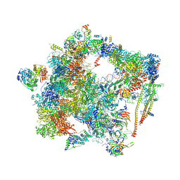 | | human Bact spliceosome core structure | | 分子名称: | 116 kDa U5 small nuclear ribonucleoprotein component, ADENOSINE-5'-DIPHOSPHATE, BUD13 homolog, ... | | 著者 | Haselbach, D, Komarov, I, Agafonov, D, Hartmuth, K, Graf, B, Kastner, B, Luehrmann, R, Stark, H. | | 登録日 | 2018-01-03 | | 公開日 | 2019-03-13 | | 最終更新日 | 2024-05-08 | | 実験手法 | ELECTRON MICROSCOPY (4.5 Å) | | 主引用文献 | Structure and Conformational Dynamics of the Human Spliceosomal BactComplex.
Cell, 172, 2018
|
|
2J3Z
 | | Crystal structure of the enzymatic component C2-I of the C2-toxin from Clostridium botulinum at pH 6.1 | | 分子名称: | C2 TOXIN COMPONENT I, COBALT (II) ION, GLYCEROL, ... | | 著者 | Schleberger, C, Hochmann, H, Barth, H, Aktories, K, Schulz, G.E. | | 登録日 | 2006-08-23 | | 公開日 | 2006-10-11 | | 最終更新日 | 2023-12-13 | | 実験手法 | X-RAY DIFFRACTION (2.3 Å) | | 主引用文献 | Structure and Action of the Binary C2 Toxin from Clostridium Botulinum.
J.Mol.Biol., 364, 2006
|
|
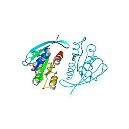
5N74
 | | Microtubule end binding protein complex | | 分子名称: | Karyogamy protein KAR9, Microtubule-associated protein RP/EB family member 1 | | 著者 | Kumar, A, Steinmetz, M. | | 登録日 | 2017-02-18 | | 公開日 | 2017-06-14 | | 最終更新日 | 2024-01-17 | | 実験手法 | X-RAY DIFFRACTION (2.3 Å) | | 主引用文献 | Short Linear Sequence Motif LxxPTPh Targets Diverse Proteins to Growing Microtubule Ends.
Structure, 25, 2017
|
|
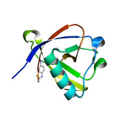
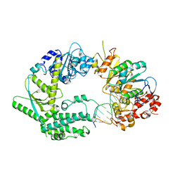
3FQ7
 | | Gabaculine complex of GSAM | | 分子名称: | 3-[O-PHOSPHONOPYRIDOXYL]--AMINO-BENZOIC ACID, Glutamate-1-semialdehyde 2,1-aminomutase | | 著者 | Stetefeld, J. | | 登録日 | 2009-01-07 | | 公開日 | 2009-11-24 | | 最終更新日 | 2024-02-21 | | 実験手法 | X-RAY DIFFRACTION (2.15 Å) | | 主引用文献 | Absence of a catalytic water confers resistance to the neurotoxin gabaculine.
Faseb J., 24, 2010
|
|
8QCW
 | |
5ME9
 | | Crystal structure of yeast Cdt1 (N terminal and middle domain), form 1. | | 分子名称: | Cell division cycle protein CDT1, GLYCEROL, SULFATE ION | | 著者 | Pye, V.E, Frigola, J, Diffley, J.F.X, Cherepanov, P. | | 登録日 | 2016-11-14 | | 公開日 | 2017-05-17 | | 最終更新日 | 2017-07-05 | | 実験手法 | X-RAY DIFFRACTION (2.7 Å) | | 主引用文献 | Cdt1 stabilizes an open MCM ring for helicase loading.
Nat Commun, 8, 2017
|
|
8S9H
 | | Crystal structure of monkey TLR7 ectodomain with compound 14 | | 分子名称: | (3S)-3-{[5-amino-1-({3-methoxy-5-[1-(oxan-4-yl)piperidin-4-yl]pyridin-2-yl}methyl)-1H-pyrazolo[4,3-d]pyrimidin-7-yl]amino}hexan-1-ol, 2-acetamido-2-deoxy-beta-D-glucopyranose, 2-acetamido-2-deoxy-beta-D-glucopyranose-(1-4)-2-acetamido-2-deoxy-beta-D-glucopyranose, ... | | 著者 | Critton, D.A. | | 登録日 | 2023-03-28 | | 公開日 | 2024-02-07 | | 最終更新日 | 2024-02-28 | | 実験手法 | X-RAY DIFFRACTION (2.437 Å) | | 主引用文献 | Identification and Optimization of Small Molecule Pyrazolopyrimidine TLR7 Agonists for Applications in Immuno-oncology.
Acs Med.Chem.Lett., 15, 2024
|
|
5MKV
 | | Crystal Structure of Human Dihydropyrimidinease-like 2 (DPYSL2A)/Collapsin Response Mediator Protein (CRMP2) residues 13-516 | | 分子名称: | 1,2-ETHANEDIOL, Dihydropyrimidinase-related protein 2 | | 著者 | Sethi, R, Zheng, Y, Krojer, T, Velupillai, S, Arrowsmith, C.H, Edwards, A.M, Bountra, C, Ahmed, A.A, von Delft, F. | | 登録日 | 2016-12-05 | | 公開日 | 2017-02-22 | | 最終更新日 | 2024-01-17 | | 実験手法 | X-RAY DIFFRACTION (1.8 Å) | | 主引用文献 | Tuning microtubule dynamics to enhance cancer therapy by modulating FER-mediated CRMP2 phosphorylation.
Nat Commun, 9, 2018
|
|
6SXB
 | | XPF-ERCC1 Cryo-EM Structure, DNA-Bound form | | 分子名称: | DNA (5'-D(*TP*CP*AP*GP*CP*AP*TP*CP*TP*G)-3'), DNA (5'-D(P*CP*AP*GP*AP*TP*GP*CP*TP*GP*A)-3'), DNA excision repair protein ERCC-1, ... | | 著者 | Jones, M.L, Briggs, D.C, McDonald, N.Q. | | 登録日 | 2019-09-25 | | 公開日 | 2020-03-11 | | 最終更新日 | 2024-05-22 | | 実験手法 | ELECTRON MICROSCOPY (7.9 Å) | | 主引用文献 | Cryo-EM structures of the XPF-ERCC1 endonuclease reveal how DNA-junction engagement disrupts an auto-inhibited conformation.
Nat Commun, 11, 2020
|
|
421P
 | | THREE-DIMENSIONAL STRUCTURES OF H-RAS P21 MUTANTS: MOLECULAR BASIS FOR THEIR INABILITY TO FUNCTION AS SIGNAL SWITCH MOLECULES | | 分子名称: | H-RAS P21 PROTEIN, MAGNESIUM ION, PHOSPHOAMINOPHOSPHONIC ACID-GUANYLATE ESTER | | 著者 | Krengel, U, John, J, Scherer, A, Kabsch, W, Wittinghofer, A, Pai, E.F. | | 登録日 | 1991-06-06 | | 公開日 | 1994-01-31 | | 最終更新日 | 2024-02-28 | | 実験手法 | X-RAY DIFFRACTION (2.2 Å) | | 主引用文献 | Three-dimensional structures of H-ras p21 mutants: molecular basis for their inability to function as signal switch molecules.
Cell(Cambridge,Mass.), 62, 1990
|
|
5PTP
 | |


