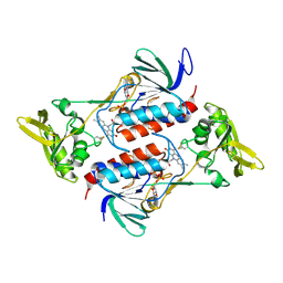
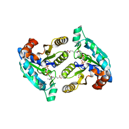
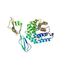
2YBA
 
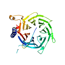 | | Crystal structure of Nurf55 in complex with histone H3 | | 分子名称: | HISTONE H3, PROBABLE HISTONE-BINDING PROTEIN CAF1 | | 著者 | Schmitges, F.W, Prusty, A.B, Faty, M, Stutzer, A, Lingaraju, G.M, Aiwazian, J, Sack, R, Hess, D, Li, L, Zhou, S, Bunker, R.D, Wirth, U, Bouwmeester, T, Bauer, A, Ly-Hartig, N, Zhao, K, Chan, H, Gu, J, Gut, H, Fischle, W, Muller, J, Thoma, N.H. | | 登録日 | 2011-03-02 | | 公開日 | 2011-05-11 | | 最終更新日 | 2024-05-01 | | 実験手法 | X-RAY DIFFRACTION (2.55 Å) | | 主引用文献 | Histone Methylation by Prc2 is Inhibited by Active Chromatin Marks
Mol.Cell, 42, 2011
|
|
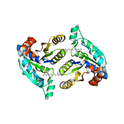
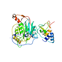
2IPF
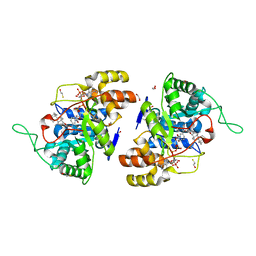 | | Crystal structure of 17alpha-hydroxysteroid dehydrogenase in complex with NADP+ and epi-testosterone | | 分子名称: | (10ALPHA,13ALPHA,14BETA,17ALPHA)-17-HYDROXYANDROST-4-EN-3-ONE, (3(17)alpha-hydroxysteroid dehydrogenase), 1,2-ETHANEDIOL, ... | | 著者 | Faucher, F, Cantin, L, Pereira de Jesus-Tran, K, Luu-the, V, Labrie, F, Breton, R. | | 登録日 | 2006-10-12 | | 公開日 | 2007-06-19 | | 最終更新日 | 2023-08-30 | | 実験手法 | X-RAY DIFFRACTION (1.85 Å) | | 主引用文献 | Mouse 17alpha-Hydroxysteroid Dehydrogenase (AKR1C21) Binds Steroids Differently from other Aldo-keto Reductases: Identification and Characterization of Amino Acid Residues Critical for Substrate Binding.
J.Mol.Biol., 369, 2007
|
|
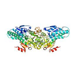
2IPG
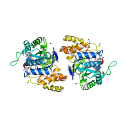 | | Crystal structure of 17alpha-hydroxysteroid dehydrogenase mutant K31A in complex with NADP+ and epi-testosterone | | 分子名称: | (10ALPHA,13ALPHA,14BETA,17ALPHA)-17-HYDROXYANDROST-4-EN-3-ONE, 1,2-ETHANEDIOL, 3(17)alpha-hydroxysteroid dehydrogenase, ... | | 著者 | Faucher, F, Cantin, L, Pereira de Jesus-Tran, K, Lemieux, M, Luu-the, V, Labrie, F, Breton, R. | | 登録日 | 2006-10-12 | | 公開日 | 2007-06-19 | | 最終更新日 | 2023-08-30 | | 実験手法 | X-RAY DIFFRACTION (1.9 Å) | | 主引用文献 | Mouse 17alpha-Hydroxysteroid Dehydrogenase (AKR1C21) Binds Steroids Differently from other Aldo-keto Reductases: Identification and Characterization of Amino Acid Residues Critical for Substrate Binding.
J.Mol.Biol., 369, 2007
|
|
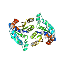
3FGN
 | |
3FMI
 | |
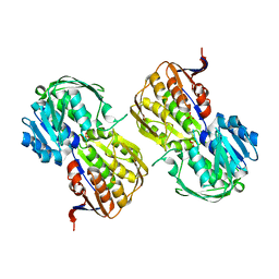
2J0U
 | | The crystal structure of eIF4AIII-Barentsz complex at 3.0 A resolution | | 分子名称: | ATP-DEPENDENT RNA HELICASE DDX48, PROTEIN CASC3 | | 著者 | Bono, F, Ebert, J, Lorentzen, E, Conti, E. | | 登録日 | 2006-08-04 | | 公開日 | 2006-09-06 | | 最終更新日 | 2023-12-13 | | 実験手法 | X-RAY DIFFRACTION (3 Å) | | 主引用文献 | The Crystal Structure of the Exon Junction Complex Reveals How It Mantains a Stable Grip on Mrna
Cell(Cambridge,Mass.), 126, 2006
|
|
2Y3I
 | | The structure of the fully closed conformation of human PGK in complex with L-ADP, 3PG and the TSA aluminium tetrafluoride | | 分子名称: | 3-PHOSPHOGLYCERIC ACID, CHLORIDE ION, L-ADENOSINE-5'-DIPHOSPHATE, ... | | 著者 | Bowler, M.W, Chaloin, L, Lionne, C. | | 登録日 | 2010-12-21 | | 公開日 | 2011-04-27 | | 最終更新日 | 2023-12-20 | | 実験手法 | X-RAY DIFFRACTION (2.9 Å) | | 主引用文献 | Interaction of Human 3-Phosphoglycerate Kinase with its Two Substrates: Is Substrate Antagonism a Kinetic Advantage?
J.Mol.Biol., 409, 2011
|
|
2F17
 | | Mouse Thiamin Pyrophosphokinase in a Ternary Complex with Pyrithiamin Pyrophosphate and AMP at 2.5 angstrom | | 分子名称: | 1-[(4-AMINO-2-METHYLPYRIMIDIN-5-YL)METHYL]-3-(2-{[HYDROXY(PHOSPHONOOXY)PHOSPHORYL]OXY}ETHYL)-2-METHYLPYRIDINIUM, 4-(2-HYDROXYETHYL)-1-PIPERAZINE ETHANESULFONIC ACID, ADENOSINE MONOPHOSPHATE, ... | | 著者 | Liu, J.Y, Timm, D.E, Hurley, T.D. | | 登録日 | 2005-11-14 | | 公開日 | 2005-11-29 | | 最終更新日 | 2023-08-23 | | 実験手法 | X-RAY DIFFRACTION (2.5 Å) | | 主引用文献 | Pyrithiamine as a substrate for thiamine pyrophosphokinase
J.Biol.Chem., 281, 2006
|
|
4NTC
 | | Crystal structure of GliT | | 分子名称: | FLAVIN-ADENINE DINUCLEOTIDE, GliT | | 著者 | Scharf, D.H, Groll, M, Habel, A, Heinekamp, T, Hertweck, C, Brakhage, A.A, Huber, E.M. | | 登録日 | 2013-12-02 | | 公開日 | 2014-03-05 | | 最終更新日 | 2024-11-06 | | 実験手法 | X-RAY DIFFRACTION (1.9 Å) | | 主引用文献 | Flavoenzyme-Catalyzed Formation of Disulfide Bonds in Natural Products
Angew.Chem.Int.Ed.Engl., 53, 2014
|
|
3FMF
 | |
2YVK
 | | Crystal structure of 5-methylthioribose 1-phosphate isomerase product complex from Bacillus subtilis | | 分子名称: | 5-S-METHYL-1-O-PHOSPHONO-5-THIO-D-RIBULOSE, Methylthioribose-1-phosphate isomerase | | 著者 | Tamura, H, Inoue, T, Kai, Y, Matsumura, H. | | 登録日 | 2007-04-13 | | 公開日 | 2008-01-22 | | 最終更新日 | 2023-10-25 | | 実験手法 | X-RAY DIFFRACTION (2.4 Å) | | 主引用文献 | Crystal structure of 5-methylthioribose 1-phosphate isomerase product complex from Bacillus subtilis: Implications for catalytic mechanism
Protein Sci., 17, 2008
|
|
4FQD
 | |
3FPA
 | |
4MEX
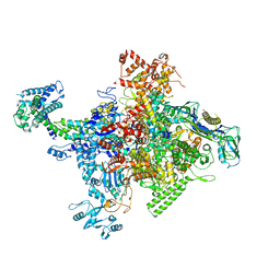 | | Crystal structure of Escherichia coli RNA polymerase in complex with salinamide A | | 分子名称: | DNA-directed RNA polymerase subunit alpha, DNA-directed RNA polymerase subunit beta, DNA-directed RNA polymerase subunit beta', ... | | 著者 | Feng, Y, Zhang, Y, Arnold, E, Ebright, R.H. | | 登録日 | 2013-08-27 | | 公開日 | 2014-05-21 | | 最終更新日 | 2025-03-26 | | 実験手法 | X-RAY DIFFRACTION (3.902 Å) | | 主引用文献 | Transcription inhibition by the depsipeptide antibiotic salinamide A.
Elife, 3, 2014
|
|
4OX2
 | |
4GLJ
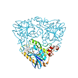 | | Crystal structure of methylthioadenosine phosphorylase in complex with rhodamine B | | 分子名称: | CHLORIDE ION, N-[9-(2-carboxyphenyl)-6-(diethylamino)-3H-xanthen-3-ylidene]-N-ethylethanaminium, PHOSPHATE ION, ... | | 著者 | Bujacz, A, Bujacz, G, Cieslinski, H, Bartasun, P. | | 登録日 | 2012-08-14 | | 公開日 | 2013-02-20 | | 最終更新日 | 2023-09-13 | | 実験手法 | X-RAY DIFFRACTION (1.9 Å) | | 主引用文献 | A study on the interaction of rhodamine B with methylthioadenosine phosphorylase protein sourced from an antarctic soil metagenomic library.
Plos One, 8, 2013
|
|
2XYQ
 | | Crystal structure of the nsp16 nsp10 SARS coronavirus complex | | 分子名称: | CHLORIDE ION, MAGNESIUM ION, NON-STRUCTURAL PROTEIN 10, ... | | 著者 | Decroly, E, Debarnot, C, Ferron, F, Bouvet, M, Coutard, B, Imbert, I, Gluais, L, Papageorgiou, N, Ortiz-Lombardia, M, Lescar, J, Canard, B. | | 登録日 | 2010-11-18 | | 公開日 | 2011-10-19 | | 最終更新日 | 2023-12-20 | | 実験手法 | X-RAY DIFFRACTION (2 Å) | | 主引用文献 | Crystal Structure and Functional Analysis of the Sars-Coronavirus RNA CAP 2'-O-Methyltransferase Nsp10/Nsp16 Complex.
Plos Pathog., 7, 2011
|
|
4MZZ
 | |
4GLF
 | |
2MJP
 | | STRUCTURE-BASED IDENTIFICATION OF THE BIOCHEMICAL FUNCTION OF A HYPOTHETICAL PROTEIN FROM METHANOCOCCUS JANNASCHII:MJ0226 | | 分子名称: | PHOSPHOAMINOPHOSPHONIC ACID-ADENYLATE ESTER, PYROPHOSPHATASE | | 著者 | Hwang, K.Y, Chung, J.H, Han, Y.S, Kim, S.H, Cho, Y, Berkeley Structural Genomics Center (BSGC) | | 登録日 | 1999-01-27 | | 公開日 | 2000-01-28 | | 最終更新日 | 2023-12-27 | | 実験手法 | X-RAY DIFFRACTION (2.2 Å) | | 主引用文献 | Structure-based identification of a novel NTPase from Methanococcus jannaschii.
Nat.Struct.Biol., 6, 1999
|
|
4O6J
 | | Crystal sturucture of T. acidophilum IdeR | | 分子名称: | FE (II) ION, Iron-dependent transcription repressor related protein | | 著者 | Lee, J.Y, Yeo, H.K. | | 登録日 | 2013-12-20 | | 公開日 | 2014-05-21 | | 最終更新日 | 2024-10-30 | | 実験手法 | X-RAY DIFFRACTION (2.3 Å) | | 主引用文献 | Structural analysis and insight into metal-ion activation of the iron-dependent regulator from Thermoplasma acidophilum.
Acta Crystallogr.,Sect.D, 70, 2014
|
|
2YRF
 | | Crystal structure of 5-methylthioribose 1-phosphate isomerase from Bacillus subtilis complexed with sulfate ion | | 分子名称: | Methylthioribose-1-phosphate isomerase, SULFATE ION | | 著者 | Tamura, H, Inoue, T, Kai, Y, Matsumura, H. | | 登録日 | 2007-04-02 | | 公開日 | 2008-01-22 | | 最終更新日 | 2024-10-30 | | 実験手法 | X-RAY DIFFRACTION (2.7 Å) | | 主引用文献 | Crystal structure of 5-methylthioribose 1-phosphate isomerase product complex from Bacillus subtilis: Implications for catalytic mechanism
Protein Sci., 17, 2008
|
|
3C9C
 | | Structural Basis of Histone H4 Recognition by p55 | | 分子名称: | CADMIUM ION, Chromatin assembly factor 1 p55 subunit, Histone H4, ... | | 著者 | Song, J.J, Garlick, J.D, Kingston, R.E. | | 登録日 | 2008-02-15 | | 公開日 | 2008-05-13 | | 最終更新日 | 2023-08-30 | | 実験手法 | X-RAY DIFFRACTION (3.2 Å) | | 主引用文献 | Structural basis of histone H4 recognition by p55.
Genes Dev., 22, 2008
|
|
4ONT
 | | Ternary host recognition complex of complement factor H, C3d, and sialic acid | | 分子名称: | Complement C3d fragment, Complement factor H, GLYCEROL, ... | | 著者 | Blaum, B.S, Stehle, T.S. | | 登録日 | 2014-01-29 | | 公開日 | 2014-11-26 | | 最終更新日 | 2024-11-20 | | 実験手法 | X-RAY DIFFRACTION (2.15 Å) | | 主引用文献 | Structural basis for sialic acid-mediated self-recognition by complement factor H.
Nat.Chem.Biol., 11, 2015
|
|
2LFA
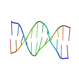 | | Oligonucleotide duplex contaning (5'S)-8,5'-cyclo-2'-deoxyguansine | | 分子名称: | DNA (5'-D(*AP*CP*AP*AP*AP*CP*AP*CP*GP*CP*AP*C)-3'), DNA (5'-D(*GP*TP*GP*CP*(2LF)P*TP*GP*TP*TP*TP*GP*T)-3') | | 著者 | Huang, H, Das, R.S, Basu, A, Stone, M.P. | | 登録日 | 2011-06-29 | | 公開日 | 2012-01-04 | | 最終更新日 | 2024-05-01 | | 実験手法 | SOLUTION NMR | | 主引用文献 | Structure of (5'S)-8,5'-Cyclo-2'-deoxyguanosine in DNA.
J.Am.Chem.Soc., 133, 2011
|
|


