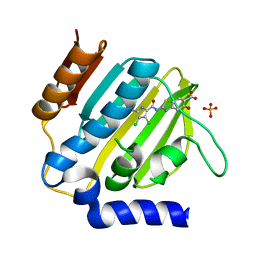
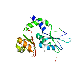
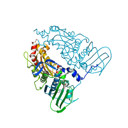
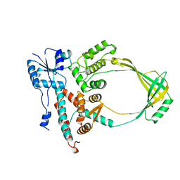
1Q3B
 
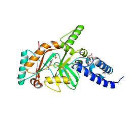
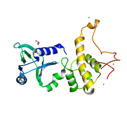 | | Crystal structure of the DNA repair enzyme endonuclease-VIII (Nei) from E. coli: The R252A mutant at 2.05 resolution. | | 分子名称: | Endonuclease VIII, GLYCEROL, MAGNESIUM ION, ... | | 著者 | Golan, G, Zharkov, D.O, Feinberg, H, Fernandes, A.S, Zaika, E.I, Kycia, J.H, Grollman, A.P, Shoham, G. | | 登録日 | 2003-07-29 | | 公開日 | 2004-08-03 | | 最終更新日 | 2023-08-16 | | 実験手法 | X-RAY DIFFRACTION (2.05 Å) | | 主引用文献 | Structure of the uncomplexed DNA repair enzyme endonuclease VIII indicates significant interdomain flexibility.
Nucleic Acids Res., 33, 2005
|
|
3IVP
 | |
3RLN
 | |
3RLO
 | |
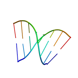
1KSE
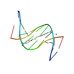
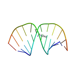
 | | Solution Structure of a quinolone-capped DNA duplex | | 分子名称: | 5'-D(*(5AT)P*GP*CP*GP*CP*A)-3', OXOLINIC ACID | | 著者 | Tuma, J, Connors, W.H, Stitelman, D.H, Richert, C. | | 登録日 | 2002-01-12 | | 公開日 | 2002-05-08 | | 最終更新日 | 2024-05-22 | | 実験手法 | SOLUTION NMR | | 主引用文献 | On the effect of covalently appended quinolones on termini of DNA duplexes.
J.Am.Chem.Soc., 124, 2002
|
|
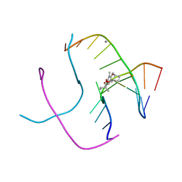
3EY0
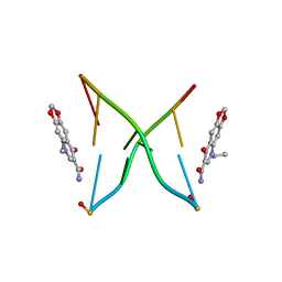
 | | A new form of DNA-drug interaction in the minor groove of a coiled coil | | 分子名称: | 1,5-BIS(4-AMIDINOPHENOXY)PENTANE, 5'-D(*DAP*DTP*DAP*DTP*DAP*DTP*DAP*DTP*DAP*DT)-3', MAGNESIUM ION | | 著者 | Pous, J, Moreno, T, Subirana, J.A, Campos, J.L. | | 登録日 | 2008-10-17 | | 公開日 | 2009-10-27 | | 最終更新日 | 2024-04-03 | | 実験手法 | X-RAY DIFFRACTION (2.52 Å) | | 主引用文献 | Coiled-coil conformation of a pentamidine-DNA complex
Acta Crystallogr.,Sect.D, 66, 2010
|
|
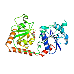
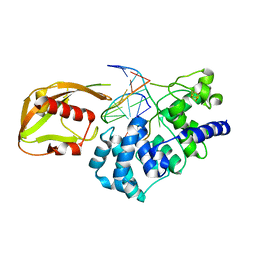
4EFB
 | | Crystal structure of DNA ligase | | 分子名称: | 4-amino-2-(cyclopentyloxy)-6-{[(1R,2S)-2-hydroxycyclopentyl]oxy}pyrimidine-5-carboxamide, BETA-NICOTINAMIDE RIBOSE MONOPHOSPHATE, DNA ligase, ... | | 著者 | Wei, Y, Wang, T, Charifson, P, Xu, W. | | 登録日 | 2012-03-29 | | 公開日 | 2013-04-03 | | 最終更新日 | 2024-02-28 | | 実験手法 | X-RAY DIFFRACTION (2.2 Å) | | 主引用文献 | Crystal structure of DNA ligase
To be Published
|
|
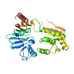
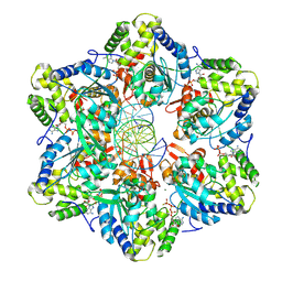
2BGU
 | | CRYSTAL STRUCTURE OF THE DNA MODIFYING ENZYME BETA-GLUCOSYLTRANSFERASE IN THE PRESENCE AND ABSENCE OF THE SUBSTRATE URIDINE DIPHOSPHOGLUCOSE | | 分子名称: | BETA-GLUCOSYLTRANSFERASE, URIDINE-5'-DIPHOSPHATE | | 著者 | Vrielink, A, Rueger, W, Driessen, H.P.C, Freemont, P.S. | | 登録日 | 1994-06-09 | | 公開日 | 1995-12-09 | | 最終更新日 | 2024-02-14 | | 実験手法 | X-RAY DIFFRACTION (2.2 Å) | | 主引用文献 | Crystal structure of the DNA modifying enzyme beta-glucosyltransferase in the presence and absence of the substrate uridine diphosphoglucose.
EMBO J., 13, 1994
|
|
5NZX
 | | Crystal structure of DNA cross-link repair protein 1A in complex with Ceftriaxone (alternative site) | | 分子名称: | Ceftriaxone, DNA cross-link repair 1A protein, NICKEL (II) ION | | 著者 | Newman, J.A, Aitkenhead, H, Kupinska, K, Burgess-Brown, N.A, Talon, R, Krojer, T, von Delft, F, Arrowsmith, C.H, Edwards, A, Bountra, C, Gileadi, O. | | 登録日 | 2017-05-15 | | 公開日 | 2017-06-14 | | 最終更新日 | 2024-01-17 | | 実験手法 | X-RAY DIFFRACTION (1.47 Å) | | 主引用文献 | Crystal structure of DNA cross-link repair protein 1A in complex with Ceftriaxone (alternative site)
To Be Published
|
|
7P2N
 | | E.coli GyrB24 with inhibitor LSJ38 (EBL2684) | | 分子名称: | 2-[[3,4-bis(chloranyl)-5-methyl-1H-pyrrol-2-yl]carbonylamino]-5-oxidanyl-1,3-benzothiazole-6-carboxylic acid, DNA gyrase subunit B, PHOSPHATE ION | | 著者 | Stevenson, C.E.M, Lawson, D.M, Maxwell, A.M, Henderson, S.R, Kikelj, D, Zega, A, Zidar, N, Ilas, J, Tomasic, T, Masic, L.P. | | 登録日 | 2021-07-06 | | 公開日 | 2022-07-20 | | 最終更新日 | 2024-01-31 | | 実験手法 | X-RAY DIFFRACTION (1.16 Å) | | 主引用文献 | Exploring the 5-Substituted 2-Aminobenzothiazole-Based DNA Gyrase B Inhibitors Active against ESKAPE Pathogens.
Acs Omega, 8, 2023
|
|
2BGT
 | |
8BGJ
 | |
2MH2
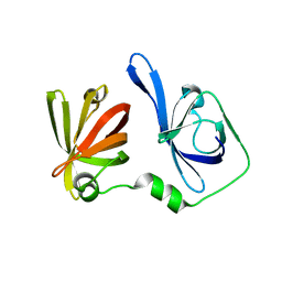
 | | Structural insights into the DNA recognition and protein interaction domains reveal fundamental homologous DNA pairing properties of HOP2 | | 分子名称: | Homologous-pairing protein 2 homolog | | 著者 | Moktan, H, Guiraldelli, M.F, Eyter, C.A, Zhao, W, Camerini-Otero, R.D, Sung, P, Zhou, D.H, Pezza, R.J. | | 登録日 | 2013-11-13 | | 公開日 | 2014-04-16 | | 最終更新日 | 2024-05-01 | | 実験手法 | SOLUTION NMR | | 主引用文献 | Solution Structure and DNA-binding Properties of the Winged Helix Domain of the Meiotic Recombination HOP2 Protein.
J.Biol.Chem., 289, 2014
|
|
7Z3X
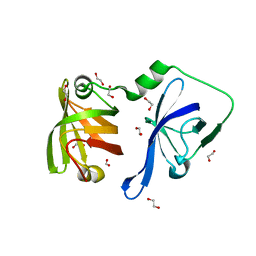
 | | Crystal structure of FIR RRM1-2 Y115F mutant bound to FUSE ssDNA | | 分子名称: | 1,2-ETHANEDIOL, DNA (5'-D(*TP*TP*T)-3'), DNA (5'-D(P*GP*TP*())-3'), ... | | 著者 | Ni, X, Chaikuad, A, Knapp, S, Structural Genomics Consortium (SGC) | | 登録日 | 2022-03-02 | | 公開日 | 2022-11-09 | | 最終更新日 | 2024-02-07 | | 実験手法 | X-RAY DIFFRACTION (1.65 Å) | | 主引用文献 | Crystal structure of FIR RRM1-2 Y115F mutant bound to FUSE ssDNA
To Be Published
|
|
8EFV
 | |
1M6R
 | |
7QFN
 | | Human Topoisomerase II Beta ATPase ADP | | 分子名称: | ADENOSINE-5'-DIPHOSPHATE, DNA topoisomerase 2-beta, MAGNESIUM ION, ... | | 著者 | Ling, E.M, Basle, A, Cowell, I.G, Blower, T.R, Austin, C.A. | | 登録日 | 2021-12-06 | | 公開日 | 2022-05-25 | | 最終更新日 | 2024-06-19 | | 実験手法 | X-RAY DIFFRACTION (2.621 Å) | | 主引用文献 | A comprehensive structural analysis of the ATPase domain of human DNA topoisomerase II beta bound to AMPPNP, ADP, and the bisdioxopiperazine, ICRF193.
Structure, 30, 2022
|
|
7QFO
 | | Human Topoisomerase II Beta ATPase AMPPNP | | 分子名称: | ALANINE, DNA topoisomerase 2-beta, MAGNESIUM ION, ... | | 著者 | Ling, E.M, Basle, A, Cowell, I.G, Blower, T.R, Austin, C.A. | | 登録日 | 2021-12-06 | | 公開日 | 2022-05-25 | | 最終更新日 | 2024-01-31 | | 実験手法 | X-RAY DIFFRACTION (1.9 Å) | | 主引用文献 | A comprehensive structural analysis of the ATPase domain of human DNA topoisomerase II beta bound to AMPPNP, ADP, and the bisdioxopiperazine, ICRF193.
Structure, 30, 2022
|
|
3HGD
 | |
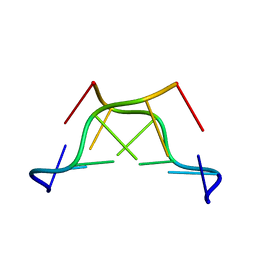
1FHY
 | | PSORALEN CROSS-LINKED D(CCGCTAGCGG) FORMS HOLLIDAY JUNCTION | | 分子名称: | 4'-HYDROXYMETHYL-4,5',8-TRIMETHYLPSORALEN, CALCIUM ION, DNA (5'-D(*CP*CP*GP*CP*TP*AP*GP*CP*GP*G)-3') | | 著者 | Eichman, B.F, Mooers, B.H.M, Alberti, M, Hearst, J.E, Ho, P.S. | | 登録日 | 2000-08-02 | | 公開日 | 2001-04-21 | | 最終更新日 | 2024-02-07 | | 実験手法 | X-RAY DIFFRACTION (2.2 Å) | | 主引用文献 | The crystal structures of psoralen cross-linked DNAs: drug-dependent formation of Holliday junctions.
J.Mol.Biol., 308, 2001
|
|
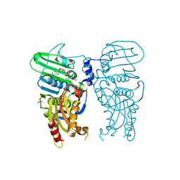
1RRQ
 | | MutY adenine glycosylase in complex with DNA containing an A:oxoG pair | | 分子名称: | 5'-D(*TP*GP*TP*CP*CP*AP*AP*GP*TP*CP*T)-3', 5'-D(AP*AP*GP*AP*CP*(8OG)P*TP*GP*GP*AP*C)-3', CALCIUM ION, ... | | 著者 | Fromme, J.C, Banerjee, A, Huang, S.J, Verdine, G.L. | | 登録日 | 2003-12-08 | | 公開日 | 2004-02-17 | | 最終更新日 | 2024-02-14 | | 実験手法 | X-RAY DIFFRACTION (2.22 Å) | | 主引用文献 | Structural basis for removal of adenine mispaired with 8-oxoguanine by MutY adenine DNA glycosylase
Nature, 427, 2004
|
|
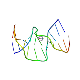
1LU5
 | | 2.4 Angstrom Crystal Structure of the Asymmetric Platinum Complex {Pt(ammine)(cyclohexylamine)}2+ Bound to a Dodecamer DNA Duplex | | 分子名称: | 5'-D(*CP*CP*TP*CP*TP*GP*GP*TP*CP*TP*CP*C)-3', 5'-D(*GP*GP*AP*GP*AP*CP*CP*AP*GP*AP*GP*G)-3', CIS-(AMMINE)(CYCLOHEXYLAMINE)PLATINUM(II) COMPLEX | | 著者 | Silverman, A.P, Bu, W, Cohen, S.M, Lippard, S.J. | | 登録日 | 2002-05-21 | | 公開日 | 2002-12-20 | | 最終更新日 | 2024-02-14 | | 実験手法 | X-RAY DIFFRACTION (2.4 Å) | | 主引用文献 | 2.4-A Crystal Structure of the Asymmetric Platinum Complex {Pt(ammine)(cyclohexylamine)}2+ Bound to
a Dodecamer DNA Duplex
J.Biol.Chem., 277, 2002
|
|
1Q3U
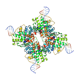 | | Crystal structure of a wild-type Cre recombinase-loxP synapse: pre-cleavage complex | | 分子名称: | Cre recombinase, IODIDE ION, MAGNESIUM ION, ... | | 著者 | Ennifar, E, Meyer, J.E.W, Buchholz, F, Stewart, A.F, Suck, D. | | 登録日 | 2003-08-01 | | 公開日 | 2003-09-16 | | 最終更新日 | 2023-08-16 | | 実験手法 | X-RAY DIFFRACTION (2.9 Å) | | 主引用文献 | Crystal structure of a wild-type Cre recombinase-loxP synapse reveals a novel spacer conformation suggesting an alternative mechanism for DNA cleavage activation
Nucleic Acids Res., 31, 2003
|
|
1ECL
 | |
6R14
 | | Structure of kiteplatinated dsDNA | | 分子名称: | Kiteplatin, Kiteplatinated DNA oligomer, chain A, ... | | 著者 | Margiotta, N, Papadia, P, Kubicek, K, Krejcikova, M, Gkionis, K, Sponer, J. | | 登録日 | 2019-03-13 | | 公開日 | 2020-04-01 | | 最終更新日 | 2024-05-15 | | 実験手法 | SOLUTION NMR | | 主引用文献 | Structural characterization of kiteplatinated DNA
To Be Published
|
|


