7RD8
 
 | | Structure of the S. cerevisiae P4B ATPase lipid flippase in the E1-ATP state | | 分子名称: | MAGNESIUM ION, PHOSPHOMETHYLPHOSPHONIC ACID ADENYLATE ESTER, Probable phospholipid-transporting ATPase NEO1 | | 著者 | Bai, L, Jain, B.K, You, Q, Duan, H.D, Graham, T.R, Li, H. | | 登録日 | 2021-07-09 | | 公開日 | 2021-09-29 | | 最終更新日 | 2024-06-05 | | 実験手法 | ELECTRON MICROSCOPY (5.64 Å) | | 主引用文献 | Structural basis of the P4B ATPase lipid flippase activity.
Nat Commun, 12, 2021
|
|
5DTD
 | |
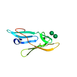
1MZH
 | | QR15, an Aldolase | | 分子名称: | Deoxyribose-phosphate aldolase, PHOSPHATE ION | | 著者 | Tan, A.Y, Smith, P.C, Shen, J, Xiao, R, Acton, T, Rost, B, Montelione, G, Hunt, J.F, Northeast Structural Genomics Consortium (NESG) | | 登録日 | 2002-10-07 | | 公開日 | 2003-02-04 | | 最終更新日 | 2024-02-14 | | 実験手法 | X-RAY DIFFRACTION (2 Å) | | 主引用文献 | Crystal Structure of Aquifex Aeolicus Aldolase,
Northeast Structural Genomics Consortium Target
QR15
To be Published
|
|
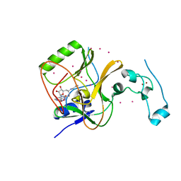
1N06
 | | Crystal Structure of Schizosaccharomyces pombe Riboflavin Kinase Reveals a Novel ATP and Riboflavin Binding Fold | | 分子名称: | ADENOSINE-5'-DIPHOSPHATE, PUTATIVE riboflavin kinase | | 著者 | Bauer, S, Kemter, K, Bacher, A, Huber, R, Fischer, M, Steinbacher, S. | | 登録日 | 2002-10-11 | | 公開日 | 2003-02-25 | | 最終更新日 | 2024-02-14 | | 実験手法 | X-RAY DIFFRACTION (2 Å) | | 主引用文献 | Crystal Structure of Schizosaccharomyces pombe Riboflavin Kinase Reveals a Novel ATP and Riboflavin Binding Fold
J.Mol.Biol., 326, 2003
|
|
7R9D
 | |
1N0E
 | | CRYSTAL STRUCTURE OF A CELL DIVISION AND CELL WALL BIOSYNTHESIS PROTEIN UPF0040 FROM MYCOPLASMA PNEUMONIAE: INDICATION OF A NOVEL FOLD WITH A POSSIBLE NEW CONSERVED SEQUENCE MOTIF | | 分子名称: | Protein mraZ | | 著者 | Chen, S, Jancrick, J, Yokota, H, Kim, R, Kim, S.-H, Berkeley Structural Genomics Center (BSGC) | | 登録日 | 2002-10-13 | | 公開日 | 2003-10-21 | | 最終更新日 | 2024-02-14 | | 実験手法 | X-RAY DIFFRACTION (2.7 Å) | | 主引用文献 | Crystal structure of a protein associated with cell division from Mycoplasma pneumoniae (GI: 13508053): a novel fold with a conserved sequence motif.
Proteins, 55, 2004
|
|
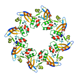
1N11
 | | D34 REGION OF HUMAN ANKYRIN-R AND LINKER | | 分子名称: | Ankyrin, BROMIDE ION, CHLORIDE ION | | 著者 | Michaely, P, Tomchick, D.R, Machius, M, Anderson, R.G.W. | | 登録日 | 2002-10-16 | | 公開日 | 2002-12-11 | | 最終更新日 | 2024-02-14 | | 実験手法 | X-RAY DIFFRACTION (2.7 Å) | | 主引用文献 | Crystal structure of a 12 ANK repeat stack from human ankyrinR
Embo J., 21, 2002
|
|
4FMU
 | | Crystal structure of Methyltransferase domain of human SET domain-containing protein 2 Compound: Pr-SNF | | 分子名称: | (2S,5S)-2-amino-6-[(2R,3S,4R,5R)-5-(6-amino-9H-purin-9-yl)-3,4-dihydroxytetrahydrofuran-2-yl]-5-(propylamino)hexanoic acid, Histone-lysine N-methyltransferase SETD2, UNKNOWN ATOM OR ION, ... | | 著者 | Dong, A, Zeng, H, Ibanez, G, Zheng, W, Tempel, W, Bountra, C, Arrowsmith, C.H, Edwards, A.M, Brown, P.J, Min, J, Luo, M, Wu, H, Structural Genomics Consortium (SGC) | | 登録日 | 2012-06-18 | | 公開日 | 2012-09-05 | | 最終更新日 | 2023-09-13 | | 実験手法 | X-RAY DIFFRACTION (2.1 Å) | | 主引用文献 | Sinefungin Derivatives as Inhibitors and Structure Probes of Protein Lysine Methyltransferase SETD2.
J.Am.Chem.Soc., 134, 2012
|
|
1MRJ
 | | STUDIES ON CRYSTAL STRUCTURES ACTIVE CENTER GEOMETRY AND DEPURINE MECHANISM OF TWO RIBOSOME-INACTIVATING PROTEINS | | 分子名称: | ADENOSINE, ALPHA-TRICHOSANTHIN | | 著者 | Huang, Q, Liu, S, Tang, Y, Jin, S, Wang, Y. | | 登録日 | 1994-07-01 | | 公開日 | 1995-02-07 | | 最終更新日 | 2024-02-14 | | 実験手法 | X-RAY DIFFRACTION (1.6 Å) | | 主引用文献 | Studies on crystal structures, active-centre geometry and depurinating mechanism of two ribosome-inactivating proteins.
Biochem.J., 309, 1995
|
|
1MZA
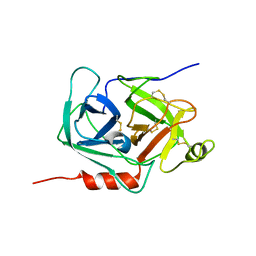 | | crystal structure of human pro-granzyme K | | 分子名称: | pro-granzyme K | | 著者 | Hink-Schauer, C, Estebanez-Perpina, E, Wilharm, E, Fuentes-Prior, P, Klinkert, W, Bode, W, Jenne, D.E. | | 登録日 | 2002-10-07 | | 公開日 | 2003-01-14 | | 最終更新日 | 2024-04-03 | | 実験手法 | X-RAY DIFFRACTION (2.23 Å) | | 主引用文献 | The 2.2-A Crystal Structure of Human Pro-granzyme K Reveals a Rigid Zymogen with Unusual Features
J.BIOL.CHEM., 277, 2002
|
|
1MUY
 | | CATALYTIC DOMAIN OF MUTY FROM ESCHERICHIA COLI | | 分子名称: | ADENINE GLYCOSYLASE, GLYCEROL, IMIDAZOLE, ... | | 著者 | Guan, Y, Tainer, J.A. | | 登録日 | 1998-08-20 | | 公開日 | 1999-08-20 | | 最終更新日 | 2024-02-14 | | 実験手法 | X-RAY DIFFRACTION (1.4 Å) | | 主引用文献 | MutY catalytic core, mutant and bound adenine structures define specificity for DNA repair enzyme superfamily.
Nat.Struct.Biol., 5, 1998
|
|
1MZI
 | | Solution ensemble structures of HIV-1 gp41 2F5 mAb epitope | | 分子名称: | 2F5 epitope of HIV-1 gp41 fusion protein | | 著者 | Barbato, G, Bianchi, E, Ingallinella, P, Hurni, W.H, Miller, M.D, Ciliberto, G, Cortese, R, Bazzo, R, Shiver, J.W, Pessi, A. | | 登録日 | 2002-10-08 | | 公開日 | 2003-09-02 | | 最終更新日 | 2024-05-29 | | 実験手法 | SOLUTION NMR | | 主引用文献 | Structural analysis of the epitope of the anti-HIV antibody 2F5 sheds light into its mechanism of neutralization and HIV fusion.
J.Mol.Biol., 330, 2003
|
|
7RTS
 | |
1N0G
 | | Crystal Structure of A Cell Division and Cell Wall Biosynthesis Protein UPF0040 from Mycoplasma pneumoniae: Indication of A Novel Fold with A Possible New Conserved Sequence Motif | | 分子名称: | Protein mraZ | | 著者 | Chen, S, Jancarik, J, Yokota, H, Kim, R, Kim, S.-H, Berkeley Structural Genomics Center (BSGC) | | 登録日 | 2002-10-13 | | 公開日 | 2003-10-21 | | 最終更新日 | 2024-02-14 | | 実験手法 | X-RAY DIFFRACTION (2.8 Å) | | 主引用文献 | Crystal structure of a protein associated with cell division from Mycoplasma pneumoniae (GI: 13508053): a novel fold with a conserved sequence motif.
Proteins, 55, 2004
|
|
5DJV
 | |
7RTB
 | | Peptide-19 bound to the Glucagon-Like Peptide-1 Receptor (GLP-1R) | | 分子名称: | Glucagon-like peptide 1 receptor, Guanine nucleotide-binding protein G(I)/G(S)/G(O) subunit gamma-2, Guanine nucleotide-binding protein G(I)/G(S)/G(T) subunit beta-1, ... | | 著者 | Johnson, R.M, Danev, R, Sexton, P.M, Wootten, D. | | 登録日 | 2021-08-12 | | 公開日 | 2021-10-06 | | 実験手法 | ELECTRON MICROSCOPY (2.14 Å) | | 主引用文献 | Cryo-EM structure of the dual incretin receptor agonist, peptide-19, in complex with the glucagon-like peptide-1 receptor.
Biochem.Biophys.Res.Commun., 578, 2021
|
|
2YP3
 | | Haemagglutinin of 2004 Human H3N2 Virus in Complex with Human Receptor Analogue 6SLN | | 分子名称: | 2-acetamido-2-deoxy-beta-D-glucopyranose, 2-acetamido-2-deoxy-beta-D-glucopyranose-(1-4)-2-acetamido-2-deoxy-beta-D-glucopyranose, 4-(2-HYDROXYETHYL)-1-PIPERAZINE ETHANESULFONIC ACID, ... | | 著者 | Xiong, X, Lin, Y.P, Wharton, S.A, Martin, S.R, Coombs, P.J, Vachieri, S.G, Christodoulou, E, Walker, P.A, Liu, J, Skehel, J.J, Gamblin, S.J, Hay, A.J, Daniels, R.S, McCauley, J.W. | | 登録日 | 2012-10-29 | | 公開日 | 2012-11-07 | | 最終更新日 | 2020-07-29 | | 実験手法 | X-RAY DIFFRACTION (1.88 Å) | | 主引用文献 | Evolution of the Receptor Binding Properties of the Influenza A(H3N2) Hemagglutinin.
Proc.Natl.Acad.Sci.USA, 109, 2012
|
|
5DQJ
 | | Structure of unliganded S55-5 Fab | | 分子名称: | S55-5 Fab (IgG1 kappa) light chain, S55-5 Fab (IgG1) heavy chain | | 著者 | Haji-Ghassemi, O, Evans, S.V. | | 登録日 | 2015-09-14 | | 公開日 | 2016-03-09 | | 最終更新日 | 2023-09-27 | | 実験手法 | X-RAY DIFFRACTION (2.61 Å) | | 主引用文献 | Lipid A-antibody structures reveal a widely-utilized pocket specific for negatively charged groups derived from unrelated V-genes
J.Biol.Chem., 2015
|
|
5DKE
 | | Crystal Structure of the ER-alpha Ligand-binding Domain in complex with a 3-naphthyl-substituted, methyl, cis-diaryl-ethylene compound 4,4'-[2-(naphthalen-2-yl)prop-1-ene-1,1-diyl]diphenol | | 分子名称: | 4,4'-[2-(naphthalen-2-yl)prop-1-ene-1,1-diyl]diphenol, Estrogen receptor, Nuclear receptor coactivator 2 | | 著者 | Nwachukwu, J.C, Srinivasan, S, Zheng, Y, Wang, S, Min, J, Dong, C, Liao, Z, Cavett, V, Nowak, J, Houtman, R, Carlson, K.E, Josan, J.S, Elemento, O, Katzenellenbogen, J.A, Zhou, H.B, Nettles, K.W. | | 登録日 | 2015-09-03 | | 公開日 | 2016-05-04 | | 最終更新日 | 2024-03-06 | | 実験手法 | X-RAY DIFFRACTION (2.6 Å) | | 主引用文献 | Predictive features of ligand-specific signaling through the estrogen receptor.
Mol.Syst.Biol., 12, 2016
|
|
2YHH
 | | Microvirin:mannobiose complex | | 分子名称: | MANNAN-BINDING LECTIN, alpha-D-mannopyranose-(1-2)-alpha-D-mannopyranose | | 著者 | Hussan, S, Bewley, C.A, Clore, G.M, Gustchina, E, Ghirlando, R. | | 登録日 | 2011-05-02 | | 公開日 | 2011-06-15 | | 最終更新日 | 2020-07-29 | | 実験手法 | SOLUTION NMR | | 主引用文献 | Solution Structure of the Monovalent Lectin Microvirin in Complex with Man(Alpha)(1-2)Man Provides a Basis for Anti-HIV Activity with Low Toxicity.
J.Biol.Chem., 286, 2011
|
|
1N36
 | | Structure of the Thermus thermophilus 30S ribosomal subunit in the presence of crystallographically disordered codon and near-cognate transfer RNA anticodon stem-loop mismatched at the second codon position | | 分子名称: | 16S RIBOSOMAL RNA, 30S RIBOSOMAL PROTEIN S10, 30S RIBOSOMAL PROTEIN S11, ... | | 著者 | Ogle, J.M, Murphy IV, F.V, Tarry, M.J, Ramakrishnan, V. | | 登録日 | 2002-10-25 | | 公開日 | 2002-11-29 | | 最終更新日 | 2024-02-14 | | 実験手法 | X-RAY DIFFRACTION (3.65 Å) | | 主引用文献 | Selection of tRNA by the Ribosome Requires a Transition from an Open to a Closed Form
Cell(Cambridge,Mass.), 111, 2002
|
|
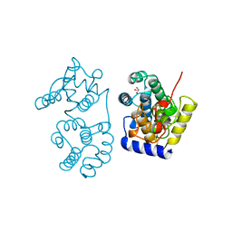
1N3K
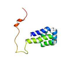 | | Solution structure of phosphoprotein enriched in astrocytes 15 kDa (PEA-15) | | 分子名称: | Astrocytic phosphoprotein PEA-15 | | 著者 | Hill, J.M, Vaidyanathan, H, Ramos, J.W, Ginsberg, M.H, Werner, M.H. | | 登録日 | 2002-10-28 | | 公開日 | 2003-01-14 | | 最終更新日 | 2024-05-22 | | 実験手法 | SOLUTION NMR | | 主引用文献 | Recognition of ERK MAP Kinase by PEA-15 Reveals a Common Docking Site Within the Death Domain and Death Effector Domain
Embo J., 21, 2002
|
|
7R96
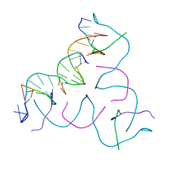 | | Self-Assembled 3D DNA Hexagonal Tensegrity Triangle | | 分子名称: | DNA (5'-D(*AP*GP*GP*CP*AP*GP*CP*CP*TP*GP*TP*AP*CP*GP*GP*AP*CP*AP*TP*CP*A)-3'), DNA (5'-D(*TP*CP*TP*GP*AP*TP*GP*T)-3'), DNA (5'-D(P*CP*CP*GP*TP*AP*CP*A)-3'), ... | | 著者 | Lu, B, Vecchioni, S, Ohayon, Y.P, Sha, R, Mao, C, Seeman, N.C. | | 登録日 | 2021-06-28 | | 公開日 | 2021-10-20 | | 最終更新日 | 2023-10-18 | | 実験手法 | X-RAY DIFFRACTION (5.68 Å) | | 主引用文献 | 3D Hexagonal Arrangement of DNA Tensegrity Triangles.
Acs Nano, 15, 2021
|
|
1N19
 | | Structure of the HSOD A4V mutant | | 分子名称: | COPPER (I) ION, SULFATE ION, Superoxide Dismutase [Cu-Zn], ... | | 著者 | Cardoso, R.M.F, Thayer, M.M, DiDonato, M, Lo, T.P, Bruns, C.K, Getzoff, E.D, Tainer, J.A. | | 登録日 | 2002-10-16 | | 公開日 | 2002-11-27 | | 最終更新日 | 2021-10-27 | | 実験手法 | X-RAY DIFFRACTION (1.86 Å) | | 主引用文献 | Insights into Lou Gehrig's disease from the structure and instability of the A4V mutant of human Cu,Zn superoxide dismutase.
J.Mol.Biol., 324, 2002
|
|
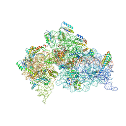
7RRO
 | | Structure of the 48-nm repeat doublet microtubule from bovine tracheal cilia | | 分子名称: | Armadillo repeat containing 4, Chromosome 3 C1orf194 homolog, Cilia and flagella associated protein 161, ... | | 著者 | Gui, M, Anderson, J.R, Botsch, J.J, Meleppattu, S, Singh, S.K, Zhang, Q, Brown, A. | | 登録日 | 2021-08-10 | | 公開日 | 2021-10-27 | | 最終更新日 | 2024-06-05 | | 実験手法 | ELECTRON MICROSCOPY (3.4 Å) | | 主引用文献 | De novo identification of mammalian ciliary motility proteins using cryo-EM.
Cell, 184, 2021
|
|


