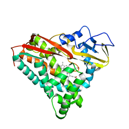
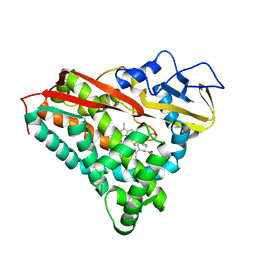
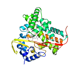
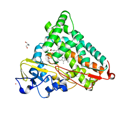
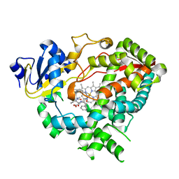
1SMJ
 
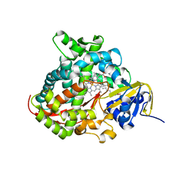 | | Structure of the A264E mutant of cytochrome P450 BM3 complexed with palmitoleate | | 分子名称: | Bifunctional P-450:NADPH-P450 reductase, PALMITOLEIC ACID, PROTOPORPHYRIN IX CONTAINING FE | | 著者 | Joyce, M.G, Girvan, H.M, Munro, A.W, Leys, D. | | 登録日 | 2004-03-09 | | 公開日 | 2004-06-08 | | 最終更新日 | 2023-08-23 | | 実験手法 | X-RAY DIFFRACTION (2.75 Å) | | 主引用文献 | A Single Mutation in Cytochrome P450 BM3 Induces the Conformational Rearrangement Seen upon Substrate Binding in the Wild-type Enzyme
J.Biol.Chem., 279, 2004
|
|
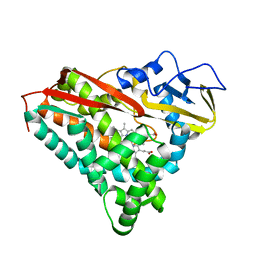
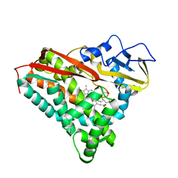
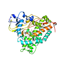
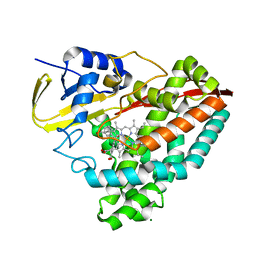
1PQ2
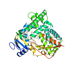 | | Crystal Structure of Human Drug Metabolizing Cytochrome P450 2C8 | | 分子名称: | Cytochrome P450 2C8, PALMITIC ACID, PHOSPHATE ION, ... | | 著者 | Schoch, G.A, Yano, J.K, Wester, M.R, Griffin, K.J, Stout, C.D, Johnson, E.F. | | 登録日 | 2003-06-17 | | 公開日 | 2004-01-13 | | 最終更新日 | 2023-08-16 | | 実験手法 | X-RAY DIFFRACTION (2.7 Å) | | 主引用文献 | Structure of human microsomal cytochrome P450 2C8. Evidence for a peripheral fatty acid binding site
J.Biol.Chem., 279, 2004
|
|
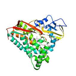
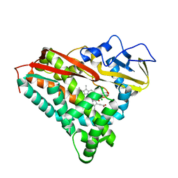
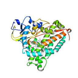
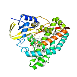
1Q5D
 | | Epothilone B-bound Cytochrome P450epoK | | 分子名称: | 7,11-DIHYDROXY-8,8,10,12,16-PENTAMETHYL-3-[1-METHYL-2-(2-METHYL-THIAZOL-4-YL)VINYL]-4,17-DIOXABICYCLO[14.1.0]HEPTADECANE-5,9-DIONE, P450 epoxidase, PROTOPORPHYRIN IX CONTAINING FE | | 著者 | Nagano, S, Li, H, Shimizu, H, Nishida, C, Ogura, H, Ortiz de Montellano, P.R, Poulos, T.L. | | 登録日 | 2003-08-06 | | 公開日 | 2003-10-28 | | 最終更新日 | 2024-04-03 | | 実験手法 | X-RAY DIFFRACTION (1.93 Å) | | 主引用文献 | Crystal structures of epothilone D-bound, epothilone B-bound, and substrate-free forms of cytochrome P450epoK
J.Biol.Chem., 278, 2003
|
|
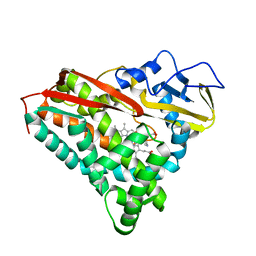
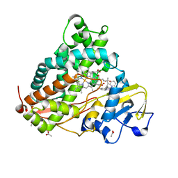
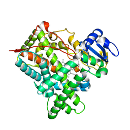
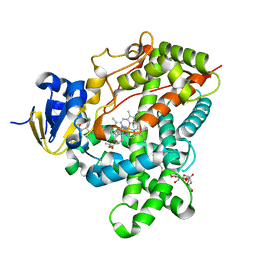
1Q5E
 | | Substrate-free Cytochrome P450epoK | | 分子名称: | P450 epoxidase, PROTOPORPHYRIN IX CONTAINING FE | | 著者 | Nagano, S, Li, H, Shimizu, H, Nishida, C, Ogura, H, Ortiz de Montellano, P.R, Poulos, T.L. | | 登録日 | 2003-08-06 | | 公開日 | 2003-10-28 | | 最終更新日 | 2024-04-03 | | 実験手法 | X-RAY DIFFRACTION (2.65 Å) | | 主引用文献 | Crystal structures of epothilone D-bound, epothilone B-bound, and substrate-free forms of cytochrome P450epoK
J.Biol.Chem., 278, 2003
|
|
1PHD
 | |
1PHG
 | |
1PHC
 | |
1PHF
 | |
1P0V
 | | F393A mutant heme domain of flavocytochrome P450 BM3 | | 分子名称: | Bifunctional P-450:NADPH-P450 reductase, PROTOPORPHYRIN IX CONTAINING FE | | 著者 | Ost, T.W.B, Clark, J, Miles, C.S, Walkinshaw, M.D, Reid, G.A, Chapman, S.K, Daff, S, Mowat, C.G. | | 登録日 | 2003-04-11 | | 公開日 | 2003-12-09 | | 最終更新日 | 2023-08-16 | | 実験手法 | X-RAY DIFFRACTION (2.05 Å) | | 主引用文献 | Oxygen Activation and Electron Transfer in Flavocytochrome P450 BM3
J.Am.Chem.Soc., 125, 2003
|
|
1T93
 | | Evidence for Multiple Substrate Recognition and Molecular Mechanism of C-C reaction by Cytochrome P450 CYP158A2 from Streptomyces Coelicolor A3(2) | | 分子名称: | FLAVIOLIN, PROTOPORPHYRIN IX CONTAINING FE, putative cytochrome P450 | | 著者 | Zhao, B, Sundaramoorthy, M, Waterman, M.R. | | 登録日 | 2004-05-14 | | 公開日 | 2005-01-25 | | 最終更新日 | 2023-08-23 | | 実験手法 | X-RAY DIFFRACTION (1.62 Å) | | 主引用文献 | Binding of Two Flaviolin Substrate Molecules, Oxidative Coupling, and Crystal Structure of Streptomyces coelicolor A3(2) Cytochrome P450 158A2.
J.Biol.Chem., 280, 2005
|
|
1PHE
 | |
1PHB
 | |
1PHA
 | |
1PKF
 | | Crystal Structure of Epothilone D-bound Cytochrome P450epoK | | 分子名称: | EPOTHILONE D, PROTOPORPHYRIN IX CONTAINING FE, cytochrome p450EpoK | | 著者 | Nagano, S, Li, H, Shimizu, H, Nishida, C, Ogura, H, Ortiz de Montellano, P.R, Poulos, T.L. | | 登録日 | 2003-06-05 | | 公開日 | 2003-10-28 | | 最終更新日 | 2024-02-14 | | 実験手法 | X-RAY DIFFRACTION (2.1 Å) | | 主引用文献 | Crystal Structures of Epothilone D-bound, Epothilone B-bound, and Substrate-free Forms of Cytochrome P450epoK
J.Biol.Chem., 278, 2003
|
|
1QMQ
 | | Optical detection of cytochrome P450 by sensitizer-linked substrates | | 分子名称: | ACETATE ION, CYTOCHROME P450, DELTA-BIS(2,2'-BIPYRIDINE)-(5-METHYL-2-2'-BIPYRIDINE)-C9-ADAMANTANE RUTHENIUM (II), ... | | 著者 | Crane, B.R, Dmochowski, I.J, Gray, H.B. | | 登録日 | 1999-10-05 | | 公開日 | 2000-10-06 | | 最終更新日 | 2023-12-13 | | 実験手法 | X-RAY DIFFRACTION (1.55 Å) | | 主引用文献 | Optical Detection of Cytochrome P450 by Sensitizer-Linked Substrates
Proc.Natl.Acad.Sci.USA, 96, 1999
|
|
1T2B
 | | Crystal Structure of cytochrome P450cin complexed with its substrate 1,8-cineole | | 分子名称: | 1,3,3-TRIMETHYL-2-OXABICYCLO[2.2.2]OCTANE, P450cin, PROTOPORPHYRIN IX CONTAINING FE | | 著者 | Meharenna, Y.T, Li, H, Hawkes, D.B, Pearson, A.G, De Voss, J, Poulos, T.L. | | 登録日 | 2004-04-20 | | 公開日 | 2005-04-26 | | 最終更新日 | 2023-08-23 | | 実験手法 | X-RAY DIFFRACTION (1.7 Å) | | 主引用文献 | Crystal structure of P450cin in a complex with its substrate, 1,8-cineole, a close structural homologue to D-camphor, the substrate for P450cam
Biochemistry, 43, 2004
|
|
1T85
 | | Crystal Structure of the Ferrous CO-bound Cytochrome P450cam Mutant (L358P/C334A) | | 分子名称: | CAMPHOR, CARBON MONOXIDE, Cytochrome P450-cam, ... | | 著者 | Nagano, S, Tosha, T, Ishimori, K, Morishima, I, Poulos, T.L. | | 登録日 | 2004-05-11 | | 公開日 | 2004-06-01 | | 最終更新日 | 2024-02-14 | | 実験手法 | X-RAY DIFFRACTION (1.8 Å) | | 主引用文献 | Crystal structure of the cytochrome p450cam mutant that exhibits the same spectral perturbations induced by putidaredoxin binding.
J.Biol.Chem., 279, 2004
|
|
1T86
 | | Crystal Structure of the Ferrous Cytochrome P450cam Mutant (L358P/C334A) | | 分子名称: | CAMPHOR, Cytochrome P450-cam, POTASSIUM ION, ... | | 著者 | Nagano, S, Tosha, T, Ishimori, K, Morishima, I, Poulos, T.L. | | 登録日 | 2004-05-11 | | 公開日 | 2004-05-25 | | 最終更新日 | 2024-02-14 | | 実験手法 | X-RAY DIFFRACTION (1.9 Å) | | 主引用文献 | Crystal structure of the cytochrome p450cam mutant that exhibits the same spectral perturbations induced by putidaredoxin binding.
J.Biol.Chem., 279, 2004
|
|
1SUO
 | | Structure of mammalian cytochrome P450 2B4 with bound 4-(4-chlorophenyl)imidazole | | 分子名称: | 4-(4-CHLOROPHENYL)IMIDAZOLE, Cytochrome P450 2B4, PROTOPORPHYRIN IX CONTAINING FE | | 著者 | Scott, E.E, White, M.A, He, Y.A, Johnson, E.F, Stout, C.D, Halpert, J.R. | | 登録日 | 2004-03-26 | | 公開日 | 2004-07-20 | | 最終更新日 | 2023-08-23 | | 実験手法 | X-RAY DIFFRACTION (1.9 Å) | | 主引用文献 | Structure of mammalian cytochrome P450 2B4 complexed with 4-(4-chlorophenyl)imidazole at 1.9 {angstrom} resolution: Insight into the range of P450 conformations and coordination of redox partner binding.
J.Biol.Chem., 279, 2004
|
|
1T88
 | | Crystal Structure of the Ferrous Cytochrome P450cam (C334A) | | 分子名称: | 2-AMINO-2-HYDROXYMETHYL-PROPANE-1,3-DIOL, CAMPHOR, Cytochrome P450-cam, ... | | 著者 | Nagano, S, Tosha, T, Ishimori, K, Morishima, I, Poulos, T.L. | | 登録日 | 2004-05-11 | | 公開日 | 2004-05-25 | | 最終更新日 | 2024-02-14 | | 実験手法 | X-RAY DIFFRACTION (1.9 Å) | | 主引用文献 | Crystal structure of the cytochrome p450cam mutant that exhibits the same spectral perturbations induced by putidaredoxin binding.
J.Biol.Chem., 279, 2004
|
|
8D39
 | |
8D1C
 | | Crystal structure of T252E-CYP199A4 in complex with 4-(Trifluoromethoxy)benzoic acid | | 分子名称: | 4-(trifluoromethoxy)benzoic acid, CHLORIDE ION, Cytochrome P450, ... | | 著者 | Lee, J.H.Z, Bruning, J.B, Bell, S.G. | | 登録日 | 2022-05-27 | | 公開日 | 2023-03-01 | | 最終更新日 | 2023-10-25 | | 実験手法 | X-RAY DIFFRACTION (1.95 Å) | | 主引用文献 | Selective Oxidations Using a Cytochrome P450 Enzyme Variant Driven with Surrogate Oxygen Donors and Light.
Chemistry, 28, 2022
|
|
3TMZ
 | | Crystal Structure of P450 2B4(H226Y) in complex with Amlodipine | | 分子名称: | 5-CYCLOHEXYL-1-PENTYL-BETA-D-MALTOSIDE, Amlodipine, Cytochrome P450 2B4, ... | | 著者 | Shah, M.B, Pascual, J, Stout, C.D, Halpert, J.R. | | 登録日 | 2011-09-01 | | 公開日 | 2012-09-12 | | 最終更新日 | 2023-09-13 | | 実験手法 | X-RAY DIFFRACTION (2.248 Å) | | 主引用文献 | Conformational Adaptation of Human Cytochrome P450 2B6 and Rabbit Cytochrome P450 2B4 Revealed upon Binding Multiple Amlodipine Molecules.
Biochemistry, 51, 2012
|
|
3TJS
 | | Crystal Structure of the complex between human cytochrome P450 3A4 and desthiazolylmethyloxycarbonyl ritonavir | | 分子名称: | Cytochrome P450 3A4, N-[(2S,4S,5S)-5-amino-4-hydroxy-1,6-diphenylhexan-2-yl]-N~2~-(methyl{[2-(propan-2-yl)-1,3-thiazol-4-yl]methyl}carbamoyl)-L-valinamide, PROTOPORPHYRIN IX CONTAINING FE | | 著者 | Sevrioukova, I.F, Poulos, T.L. | | 登録日 | 2011-08-24 | | 公開日 | 2012-03-07 | | 最終更新日 | 2023-09-13 | | 実験手法 | X-RAY DIFFRACTION (2.25 Å) | | 主引用文献 | Interaction of human cytochrome P4503A4 with ritonavir analogs.
Arch.Biochem.Biophys., 520, 2012
|
|
3TK3
 | | Cytochrome P450 2B4 mutant L437A in complex with 4-(4-chlorophenyl)imidazole | | 分子名称: | 4-(4-CHLOROPHENYL)IMIDAZOLE, Cytochrome P450 2B4, PROTOPORPHYRIN IX CONTAINING FE | | 著者 | Gay, S.C, Jang, H.H, Wilderman, P.R, Zhang, Q, Stout, C.D, Halpert, J.R. | | 登録日 | 2011-08-25 | | 公開日 | 2011-11-16 | | 最終更新日 | 2023-09-13 | | 実験手法 | X-RAY DIFFRACTION (2.8001 Å) | | 主引用文献 | Investigation by site-directed mutagenesis of the role of cytochrome P450 2B4 non-active-site residues in protein-ligand interactions based on crystal structures of the ligand-bound enzyme.
Febs J., 279, 2012
|
|


