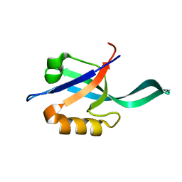
1V6N
 
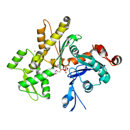
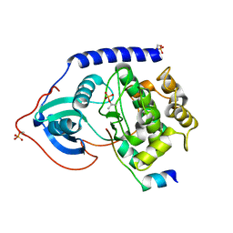
 | | Peanut lectin with 9mer peptide (PVIWSSATG) | | 分子名称: | CALCIUM ION, Galactose-binding lectin, MANGANESE (II) ION | | 著者 | Kundhavai Natchiar, S, Arockia Jeyaprakash, A, Ramya, T.N.C, Thomas, C.J, Suguna, K, Surolia, A, Vijayan, M. | | 登録日 | 2003-12-02 | | 公開日 | 2004-02-10 | | 最終更新日 | 2023-10-25 | | 実験手法 | X-RAY DIFFRACTION (3.5 Å) | | 主引用文献 | Structural plasticity of peanut lectin: an X-ray analysis involving variation in pH, ligand binding and crystal structure.
Acta Crystallogr.,Sect.D, 60, 2004
|
|
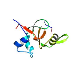
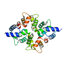
2HF4
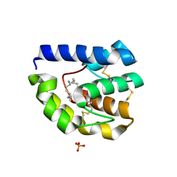
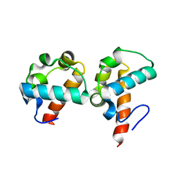
 | | Crystal structure of Monomeric Actin in its ATP-bound state | | 分子名称: | ADENOSINE-5'-TRIPHOSPHATE, Actin-5C, CALCIUM ION | | 著者 | Rould, M.A, Wan, Q, Joel, P.B, Lowey, S, Trybus, K.M. | | 登録日 | 2006-06-22 | | 公開日 | 2006-08-29 | | 最終更新日 | 2023-08-30 | | 実験手法 | X-RAY DIFFRACTION (1.8 Å) | | 主引用文献 | Crystal Structures of Expressed Non-polymerizable Monomeric Actin in the ADP and ATP States.
J.Biol.Chem., 281, 2006
|
|
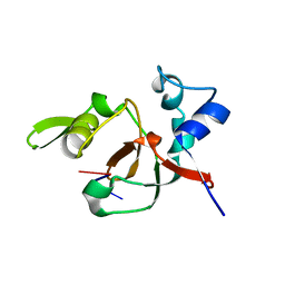
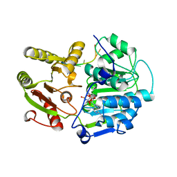
2GTE
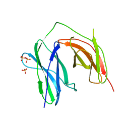
 | | Drosophila OBP LUSH bound to attractant pheromone 11-cis-vaccenyl acetate | | 分子名称: | (Z)-OCTADEC-11-ENYL ACETATE, General odorant-binding protein lush, PHOSPHATE ION | | 著者 | Laughlin, J.D, Ha, T, Smith, D.P, Jones, D.N.M. | | 登録日 | 2006-04-27 | | 公開日 | 2007-06-12 | | 最終更新日 | 2024-04-03 | | 実験手法 | X-RAY DIFFRACTION (1.4 Å) | | 主引用文献 | Activation of pheromone-sensitive neurons is mediated by conformational activation of pheromone-binding protein
Cell(Cambridge,Mass.), 133, 2008
|
|
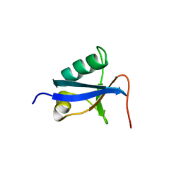
1V6O
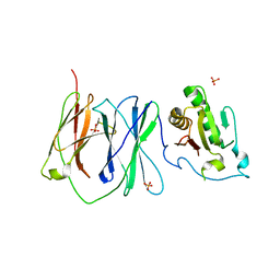
 | | Peanut lectin complexed with 10mer peptide (PVRIWSSATG) | | 分子名称: | CALCIUM ION, Galactose-binding lectin, MANGANESE (II) ION | | 著者 | Kundhavai Natchiar, S, Arockia Jeyaprakash, A, Ramya, T.N.C, Thomas, C.J, Suguna, K, Surolia, A, Vijayan, M. | | 登録日 | 2003-12-02 | | 公開日 | 2004-02-10 | | 最終更新日 | 2023-10-25 | | 実験手法 | X-RAY DIFFRACTION (3 Å) | | 主引用文献 | Structural plasticity of peanut lectin: an X-ray analysis involving variation in pH, ligand binding and crystal structure.
Acta Crystallogr.,Sect.D, 60, 2004
|
|
1V6J
 | | peanut lectin-lactose complex crystallized in orthorhombic form at acidic pH | | 分子名称: | CALCIUM ION, Galactose-binding lectin, MANGANESE (II) ION, ... | | 著者 | Kundhavai Natchiar, S, Arockia Jeyaprakash, A, Ramya, T.N.C, Thomas, C.J, Suguna, K, Surolia, A, Vijayan, M. | | 登録日 | 2003-12-01 | | 公開日 | 2004-02-10 | | 最終更新日 | 2023-12-27 | | 実験手法 | X-RAY DIFFRACTION (2.9 Å) | | 主引用文献 | Structural plasticity of peanut lectin: an X-ray analysis involving variation in pH, ligand binding and crystal structure.
Acta Crystallogr.,Sect.D, 60, 2004
|
|
1V6M
 | | Peanut Lectin with 9mer peptide (IWSSAGNVA) | | 分子名称: | CALCIUM ION, Galactose-binding lectin, MANGANESE (II) ION | | 著者 | Kundhavai Natchiar, S, Arockia Jeyaprakash, A, Ramya, T.N.C, Thomas, C.J, Suguna, K, Surolia, A, Vijayan, M. | | 登録日 | 2003-12-02 | | 公開日 | 2004-02-10 | | 最終更新日 | 2023-10-25 | | 実験手法 | X-RAY DIFFRACTION (2.7 Å) | | 主引用文献 | Structural plasticity of peanut lectin: an X-ray analysis involving variation in pH, ligand binding and crystal structure.
Acta Crystallogr.,Sect.D, 60, 2004
|
|
2IBB
 | |
2IBG
 | |
1TK7
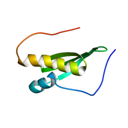
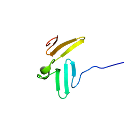 | | NMR structure of WW domains (WW3-4) from Suppressor of Deltex | | 分子名称: | CG4244-PB | | 著者 | Fedoroff, O.Y, Avis, J.M, Golovanov, A.P, Baron, M, Townson, S.A. | | 登録日 | 2004-06-08 | | 公開日 | 2004-07-20 | | 最終更新日 | 2024-05-22 | | 実験手法 | SOLUTION NMR | | 主引用文献 | The structure and dynamics of tandem WW domains in a negative regulator of notch signaling, Suppressor of deltex
J.Biol.Chem., 279, 2004
|
|
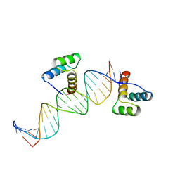
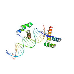
2GLO
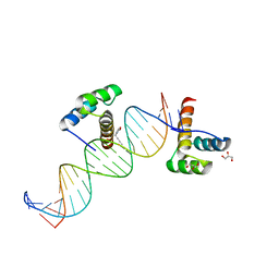
 | | Solution structure of the Brinker DNA binding domain in complex with the omb enhancer | | 分子名称: | 5'-D(*GP*TP*TP*GP*AP*CP*GP*CP*CP*TP*CP*A)-3', 5'-D(*TP*GP*AP*GP*GP*CP*GP*TP*CP*AP*AP*C)-3', brinker CG9653-PA | | 著者 | Cordier, F, Hartmann, B, Rogowski, M, Affolter, M, Grzesiek, S. | | 登録日 | 2006-04-05 | | 公開日 | 2006-08-29 | | 最終更新日 | 2024-05-29 | | 実験手法 | SOLUTION NMR | | 主引用文献 | DNA recognition by the brinker repressor - an extreme case of coupling between binding and folding
J.Mol.Biol., 361, 2006
|
|
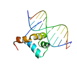
2HDD
 | | ENGRAILED HOMEODOMAIN Q50K VARIANT DNA COMPLEX | | 分子名称: | DNA (5'-D(*AP*TP*CP*CP*GP*GP*GP*GP*AP*TP*TP*AP*CP*AP*TP*GP*G P*CP*AP*AP*A)-3'), DNA (5'-D(*TP*TP*TP*TP*GP*CP*CP*AP*TP*GP*TP*AP*AP*TP*CP*CP*C P*CP*GP*GP*A)-3'), PROTEIN (ENGRAILED HOMEODOMAIN Q50K) | | 著者 | Tucker-Kellogg, L, Rould, M.A, Chambers, K.A, Ades, S.E, Sauer, R.T, Pabo, C.O. | | 登録日 | 1998-02-10 | | 公開日 | 1998-05-27 | | 最終更新日 | 2024-02-14 | | 実験手法 | X-RAY DIFFRACTION (1.9 Å) | | 主引用文献 | Engrailed (Gln50-->Lys) homeodomain-DNA complex at 1.9 A resolution: structural basis for enhanced affinity and altered specificity.
Structure, 5, 1997
|
|
2GNF
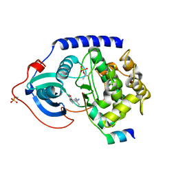 | | Protein kinase A fivefold mutant model of Rho-kinase with Y-27632 | | 分子名称: | (R)-TRANS-4-(1-AMINOETHYL)-N-(4-PYRIDYL) CYCLOHEXANECARBOXAMIDE, cAMP-dependent protein kinase inhibitor alpha, cAMP-dependent protein kinase, ... | | 著者 | Bonn, S, Herrero, S, Breitenlechner, C.B, Engh, R.A, Gassel, M, Bossemeyer, D. | | 登録日 | 2006-04-10 | | 公開日 | 2006-05-23 | | 最終更新日 | 2024-04-03 | | 実験手法 | X-RAY DIFFRACTION (2.28 Å) | | 主引用文献 | Structural analysis of protein kinase A mutants with Rho-kinase inhibitor specificity
J.Biol.Chem., 281, 2006
|
|
2GNG
 | | Protein kinase A fivefold mutant model of Rho-kinase | | 分子名称: | cAMP-dependent protein kinase inhibitor alpha, cAMP-dependent protein kinase, alpha-catalytic subunit | | 著者 | Bonn, S, Herrero, S, Breitenlechner, C.B, Engh, R.A, Gassel, M, Bossemeyer, D. | | 登録日 | 2006-04-10 | | 公開日 | 2006-05-23 | | 最終更新日 | 2024-04-03 | | 実験手法 | X-RAY DIFFRACTION (1.87 Å) | | 主引用文献 | Structural analysis of protein kinase A mutants with Rho-kinase inhibitor specificity
J.Biol.Chem., 281, 2006
|
|
1T2S
 | | Structural basis for 3' end recognition of nucleic acids by the Drosophila Argonaute 2 PAZ domain | | 分子名称: | 5'-D(*CP*TP*CP*AP*C)-3', Argonaute 2 | | 著者 | Lingel, A, Simon, B, Izaurralde, E, Sattler, M. | | 登録日 | 2004-04-22 | | 公開日 | 2004-06-01 | | 最終更新日 | 2024-05-22 | | 実験手法 | SOLUTION NMR | | 主引用文献 | Nucleic acid 3'-end recognition by the Argonaute2 PAZ domain.
Nat.Struct.Mol.Biol., 11, 2004
|
|
1T2R
 | | Structural basis for 3' end recognition of nucleic acids by the Drosophila Argonaute 2 PAZ domain | | 分子名称: | 5'-R(*CP*UP*CP*AP*C)-3', Argonaute 2 | | 著者 | Lingel, A, Simon, B, Izaurralde, E, Sattler, M. | | 登録日 | 2004-04-22 | | 公開日 | 2004-06-01 | | 最終更新日 | 2024-05-22 | | 実験手法 | SOLUTION NMR | | 主引用文献 | Nucleic acid 3'-end recognition by the Argonaute2 PAZ domain.
Nat.Struct.Mol.Biol., 11, 2004
|
|
2HOS
 | | Phage-Selected Homeodomain Bound to Unmodified DNA | | 分子名称: | 3-METHYL-1,3-OXAZOLIDIN-2-ONE, 5'-D(*AP*TP*CP*CP*GP*GP*GP*GP*AP*TP*TP*AP*CP*AP*TP*GP*GP*CP*AP*AP*A)-3', 5'-D(*TP*TP*TP*TP*GP*CP*CP*AP*TP*GP*TP*AP*AP*TP*CP*CP*CP*CP*GP*GP*A)-3', ... | | 著者 | Shokat, K.M, Feldman, M.E, Simon, M.D. | | 登録日 | 2006-07-16 | | 公開日 | 2006-12-12 | | 最終更新日 | 2023-08-30 | | 実験手法 | X-RAY DIFFRACTION (1.9 Å) | | 主引用文献 | Structure and properties of a re-engineered homeodomain protein-DNA interface.
Acs Chem.Biol., 1, 2006
|
|
2HOT
 | | Phage selected homeodomain bound to modified DNA | | 分子名称: | 3-PROP-2-YN-1-YL-1,3-OXAZOLIDIN-2-ONE, 5'-D(*AP*TP*CP*CP*GP*GP*GP*GP*AP*TP*TP*AP*CP*AP*TP*GP*GP*CP*AP*AP*A)-3', 5'-D(*TP*TP*TP*TP*GP*CP*CP*AP*TP*GP*TP*AP*AP*TP*CP*CP*CP*CP*GP*GP*A)-3', ... | | 著者 | Feldman, M.E, Simon, M.D, Shokat, K.M. | | 登録日 | 2006-07-16 | | 公開日 | 2006-12-12 | | 最終更新日 | 2024-02-14 | | 実験手法 | X-RAY DIFFRACTION (2.19 Å) | | 主引用文献 | Structure and properties of a re-engineered homeodomain protein-DNA interface.
Acs Chem.Biol., 1, 2006
|
|
1SV0
 | | Crystal Structure Of Yan-SAM/Mae-SAM Complex | | 分子名称: | Ets DNA-binding protein pokkuri, modulator of the activity of Ets CG15085-PA | | 著者 | Qiao, F, Song, H, Kim, C.A, Sawaya, M.R, Hunter, J.B, Gingery, M, Rebay, I, Courey, A.J, Bowie, J.U. | | 登録日 | 2004-03-26 | | 公開日 | 2004-07-27 | | 最終更新日 | 2024-02-14 | | 実験手法 | X-RAY DIFFRACTION (2.07 Å) | | 主引用文献 | Derepression by depolymerization; structural insights into the regulation of yan by mae.
Cell(Cambridge,Mass.), 118, 2004
|
|
2L6Z
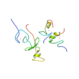 | | haddock model of GATA1NF:Lmo2LIM2-Ldb1LID with FOG | | 分子名称: | Erythroid transcription factor, LIM domain only 2, linker, ... | | 著者 | Wilkinson-White, L, Gamsjaeger, R, Dastmalchi, S, Wienert, B, Stokes, P.H, Crossley, M, Mackay, J.P, Matthews, J.M. | | 登録日 | 2010-12-01 | | 公開日 | 2011-08-31 | | 最終更新日 | 2024-05-01 | | 実験手法 | SOLUTION NMR | | 主引用文献 | Structural basis of simultaneous recruitment of the transcriptional regulators LMO2 and FOG1/ZFPM1 by the transcription factor GATA1
Proc.Natl.Acad.Sci.USA, 108, 2011
|
|
2LJH
 | |
2LC7
 | |
4BY4
 | |
2DB3
 | | Structural basis for RNA unwinding by the DEAD-box protein Drosophila Vasa | | 分子名称: | 5'-R(*UP*UP*UP*UP*UP*UP*UP*UP*UP*U)-3', ATP-dependent RNA helicase vasa, MAGNESIUM ION, ... | | 著者 | Sengoku, T, Nureki, O, Nakamura, A, Kobayashi, S, Yokoyama, S, RIKEN Structural Genomics/Proteomics Initiative (RSGI) | | 登録日 | 2005-12-14 | | 公開日 | 2006-05-02 | | 最終更新日 | 2024-03-13 | | 実験手法 | X-RAY DIFFRACTION (2.2 Å) | | 主引用文献 | Structural basis for RNA unwinding by the DEAD-box protein Drosophila Vasa.
Cell(Cambridge,Mass.), 125, 2006
|
|
4D2K
 | |
2DH1
 | | Crystal structure of peanut lectin lactose-azobenzene-4,4'-dicarboxylic acid-lactose complex | | 分子名称: | Galactose-binding lectin | | 著者 | Natchiar, S.K, Srinivas, O, Nivedita, M, Sagarika, D, Jayaraman, N, Surolia, A, Vijayan, M. | | 登録日 | 2006-03-17 | | 公開日 | 2006-08-15 | | 最終更新日 | 2023-10-25 | | 実験手法 | X-RAY DIFFRACTION (7.65 Å) | | 主引用文献 | Multivalency in lectins - A crystallographic, modelling and light-scattering study involving peanut lectin and a bivalent ligand
Curr.Sci., 90, 2006
|
|


