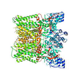
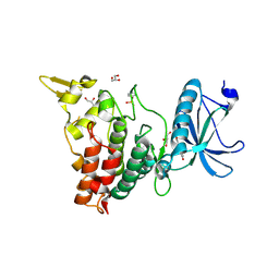
9AYF
 
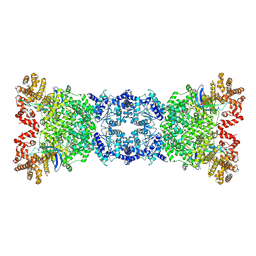
 | | Structure of human calcium-sensing receptor in complex with Gi1 (miniGi1) protein in detergent | | 分子名称: | 2-acetamido-2-deoxy-beta-D-glucopyranose, 2-acetamido-2-deoxy-beta-D-glucopyranose-(1-4)-2-acetamido-2-deoxy-beta-D-glucopyranose, 3-(2-chlorophenyl)-N-[(1R)-1-(3-methoxyphenyl)ethyl]propan-1-amine, ... | | 著者 | Zuo, H, Park, J, Frangaj, A, Ye, J, Lu, G, Manning, J.J, Asher, W.B, Lu, Z, Hu, G, Wang, L, Mendez, J, Eng, E, Zhang, Z, Lin, X, Grasucci, R, Hendrickson, W.A, Clarke, O.B, Javitch, J.A, Conigrave, A.D, Fan, Q.R. | | 登録日 | 2024-03-07 | | 公開日 | 2024-04-17 | | 最終更新日 | 2024-05-22 | | 実験手法 | ELECTRON MICROSCOPY (3.6 Å) | | 主引用文献 | Promiscuous G-protein activation by the calcium-sensing receptor.
Nature, 629, 2024
|
|
8X94
 | |
8YLN
 | | The structure of DSR2-Tail tube complex | | 分子名称: | Bacillus phage SPR Tube protein, SIR2-like domain-containing protein | | 著者 | Zheng, J, Yang, X. | | 登録日 | 2024-03-06 | | 公開日 | 2024-08-14 | | 実験手法 | ELECTRON MICROSCOPY (3.53 Å) | | 主引用文献 | Structural insights into autoinhibition and activation of defense-associated sirtuin protein.
Int.J.Biol.Macromol., 277, 2024
|
|
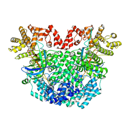
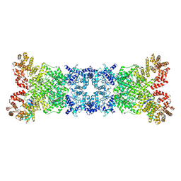
9AVG
 | | Structure of human calcium-sensing receptor in complex with chimeric Gs (miniGis) protein in nanodiscs | | 分子名称: | (19R,22S,25R)-22,25,26-trihydroxy-16,22-dioxo-17,21,23-trioxa-22lambda~5~-phosphahexacosan-19-yl (9Z)-octadec-9-enoate, 2-acetamido-2-deoxy-beta-D-glucopyranose, 2-acetamido-2-deoxy-beta-D-glucopyranose-(1-4)-2-acetamido-2-deoxy-beta-D-glucopyranose, ... | | 著者 | Zuo, H, Park, J, Frangaj, A, Ye, J, Lu, G, Manning, J.J, Asher, W.B, Lu, Z, Hu, G, Wang, L, Mendez, J, Eng, E, Zhang, Z, Lin, X, Grasucci, R, Hendrickson, W.A, Clarke, O.B, Javitch, J.A, Conigrave, A.D, Fan, Q.R. | | 登録日 | 2024-03-02 | | 公開日 | 2024-04-17 | | 最終更新日 | 2024-05-22 | | 実験手法 | ELECTRON MICROSCOPY (3.6 Å) | | 主引用文献 | Promiscuous G-protein activation by the calcium-sensing receptor.
Nature, 629, 2024
|
|
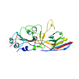
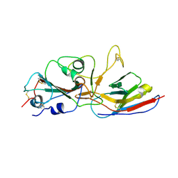
5WJ8
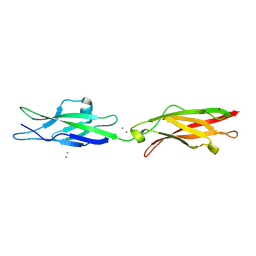 | | Crystal Structure of Human Cadherin-23 EC13-14 | | 分子名称: | CALCIUM ION, CHLORIDE ION, Cadherin-23, ... | | 著者 | Velez-Cortes, F, Conghui, C, De-la-Torre, P, Sotomayor, M. | | 登録日 | 2017-07-21 | | 公開日 | 2018-07-04 | | 最終更新日 | 2023-11-15 | | 実験手法 | X-RAY DIFFRACTION (1.86 Å) | | 主引用文献 | Zooming in on Cadherin-23: Structural Diversity and Potential Mechanisms of Inherited Deafness.
Structure, 26, 2018
|
|
9EO8
 | |
8Z18
 | |
8ZVI
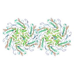 | | Structure of the bacteriophage T5 capsid | | 分子名称: | Decoration protein, Major capsid protein | | 著者 | Peng, Y, Liu, H.R. | | 登録日 | 2024-06-11 | | 公開日 | 2024-09-04 | | 実験手法 | ELECTRON MICROSCOPY (3.4 Å) | | 主引用文献 | Structures of Mature and Urea-Treated Empty Bacteriophage T5: Insights into Siphophage Infection and DNA Ejection.
Int J Mol Sci, 25, 2024
|
|
8YKF
 | |
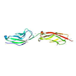
8ZES
 | | Crystal structure of the Wuhan SARS-CoV-2 RBD (333-541) complexed with P2C5 nanobody | | 分子名称: | 2-acetamido-2-deoxy-beta-D-glucopyranose, 2-acetamido-2-deoxy-beta-D-glucopyranose-(1-4)-2-acetamido-2-deoxy-beta-D-glucopyranose, Nanobody P2C5, ... | | 著者 | Sluchanko, N.N, Varfolomeeva, L.A, Shcheblyakov, D.V, Logunov, D.Y, Gintsburg, A.L, Popov, V.O, Boyko, K.M. | | 登録日 | 2024-05-06 | | 公開日 | 2024-09-04 | | 最終更新日 | 2024-09-18 | | 実験手法 | X-RAY DIFFRACTION (3.7 Å) | | 主引用文献 | Structural Basis for Evasion of New SARS-CoV-2 Variants from the Potent Virus-Neutralizing Nanobody Targeting the S-Protein Receptor-Binding Domain.
Biochemistry Mosc., 89, 2024
|
|
8ZER
 | | Crystal structure of the complex of Wuhan SARS-CoV-2 RBD (319-541) with P2C5 nanobody | | 分子名称: | 2-acetamido-2-deoxy-beta-D-glucopyranose-(1-4)-2-acetamido-2-deoxy-beta-D-glucopyranose, Nanobody P2C5, Spike protein S1, ... | | 著者 | Sluchanko, N.N, Varfolomeeva, L.A, Shcheblyakov, D.V, Logunov, D.Y, Gintsburg, A.L, Popov, V.O, Boyko, K.M. | | 登録日 | 2024-05-06 | | 公開日 | 2024-09-04 | | 最終更新日 | 2024-09-18 | | 実験手法 | X-RAY DIFFRACTION (3.1 Å) | | 主引用文献 | Structural Basis for Evasion of New SARS-CoV-2 Variants from the Potent Virus-Neutralizing Nanobody Targeting the S-Protein Receptor-Binding Domain.
Biochemistry Mosc., 89, 2024
|
|
5WJM
 | |
7ESR
 | |
7FHT
 | | Crystal structure of DYRK1A in complex with RD0448 | | 分子名称: | (5~{Z})-5-[(3-ethynyl-4-methoxy-phenyl)methylidene]-2-sulfanylidene-1,3-thiazolidin-4-one, Dual specificity tyrosine-phosphorylation-regulated kinase 1A | | 著者 | Kikuchi, M, Sumida, Y, Hosoya, T, Kii, I, Umehara, T. | | 登録日 | 2021-07-30 | | 公開日 | 2022-03-23 | | 最終更新日 | 2023-11-29 | | 実験手法 | X-RAY DIFFRACTION (2.68 Å) | | 主引用文献 | Structure-activity relationship for the folding intermediate-selective inhibition of DYRK1A.
Eur.J.Med.Chem., 227, 2022
|
|
7FHS
 | | Crystal structure of DYRK1A in complex with RD0392 | | 分子名称: | (5~{Z})-5-[(3-ethoxy-4-oxidanyl-phenyl)methylidene]-2-sulfanylidene-1,3-thiazolidin-4-one, Dual specificity tyrosine-phosphorylation-regulated kinase 1A, GLYCEROL | | 著者 | Kikuchi, M, Sumida, T, Hosoya, T, Kii, I, Umehara, T. | | 登録日 | 2021-07-30 | | 公開日 | 2022-03-23 | | 最終更新日 | 2023-11-29 | | 実験手法 | X-RAY DIFFRACTION (2.42 Å) | | 主引用文献 | Structure-activity relationship for the folding intermediate-selective inhibition of DYRK1A.
Eur.J.Med.Chem., 227, 2022
|
|
4EOG
 | | Crystal structure of Csx1 of Pyrococcus furiosus | | 分子名称: | Putative uncharacterized protein, SULFATE ION, ZINC ION | | 著者 | Kim, Y.K, Oh, B.H. | | 登録日 | 2012-04-14 | | 公開日 | 2013-01-02 | | 最終更新日 | 2013-07-24 | | 実験手法 | X-RAY DIFFRACTION (2.3 Å) | | 主引用文献 | Crystal structure and nucleic acid-binding activity of the CRISPR-associated protein Csx1 of Pyrococcus furiosus.
Proteins, 81, 2013
|
|
6DII
 | | Structure of Arabidopsis Fatty Acid Amide Hydrolase in Complex with methyl linolenyl fluorophosphonate | | 分子名称: | Fatty acid amide hydrolase, methyl-9Z,12Z,15Z-octadecatrienylphosphonofluoridate | | 著者 | Aziz, M, Wang, X, Tripathi, A, Bankaitis, V, Chapman, K.D. | | 登録日 | 2018-05-23 | | 公開日 | 2019-03-27 | | 最終更新日 | 2023-10-11 | | 実験手法 | X-RAY DIFFRACTION (3.2 Å) | | 主引用文献 | Structural analysis of a plant fatty acid amide hydrolase provides insights into the evolutionary diversity of bioactive acylethanolamides.
J.Biol.Chem., 294, 2019
|
|
4Q71
 | | Crystal Structure of Bradyrhizobium japonicum Proline Utilization A (PutA) Mutant D779W | | 分子名称: | FLAVIN-ADENINE DINUCLEOTIDE, GLYCEROL, Proline dehydrogenase, ... | | 著者 | Tanner, J.J, Luo, M, Pemberton, T.A. | | 登録日 | 2014-04-23 | | 公開日 | 2014-08-06 | | 最終更新日 | 2023-09-20 | | 実験手法 | X-RAY DIFFRACTION (2.2 Å) | | 主引用文献 | Kinetic and Structural Characterization of Tunnel-Perturbing Mutants in Bradyrhizobium japonicum Proline Utilization A.
Biochemistry, 53, 2014
|
|
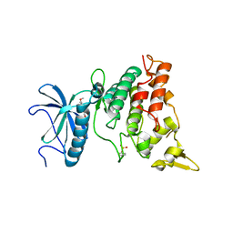
7X4H
 | | Crystal structure of CK2a1 complexed with AG1112 | | 分子名称: | 5-azanyl-3-[(~{Z})-1-cyano-2-(1~{H}-indol-3-yl)ethenyl]-1~{H}-pyrazole-4-carbonitrile, Casein Kinase 2 subunit alpha | | 著者 | Ikeda, A, Kinoshita, T, Tsuyuguchi, M. | | 登録日 | 2022-03-02 | | 公開日 | 2023-01-18 | | 最終更新日 | 2023-11-29 | | 実験手法 | X-RAY DIFFRACTION (1.77 Å) | | 主引用文献 | Bivalent binding mode of an amino-pyrazole inhibitor indicates the potentials for CK2 alpha 1-selective inhibitors.
Biochem.Biophys.Res.Commun., 630, 2022
|
|
6DHV
 | | Structure of Arabidopsis Fatty Acid Amide Hydrolase | | 分子名称: | Fatty acid amide hydrolase | | 著者 | Aziz, M, Wang, X, Tripathi, A, Bankaitis, V, Chapman, K.D. | | 登録日 | 2018-05-21 | | 公開日 | 2019-03-27 | | 最終更新日 | 2023-10-11 | | 実験手法 | X-RAY DIFFRACTION (2.099 Å) | | 主引用文献 | Structural analysis of a plant fatty acid amide hydrolase provides insights into the evolutionary diversity of bioactive acylethanolamides.
J.Biol.Chem., 294, 2019
|
|
3NZ4
 | |
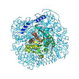
7F04
 | | Cytochrome c-type biogenesis protein CcmABCD from E. coli in complex with Heme and ATP. | | 分子名称: | 1,2-Distearoyl-sn-glycerophosphoethanolamine, ADENOSINE-5'-TRIPHOSPHATE, Cytochrome c biogenesis ATP-binding export protein CcmA, ... | | 著者 | Li, J, Zheng, W, Gu, M, Zhang, K, Zhu, J.P. | | 登録日 | 2021-06-03 | | 公開日 | 2022-11-09 | | 最終更新日 | 2024-06-12 | | 実験手法 | ELECTRON MICROSCOPY (2.86 Å) | | 主引用文献 | Structures of the CcmABCD heme release complex at multiple states.
Nat Commun, 13, 2022
|
|
6E8I
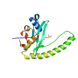 | |
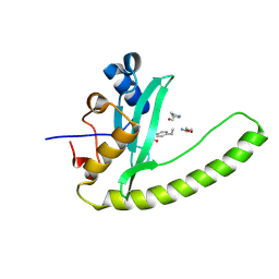
7FJR
 | | Structure of a mutant of OspA | | 分子名称: | DI(HYDROXYETHYL)ETHER, Outer surface protein A | | 著者 | Shiga, S, Makabe, K. | | 登録日 | 2021-08-04 | | 公開日 | 2022-08-10 | | 最終更新日 | 2024-02-21 | | 実験手法 | X-RAY DIFFRACTION (2.6 Å) | | 主引用文献 | beta-Strand-mediated Domain-swapping in the Absence of Hydrophobic Core Repacking.
J.Mol.Biol., 436, 2024
|
|
6E8K
 | |


