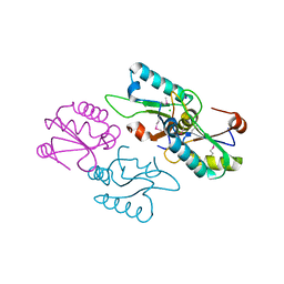
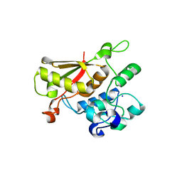
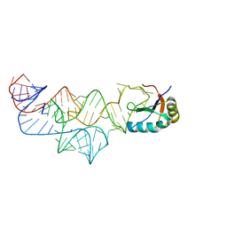
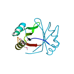
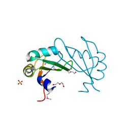
2LKZ
 
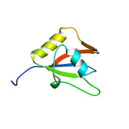 | | Solution structure of the second RRM domain of RBM5 | | 分子名称: | RNA-binding protein 5 | | 著者 | Song, Z, Wu, P, Zhang, J, Wu, J, Shi, Y. | | 登録日 | 2011-10-23 | | 公開日 | 2012-08-08 | | 最終更新日 | 2024-05-29 | | 実験手法 | SOLUTION NMR | | 主引用文献 | Solution structure of the second RRM domain of RBM5 and its unusual binding characters for different RNA targets
Biochemistry, 2012
|
|
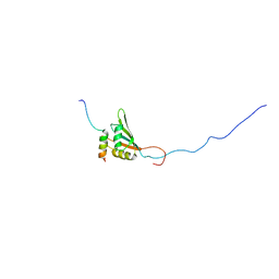
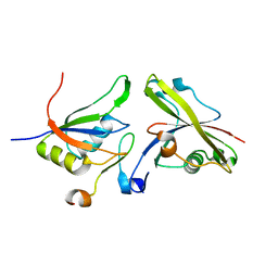
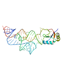
4F26
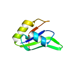 | |
4F25
 | |
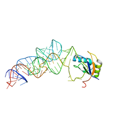
3VF0
 | | Raver1 in complex with metavinculin L954 deletion mutant | | 分子名称: | 4-(2-HYDROXYETHYL)-1-PIPERAZINE ETHANESULFONIC ACID, GLYCEROL, Ribonucleoprotein PTB-binding 1, ... | | 著者 | Lee, J.H, Vonrhein, C, Bricogne, G, Izard, T. | | 登録日 | 2012-01-09 | | 公開日 | 2012-07-25 | | 最終更新日 | 2012-09-19 | | 実験手法 | X-RAY DIFFRACTION (2.54 Å) | | 主引用文献 | The Metavinculin Tail Domain Directs Constitutive Interactions with Raver1 and vinculin RNA.
J.Mol.Biol., 422, 2012
|
|
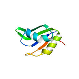
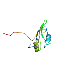
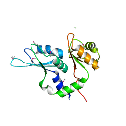
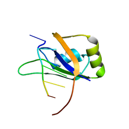
3V4M
 | |
2YKA
 | | RRM domain of mRNA export adaptor REF2-I bound to HVS ORF57 peptide | | 分子名称: | 52 KDA IMMEDIATE-EARLY PHOSPHOPROTEIN, RNA AND EXPORT FACTOR-BINDING PROTEIN 2 | | 著者 | Tunnicliffe, R.B, Hautbergue, G.M, Kalra, P, Wilson, S.A, Golovanov, A.P. | | 登録日 | 2011-05-26 | | 公開日 | 2012-06-13 | | 最終更新日 | 2024-05-15 | | 実験手法 | SOLUTION NMR | | 主引用文献 | Competitive and Cooperative Interactions Mediate RNA Transfer from Herpesvirus Saimiri Orf57 to the Mammalian Export Adaptor Alyref.
Plos Pathog., 10, 2014
|
|
2LCW
 | |
4EGL
 | | Crystal structure of two tandem RNA recognition motifs of Human antigen R | | 分子名称: | ELAV-like protein 1, GLYCEROL, SULFATE ION | | 著者 | Wang, H, Zeng, F, Liu, H, Teng, M, Li, X. | | 登録日 | 2012-03-31 | | 公開日 | 2012-05-30 | | 最終更新日 | 2023-11-08 | | 実験手法 | X-RAY DIFFRACTION (2.9 Å) | | 主引用文献 | Crystal structure of two tandem RNA recognition motifs of Human antigen R
To be Published
|
|
4ED5
 | | Crystal structure of the two N-terminal RRM domains of HuR complexed with RNA | | 分子名称: | 1,2-ETHANEDIOL, 1-METHOXY-2-(2-METHOXYETHOXY)ETHANE, 5'-R(*A*UP*UP*UP*UP*UP*AP*UP*UP*UP*U)-3', ... | | 著者 | Wang, H, Zeng, F, Liu, Q, Niu, L, Teng, M, Li, X. | | 登録日 | 2012-03-27 | | 公開日 | 2012-05-23 | | 最終更新日 | 2024-03-20 | | 実験手法 | X-RAY DIFFRACTION (2 Å) | | 主引用文献 | The structure of the ARE-binding domains of Hu antigen R (HuR) undergoes conformational changes during RNA binding.
Acta Crystallogr.,Sect.D, 69, 2013
|
|
3SDE
 | | Crystal structure of a paraspeckle-protein heterodimer, PSPC1/NONO | | 分子名称: | 1,2-ETHANEDIOL, Non-POU domain-containing octamer-binding protein, Paraspeckle component 1 | | 著者 | Passon, D.M, Lee, M, Bond, C.S. | | 登録日 | 2011-06-09 | | 公開日 | 2012-03-14 | | 最終更新日 | 2024-02-28 | | 実験手法 | X-RAY DIFFRACTION (1.9 Å) | | 主引用文献 | Structure of the heterodimer of human NONO and paraspeckle protein component 1 and analysis of its role in subnuclear body formation.
Proc.Natl.Acad.Sci.USA, 109, 2012
|
|
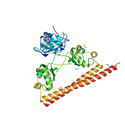
3TP2
 | |
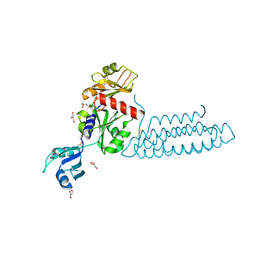
4A8X
 | | Structure of the core ASAP complex | | 分子名称: | HISTONE DEACETYLASE COMPLEX SUBUNIT SAP18, HOOK-LIKE, ISOFORM A, ... | | 著者 | Murachelli, A.G, Ebert, J, Basquin, C, Le Hir, H, Conti, E. | | 登録日 | 2011-11-21 | | 公開日 | 2012-03-07 | | 最終更新日 | 2023-12-20 | | 実験手法 | X-RAY DIFFRACTION (1.9 Å) | | 主引用文献 | The Structure of the Asap Core Complex Reveals the Existence of a Pinin-Containing Psap Complex
Nat.Struct.Mol.Biol., 19, 2012
|
|
3UWT
 | |
3UCU
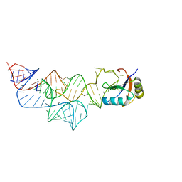 | | The c-di-GMP-I riboswitch bound to pGpG | | 分子名称: | MAGNESIUM ION, RNA (92-MER), U1 small nuclear ribonucleoprotein A, ... | | 著者 | Smith, K.D, Strobel, S.A. | | 登録日 | 2011-10-27 | | 公開日 | 2012-01-04 | | 最終更新日 | 2024-02-28 | | 実験手法 | X-RAY DIFFRACTION (2.8 Å) | | 主引用文献 | Structural and biochemical characterization of linear dinucleotide analogues bound to the c-di-GMP-I aptamer.
Biochemistry, 51, 2012
|
|
3UD4
 | |
3UD3
 | |
3UCZ
 | | The c-di-GMP-I riboswitch bound to GpG | | 分子名称: | MAGNESIUM ION, RNA (5'-R(*GP*G)-3'), RNA (92-MER), ... | | 著者 | Smith, K.D, Strobel, S.A. | | 登録日 | 2011-10-27 | | 公開日 | 2012-01-04 | | 最終更新日 | 2023-09-13 | | 実験手法 | X-RAY DIFFRACTION (2.8 Å) | | 主引用文献 | Structural and biochemical characterization of linear dinucleotide analogues bound to the c-di-GMP-I aptamer.
Biochemistry, 51, 2012
|
|
2RS2
 | | 1H, 13C, and 15N Chemical Shift Assignments for Musashi1 RBD1:r(GUAGU) complex | | 分子名称: | RNA (5'-R(*GP*UP*AP*GP*U)-3'), RNA-binding protein Musashi homolog 1 | | 著者 | Ohyama, T, Nagata, T, Tsuda, K, Imai, T, Okano, H, Yamazaki, T, Katahira, M. | | 登録日 | 2011-06-27 | | 公開日 | 2011-12-28 | | 最終更新日 | 2024-05-01 | | 実験手法 | SOLUTION NMR | | 主引用文献 | Structure of Musashi1 in a complex with target RNA: the role of aromatic stacking interactions
Nucleic Acids Res., 2011
|
|
3ULH
 | |
2LEA
 | | Solution structure of human SRSF2 (SC35) RRM | | 分子名称: | Serine/arginine-rich splicing factor 2 | | 著者 | Daubner, G.M, Clery, A, Jayne, S, Stevenin, J, Allain, F.H.-T. | | 登録日 | 2011-06-15 | | 公開日 | 2011-11-23 | | 最終更新日 | 2024-05-15 | | 実験手法 | SOLUTION NMR | | 主引用文献 | A syn-anti conformational difference allows SRSF2 to recognize guanines and cytosines equally well.
Embo J., 31, 2012
|
|
2LEC
 | | Solution structure of human SRSF2 (SC35) RRM in complex with 5'-UGGAGU-3' | | 分子名称: | RNA (5'-R(*UP*GP*GP*AP*GP*U)-3'), Serine/arginine-rich splicing factor 2 | | 著者 | Daubner, G.M, Clery, A, Jayne, S, Stevenin, J, Allain, F.H.-T. | | 登録日 | 2011-06-15 | | 公開日 | 2011-11-23 | | 最終更新日 | 2024-05-08 | | 実験手法 | SOLUTION NMR | | 主引用文献 | A syn-anti conformational difference allows SRSF2 to recognize guanines and cytosines equally well.
Embo J., 31, 2012
|
|
2LEB
 | | Solution structure of human SRSF2 (SC35) RRM in complex with 5'-UCCAGU-3' | | 分子名称: | RNA (5'-R(*UP*CP*CP*AP*GP*U)-3'), Serine/arginine-rich splicing factor 2 | | 著者 | Daubner, G.M, Clery, A, Jayne, S, Stevenin, J, Allain, F.H.-T. | | 登録日 | 2011-06-15 | | 公開日 | 2011-11-23 | | 最終更新日 | 2024-05-08 | | 実験手法 | SOLUTION NMR | | 主引用文献 | A syn-anti conformational difference allows SRSF2 to recognize guanines and cytosines equally well.
Embo J., 31, 2012
|
|
3UCG
 | |
3U1M
 | | Structure of the mRNA splicing complex component Cwc2 | | 分子名称: | Pre-mRNA-splicing factor CWC2, ZINC ION | | 著者 | Lu, P, Lu, G, Yan, C, Wang, L, Li, W, Yin, P. | | 登録日 | 2011-09-30 | | 公開日 | 2011-11-16 | | 最終更新日 | 2023-11-01 | | 実験手法 | X-RAY DIFFRACTION (1.95 Å) | | 主引用文献 | Structure of the mRNA splicing complex component Cwc2: insights into RNA recognition
Biochem.J., 441, 2012
|
|
3U1L
 | | Structure of the mRNA splicing complex component Cwc2 | | 分子名称: | Pre-mRNA-splicing factor CWC2, ZINC ION | | 著者 | Lu, P, Lu, G, Yan, C, Wang, L, Li, W, Yin, P. | | 登録日 | 2011-09-30 | | 公開日 | 2011-11-16 | | 最終更新日 | 2024-03-20 | | 実験手法 | X-RAY DIFFRACTION (1.64 Å) | | 主引用文献 | Structure of the mRNA splicing complex component Cwc2: insights into RNA recognition
Biochem.J., 441, 2012
|
|


