Loading
PDBj
メニューPDBj@FacebookPDBj@TwitterPDBj@YouTubewwPDB FoundationwwPDB
RCSB PDBPDBeBMRBAdv. SearchSearch help

7SXM
DownloadVisualize
BU of 7sxm by Molmil
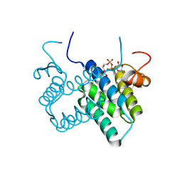
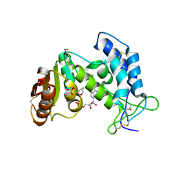
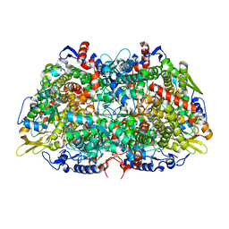
Structure of Xenon-derivatized Methyl-Coenzyme M Reductase from Methanothermobacter marburgensis
分子名称: 1-THIOETHANESULFONIC ACID, ACETATE ION, Coenzyme B, ...
著者Chen, P.Y.-T, Drennan, C.L.
登録日2021-11-23
公開日2022-04-06
最終更新日2023-11-15
実験手法X-RAY DIFFRACTION (2.503 Å)
主引用文献XFEL serial crystallography reveals the room temperature structure of methyl-coenzyme M reductase.
J.Inorg.Biochem., 230, 2022
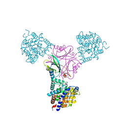
3NUH
DownloadVisualize
BU of 3nuh by Molmil
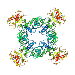
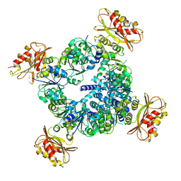
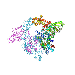
A domain insertion in E. coli GyrB adopts a novel fold that plays a critical role in gyrase function
分子名称: DNA gyrase subunit A, DNA gyrase subunit B, MAGNESIUM ION
著者Schoeffler, A.J, May, A.P, Berger, J.M.
登録日2010-07-06
公開日2010-08-11
最終更新日2023-11-22
実験手法X-RAY DIFFRACTION (3.103 Å)
主引用文献A domain insertion in Escherichia coli GyrB adopts a novel fold that plays a critical role in gyrase function.
Nucleic Acids Res., 38, 2010
3O5T
DownloadVisualize
BU of 3o5t by Molmil
Structure of DraG-GlnZ complex with ADP
分子名称: ADENOSINE-5'-DIPHOSPHATE, Dinitrogenase reductase activacting glicohydrolase, MAGNESIUM ION, ...
著者Rajendran, C, Li, X.-D, Winkler, F.K.
登録日2010-07-28
公開日2011-10-05
最終更新日2023-11-01
実験手法X-RAY DIFFRACTION (2.09 Å)
主引用文献Crystal structure of the GlnZ-DraG complex reveals a different form of PII-target interaction
Proc.Natl.Acad.Sci.USA, 108, 2011
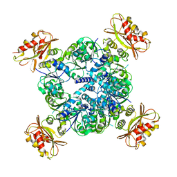
6UXA
DownloadVisualize
BU of 6uxa by Molmil
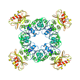
MthK N-terminal truncation state 2 bound with calcium
分子名称: Calcium-gated potassium channel MthK
著者Chen, F, Crina, N.
登録日2019-11-07
公開日2020-03-18
最終更新日2024-03-06
実験手法ELECTRON MICROSCOPY (4.5 Å)
主引用文献Ball-and-chain inactivation in a calcium-gated potassium channel.
Nature, 580, 2020
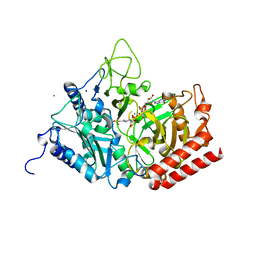
5FH0
DownloadVisualize
BU of 5fh0 by Molmil
The structure of rat cytosolic PEPCK variant E89A complex with GTP
分子名称: 4-(2-HYDROXYETHYL)-1-PIPERAZINE ETHANESULFONIC ACID, GUANOSINE-5'-TRIPHOSPHATE, MANGANESE (II) ION, ...
著者Johnson, T.A, Holyoak, T.
登録日2015-12-21
公開日2016-11-09
最終更新日2023-09-27
実験手法X-RAY DIFFRACTION (1.6 Å)
主引用文献Utilization of Substrate Intrinsic Binding Energy for Conformational Change and Catalytic Function in Phosphoenolpyruvate Carboxykinase.
Biochemistry, 55, 2016
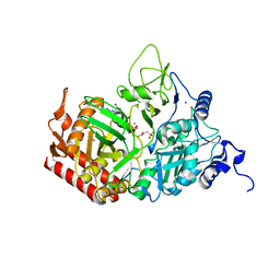
5FH4
DownloadVisualize
BU of 5fh4 by Molmil
The structure of rat cytosolic PEPCK variant E89D in complex with beta-sulfopyruvate and GTP
分子名称: 1,2-ETHANEDIOL, GUANOSINE-5'-TRIPHOSPHATE, MANGANESE (II) ION, ...
著者Johnson, T.A, Holyoak, T.
登録日2015-12-21
公開日2016-11-09
最終更新日2023-09-27
実験手法X-RAY DIFFRACTION (1.49 Å)
主引用文献Utilization of Substrate Intrinsic Binding Energy for Conformational Change and Catalytic Function in Phosphoenolpyruvate Carboxykinase.
Biochemistry, 55, 2016
3O55
DownloadVisualize
BU of 3o55 by Molmil
Crystal structure of human FAD-linked augmenter of liver regeneration (ALR)
分子名称: Augmenter of liver regeneration, FLAVIN-ADENINE DINUCLEOTIDE
著者Banci, L, Bertini, I, Calderone, V, Cefaro, C, Ciofi-Baffoni, S, Gallo, A, Kallergi, E, Lionaki, E, Pozidis, C, Tokatlidis, K.
登録日2010-07-28
公開日2011-04-13
最終更新日2023-09-06
実験手法X-RAY DIFFRACTION (1.9 Å)
主引用文献Molecular recognition and substrate mimicry drive the electron-transfer process between MIA40 and ALR.
Proc.Natl.Acad.Sci.USA, 108, 2011
3NVE
DownloadVisualize
BU of 3nve by Molmil
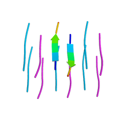
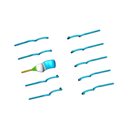
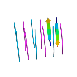
MMHFGN segment 138-143 from Syrian Hamster prion
分子名称: Major prion protein
著者Apostol, M.I, Sawaya, M.R, Eisenberg, D.
登録日2010-07-08
公開日2011-03-02
最終更新日2024-02-21
実験手法X-RAY DIFFRACTION (1.7 Å)
主引用文献Atomic structures suggest determinants of transmission barriers in Mammalian prion disease.
Biochemistry, 50, 2011
3OAP
DownloadVisualize
BU of 3oap by Molmil
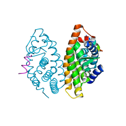
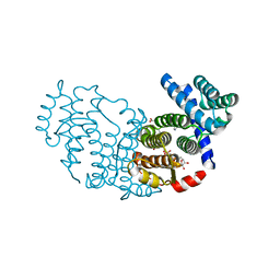
Crystal structure of human Retinoid X Receptor alpha-ligand binding domain complex with 9-cis retinoic acid and the coactivator peptide GRIP-1
分子名称: (9cis)-retinoic acid, Nuclear receptor coactivator 2, Retinoic acid receptor RXR-alpha
著者Xia, G, Smith, C.D, Muccio, D.D.
登録日2010-08-05
公開日2010-11-17
最終更新日2024-02-21
実験手法X-RAY DIFFRACTION (2.05 Å)
主引用文献Structure, Energetics and Dynamics of Binding Coactivator Peptide to Human Retinoid X Receptor Alpha Ligand Binding Domain Complex with 9-cis-Retinoic Acid.
Biochemistry, 50, 2011
3NVF
DownloadVisualize
BU of 3nvf by Molmil
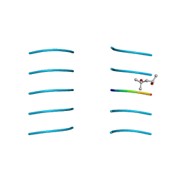
IIHFGS segment 138-143 from human prion
分子名称: (4S)-2-METHYL-2,4-PENTANEDIOL, Major prion protein
著者Apostol, M.I, Sawaya, M.R, Eisenberg, D.
登録日2010-07-08
公開日2011-03-02
最終更新日2024-02-21
実験手法X-RAY DIFFRACTION (1.8 Å)
主引用文献Atomic structures suggest determinants of transmission barriers in Mammalian prion disease.
Biochemistry, 50, 2011
6UWN
DownloadVisualize
BU of 6uwn by Molmil
MthK N-terminal truncation RCK domain state 1 bound with calcium
分子名称: CALCIUM ION, Calcium-gated potassium channel MthK
著者Chen, F, Crina, N.
登録日2019-11-05
公開日2020-03-18
最終更新日2024-03-06
実験手法ELECTRON MICROSCOPY (3.5 Å)
主引用文献Ball-and-chain inactivation in a calcium-gated potassium channel.
Nature, 580, 2020
6UX7
DownloadVisualize
BU of 6ux7 by Molmil
MthK N-terminal truncation state 1 bound with calcium
分子名称: Calcium-gated potassium channel MthK
著者Chen, F, Crina, N.
登録日2019-11-06
公開日2020-03-18
最終更新日2024-03-06
実験手法ELECTRON MICROSCOPY (6.7 Å)
主引用文献Ball-and-chain inactivation in a calcium-gated potassium channel.
Nature, 580, 2020
3NVG
DownloadVisualize
BU of 3nvg by Molmil
MIHFGN segment 137-142 from mouse prion
分子名称: Major prion protein
著者Apostol, M.I, Sawaya, M.R, Eisenberg, D.
登録日2010-07-08
公開日2011-03-02
最終更新日2024-02-21
実験手法X-RAY DIFFRACTION (1.48 Å)
主引用文献Atomic structures suggest determinants of transmission barriers in Mammalian prion disease.
Biochemistry, 50, 2011
6UX4
DownloadVisualize
BU of 6ux4 by Molmil
MthK N-terminal truncation RCK domain state 2 bound with calcium
分子名称: CALCIUM ION, Calcium-gated potassium channel MthK
著者Chen, F, Crina, N.
登録日2019-11-06
公開日2020-03-18
最終更新日2024-03-06
実験手法ELECTRON MICROSCOPY (3.5 Å)
主引用文献Ball-and-chain inactivation in a calcium-gated potassium channel.
Nature, 580, 2020
3OGU
DownloadVisualize
BU of 3ogu by Molmil
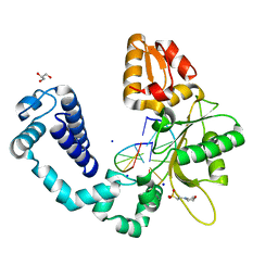
DNA Polymerase beta mutant 5P20 complexed with 6bp of DNA
分子名称: 2-(N-MORPHOLINO)-ETHANESULFONIC ACID, 5'-D(*CP*AP*TP*CP*TP*G)-3', 5'-D(P*CP*AP*GP*AP*TP*G)-3', ...
著者Marx, A, Diederichs, K, Bergen, K.
登録日2010-08-17
公開日2010-11-24
最終更新日2023-09-06
実験手法X-RAY DIFFRACTION (1.845 Å)
主引用文献Human DNA polymerase beta mutations allowing efficient abasic site bypass.
J.Biol.Chem., 286, 2011
3OG9
DownloadVisualize
BU of 3og9 by Molmil
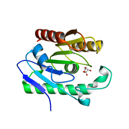
Structure of YahD with Malic acid
分子名称: D-MALATE, protein yahD a copper inducible hydrolase
著者Martinez Font, J, Mancini, S, Tauberger, E, Moniot, S.
登録日2010-08-16
公開日2010-12-15
最終更新日2023-09-06
実験手法X-RAY DIFFRACTION (1.88 Å)
主引用文献Regulation and structure of YahD, a copper inducible alpha/beta hydrolase of Lactococcus lactis IL1403
Fems Microbiol.Lett., 314, 2011
3OGI
DownloadVisualize
BU of 3ogi by Molmil
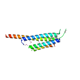
Crystal structure of the Mycobacterium tuberculosis H37Rv EsxOP complex (Rv2346c-Rv2347c)
分子名称: Putative ESAT-6-like protein 6, Putative ESAT-6-like protein 7
著者Arbing, M.A, Chan, S, Zhou, T.T, Ahn, C, Harris, L, Kuo, E, Sawaya, M.R, Cascio, D, Eisenberg, D, Integrated Center for Structure and Function Innovation (ISFI), TB Structural Genomics Consortium (TBSGC)
登録日2010-08-16
公開日2010-08-25
最終更新日2014-05-21
実験手法X-RAY DIFFRACTION (2.549 Å)
主引用文献Heterologous expression of mycobacterial Esx complexes in Escherichia coli for structural studies is facilitated by the use of maltose binding protein fusions.
Plos One, 8, 2013
3OWI
DownloadVisualize
BU of 3owi by Molmil
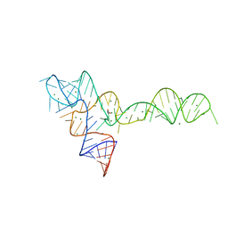
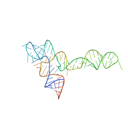
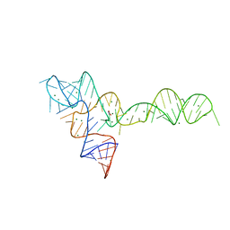
Crystal structure of the glycine riboswitch bound to glycine
分子名称: Domain II of glycine riboswitch, GLYCINE, MAGNESIUM ION
著者Huang, L, Serganov, A, Patel, D.J.
登録日2010-09-19
公開日2010-12-29
最終更新日2023-09-06
実験手法X-RAY DIFFRACTION (2.845 Å)
主引用文献Structural insights into ligand recognition by a sensing domain of the cooperative glycine riboswitch.
Mol.Cell, 40, 2010
3OXD
DownloadVisualize
BU of 3oxd by Molmil
Crystal structure of glycine riboswitch with two mutations
分子名称: MAGNESIUM ION, domain II of glycine riboswitch
著者Huang, L, Serganov, A, Patel, D.J.
登録日2010-09-21
公開日2010-12-29
最終更新日2023-09-06
実験手法X-RAY DIFFRACTION (2.999 Å)
主引用文献Structural insights into ligand recognition by a sensing domain of the cooperative glycine riboswitch.
Mol.Cell, 40, 2010
3OTO
DownloadVisualize
BU of 3oto by Molmil
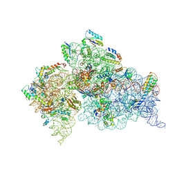
Crystal Structure of the 30S ribosomal subunit from a KsgA mutant of Thermus thermophilus (HB8)
分子名称: 16S rRNA, 30S RIBOSOMAL PROTEIN S10, 30S RIBOSOMAL PROTEIN S11, ...
著者Demirci, H, Murphy IV, F, Belardinelli, R, Kelley, A.C, Ramakrishnan, V, Gregory, S.T, Dahlberg, A.E, Jogl, G.
登録日2010-09-13
公開日2010-09-29
最終更新日2023-09-06
実験手法X-RAY DIFFRACTION (3.69 Å)
主引用文献Modification of 16S ribosomal RNA by the KsgA methyltransferase restructures the 30S subunit to optimize ribosome function.
Rna, 16, 2010
3P0K
DownloadVisualize
BU of 3p0k by Molmil
Structure of Baculovirus Sulfhydryl Oxidase Ac92
分子名称: ACETATE ION, FLAVIN-ADENINE DINUCLEOTIDE, IMIDAZOLE, ...
著者Hakim, M, Fass, D.
登録日2010-09-29
公開日2011-12-07
実験手法X-RAY DIFFRACTION (1.47 Å)
主引用文献Structure of a baculovirus sulfhydryl oxidase, a highly divergent member of the erv flavoenzyme family.
J.Virol., 85, 2011
7N71
DownloadVisualize
BU of 7n71 by Molmil
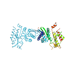
Crystal Structure of PI5P4KIIAlpha
分子名称: Phosphatidylinositol 5-phosphate 4-kinase type-2 alpha, SULFATE ION
著者Chen, S, Ha, Y.
登録日2021-06-09
公開日2021-07-07
最終更新日2023-10-18
実験手法X-RAY DIFFRACTION (2.5 Å)
主引用文献Pharmacological inhibition of PI5P4K alpha / beta disrupts cell energy metabolism and selectively kills p53-null tumor cells.
Proc.Natl.Acad.Sci.USA, 118, 2021
3P5S
DownloadVisualize
BU of 3p5s by Molmil
Structural insights into the catalytic mechanism of CD38: Evidence for a conformationally flexible covalent enzyme-substrate complex
分子名称: 2-acetamido-2-deoxy-beta-D-glucopyranose, CD38 molecule, SULFATE ION, ...
著者Egea, P.F, Muller-Stauffler, H, Kohn, I, Cakou-Kefir, C, Stroud, R.M, Kellenberburger, E, Schuber, F.
登録日2010-10-10
公開日2011-10-19
最終更新日2023-09-06
実験手法X-RAY DIFFRACTION (1.95 Å)
主引用文献Insights into the mechanism of bovine CD38/NAD+glycohydrolase from the X-ray structures of its Michaelis complex and covalently-trapped intermediates.
Plos One, 7, 2012
3OW9
DownloadVisualize
BU of 3ow9 by Molmil
Structure of an amyloid forming peptide KLVFFA from amyloid beta, alternate polymorph II
分子名称: KLVFFA hexapeptide segment from Amyloid beta
著者Landau, M, Eisenberg, D.
登録日2010-09-17
公開日2011-08-31
最終更新日2024-02-21
実験手法X-RAY DIFFRACTION (1.8 Å)
主引用文献Molecular basis for amyloid-{beta} polymorphism.
Proc.Natl.Acad.Sci.USA, 108, 2011
3OWW
DownloadVisualize
BU of 3oww by Molmil
Crystal structure of the glycine riboswitch bound to glycine
分子名称: GLYCINE, MAGNESIUM ION, domain II of glycine riboswitch
著者Huang, L, Serganov, A, Patel, D.J.
登録日2010-09-20
公開日2010-12-29
最終更新日2023-09-06
実験手法X-RAY DIFFRACTION (2.802 Å)
主引用文献Structural insights into ligand recognition by a sensing domain of the cooperative glycine riboswitch.
Mol.Cell, 40, 2010

223790

件を2024-08-14に公開中

PDB statisticsPDBj update infoContact PDBjnumon