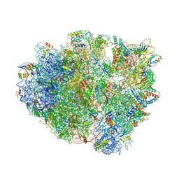
4V7J
 
 | | Structure of RelE nuclease bound to the 70S ribosome (precleavage state) | | 分子名称: | 30S ribosomal protein S10, 30S ribosomal protein S11, 30S ribosomal protein S12, ... | | 著者 | Neubauer, C, Gao, Y.-G, Andersen, K.R, Dunham, C.M, Kelley, A.C, Hentschel, J, Gerdes, K, Ramakrishnan, V, Brodersen, D.E. | | 登録日 | 2009-11-02 | | 公開日 | 2014-07-09 | | 最終更新日 | 2024-10-16 | | 実験手法 | X-RAY DIFFRACTION (3.3 Å) | | 主引用文献 | The structural basis for mRNA recognition and cleavage by the ribosome-dependent endonuclease RelE.
Cell(Cambridge,Mass.), 139, 2009
|
|
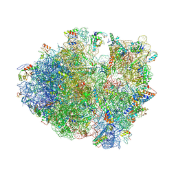
4V9B
 | | Crystal Structure of the 70S ribosome with tigecycline. | | 分子名称: | 16S ribosomal RNA, 23S ribosomal RNA, 30S RIBOSOMAL PROTEIN S10, ... | | 著者 | Jenner, L, Yusupov, M, Yusupova, G. | | 登録日 | 2012-07-18 | | 公開日 | 2014-07-09 | | 最終更新日 | 2023-09-20 | | 実験手法 | X-RAY DIFFRACTION (3.1 Å) | | 主引用文献 | Structural basis for potent inhibitory activity of the antibiotic tigecycline during protein synthesis.
Proc.Natl.Acad.Sci.USA, 110, 2013
|
|
4V84
 | | Crystal structure of a complex containing domain 3 of CrPV IGR IRES RNA bound to the 70S ribosome. | | 分子名称: | 23S ribosomal RNA, 30S ribosomal protein S10, 30S ribosomal protein S11, ... | | 著者 | Zhu, J, Korostelev, A, Costantino, D, Noller, H.F, Kieft, J.S. | | 登録日 | 2010-12-13 | | 公開日 | 2014-07-09 | | 最終更新日 | 2019-07-17 | | 実験手法 | X-RAY DIFFRACTION (3.4 Å) | | 主引用文献 | Crystal structures of complexes containing domains from two viral internal ribosome entry site (IRES) RNAs bound to the 70S ribosome.
Proc.Natl.Acad.Sci.USA, 108, 2011
|
|
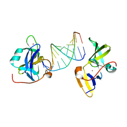
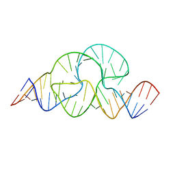
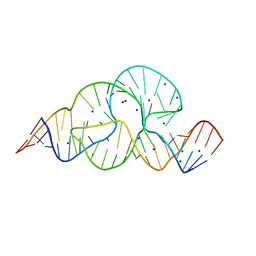
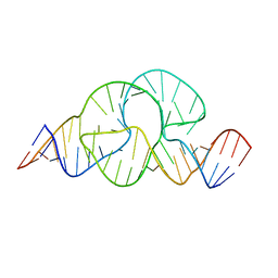
1D4R
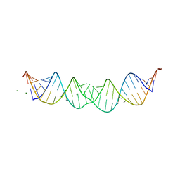 | | 29-mer fragment of human srp rna helix 6 | | 分子名称: | 29-MER OF MODIFIED SRP RNA HELIX 6, MAGNESIUM ION | | 著者 | Wild, K, Weichenrieder, O, Leonard, G.A, Cusack, S. | | 登録日 | 1999-10-05 | | 公開日 | 1999-12-02 | | 最終更新日 | 2024-02-07 | | 実験手法 | X-RAY DIFFRACTION (2 Å) | | 主引用文献 | The 2 A structure of helix 6 of the human signal recognition particle RNA
Structure Fold.Des., 7, 1999
|
|
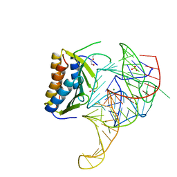
1DLM
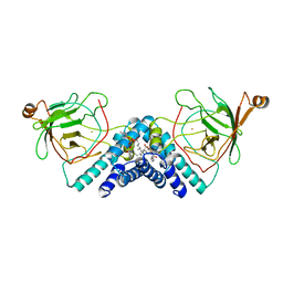 | | STRUCTURE OF CATECHOL 1,2-DIOXYGENASE FROM ACINETOBACTER CALCOACETICUS NATIVE DATA | | 分子名称: | CATECHOL 1,2-DIOXYGENASE, FE (III) ION, [1-PENTADECANOYL-2-DECANOYL-GLYCEROL-3-YL]PHOSPHONYL CHOLINE | | 著者 | Vetting, M.W, Ohlendorf, D.H. | | 登録日 | 1999-12-11 | | 公開日 | 2000-05-23 | | 最終更新日 | 2024-02-07 | | 実験手法 | X-RAY DIFFRACTION (2 Å) | | 主引用文献 | The 1.8 A crystal structure of catechol 1,2-dioxygenase reveals a novel hydrophobic helical zipper as a subunit linker.
Structure Fold.Des., 8, 2000
|
|
7BAI
 | |
4MQ7
 | | Structure of human CD1d-sulfatide | | 分子名称: | (15Z)-N-((1S,2R,3E)-2-HYDROXY-1-{[(3-O-SULFO-BETA-D-GALACTOPYRANOSYL)OXY]METHYL}HEPTADEC-3-ENYL)TETRACOS-15-ENAMIDE, 2-acetamido-2-deoxy-beta-D-glucopyranose, Antigen-presenting glycoprotein CD1d, ... | | 著者 | Luoma, A.M, Adams, E.J. | | 登録日 | 2013-09-15 | | 公開日 | 2013-12-18 | | 最終更新日 | 2023-09-20 | | 実験手法 | X-RAY DIFFRACTION (2.6032 Å) | | 主引用文献 | Crystal Structure of V delta 1 T Cell Receptor in Complex with CD1d-Sulfatide Shows MHC-like Recognition of a Self-Lipid by Human gamma delta T Cells.
Immunity, 39, 2013
|
|
3NCU
 | |
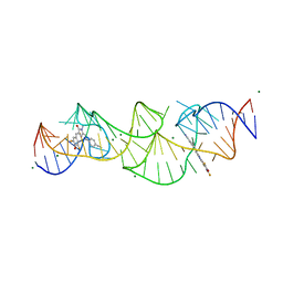
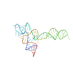
3OXJ
 | | crystal structure of glycine riboswitch, soaked in Ba2+ | | 分子名称: | BARIUM ION, GLYCINE, MAGNESIUM ION, ... | | 著者 | Huang, L, Serganov, A, Patel, D.J. | | 登録日 | 2010-09-21 | | 公開日 | 2010-12-29 | | 最終更新日 | 2024-03-13 | | 実験手法 | X-RAY DIFFRACTION (3.2 Å) | | 主引用文献 | Structural insights into ligand recognition by a sensing domain of the cooperative glycine riboswitch.
Mol.Cell, 40, 2010
|
|
5AOX
 | |
3ZD4
 | |
3ZP8
 | | HIGH-RESOLUTION FULL-LENGTH HAMMERHEAD RIBOZYME | | 分子名称: | HAMMERHEAD RIBOZYME, ENZYME STRAND, SUBSTRATE STRAND, ... | | 著者 | Anderson, M, Schultz, E, Martick, M, Scott, W.G. | | 登録日 | 2013-02-26 | | 公開日 | 2013-03-06 | | 最終更新日 | 2023-12-20 | | 実験手法 | X-RAY DIFFRACTION (1.55 Å) | | 主引用文献 | Active-Site Monovalent Cations Revealed in a 1.55 A Resolution Hammerhead Ribozyme Structure
J.Mol.Biol., 425, 2013
|
|
3ZD5
 | |
3DIY
 | |
8HZD
 | | A new fluorescent RNA aptamer bound with N618 | | 分子名称: | 4-[(~{E})-2-[(4~{Z})-4-[[3,5-bis(fluoranyl)-4-oxidanyl-phenyl]methylidene]-1-methyl-5-oxidanylidene-imidazol-2-yl]ethenyl]benzenecarbonitrile, MAGNESIUM ION, RNA (36-MER) | | 著者 | Song, Q.Q, Ren, A.M. | | 登録日 | 2023-01-09 | | 公開日 | 2024-06-19 | | 実験手法 | X-RAY DIFFRACTION (1.87 Å) | | 主引用文献 | Structural basis of a small monomeric Clivia fluorogenic RNA with a large Stokes shift.
Nat.Chem.Biol., 2024
|
|
7EOH
 | |
7EOJ
 | |
6XN9
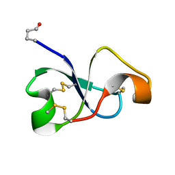 | | Solution NMR structure of recifin, a cysteine-rich tyrosyl-DNA Phosphodiesterase I modulatory peptide from the marine sponge Axinella sp. | | 分子名称: | Recifin modulatory peptide | | 著者 | Schroeder, C.I, Rosengren, K.J, O'Keefe, B.R. | | 登録日 | 2020-07-02 | | 公開日 | 2021-02-10 | | 実験手法 | SOLUTION NMR | | 主引用文献 | Recifin A, Initial Example of the Tyr-Lock Peptide Structural Family, Is a Selective Allosteric Inhibitor of Tyrosyl-DNA Phosphodiesterase I.
J.Am.Chem.Soc., 142, 2020
|
|
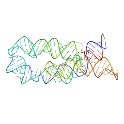
8XZK
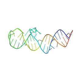 | | Crystal structure of folE riboswitch | | 分子名称: | RNA (53-MER) | | 著者 | Li, C.Y, Ren, A.M. | | 登録日 | 2024-01-21 | | 公開日 | 2024-07-24 | | 最終更新日 | 2024-08-21 | | 実験手法 | X-RAY DIFFRACTION (2.58 Å) | | 主引用文献 | Structure-based characterization and compound identification of the wild-type THF class-II riboswitch.
Nucleic Acids Res., 52, 2024
|
|
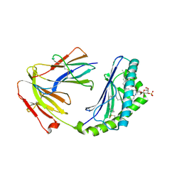
1RYD
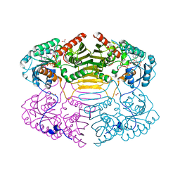 | | Crystal Structure of Glucose-Fructose Oxidoreductase from Zymomonas mobilis | | 分子名称: | ACETATE ION, BETA-MERCAPTOETHANOL, NADPH DIHYDRO-NICOTINAMIDE-ADENINE-DINUCLEOTIDE PHOSPHATE, ... | | 著者 | Kim, Y, Arora, M, Straza, M, Joachimiak, A. | | 登録日 | 2003-12-22 | | 公開日 | 2005-02-15 | | 最終更新日 | 2023-08-23 | | 実験手法 | X-RAY DIFFRACTION (2.2 Å) | | 主引用文献 | Crystal Structure of Glucose-Fructose Oxidoreductase from Zymomonas mobilis
To be Published
|
|
3OWI
 | |
3OXD
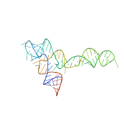 | |
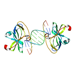
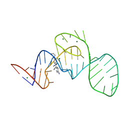
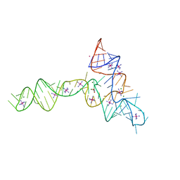
3OXE
 | | crystal structure of glycine riboswitch, Mn2+ soaked | | 分子名称: | GLYCINE, MAGNESIUM ION, MANGANESE (II) ION, ... | | 著者 | Huang, L, Serganov, A, Patel, D.J. | | 登録日 | 2010-09-21 | | 公開日 | 2010-12-29 | | 最終更新日 | 2023-09-06 | | 実験手法 | X-RAY DIFFRACTION (2.899 Å) | | 主引用文献 | Structural insights into ligand recognition by a sensing domain of the cooperative glycine riboswitch.
Mol.Cell, 40, 2010
|
|
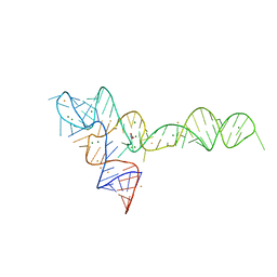
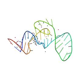
3OWZ
 | | Crystal structure of glycine riboswitch, soaked in Iridium | | 分子名称: | Domain II of glycine riboswitch, GLYCINE, IRIDIUM HEXAMMINE ION, ... | | 著者 | Huang, L, Serganov, A, Patel, D.J. | | 登録日 | 2010-09-20 | | 公開日 | 2010-12-29 | | 最終更新日 | 2024-02-21 | | 実験手法 | X-RAY DIFFRACTION (2.949 Å) | | 主引用文献 | Structural insights into ligand recognition by a sensing domain of the cooperative glycine riboswitch.
Mol.Cell, 40, 2010
|
|
3OXB
 | |


