2MR7
 
 | |
2MR8
 | |
2MR9
 | |
2MRA
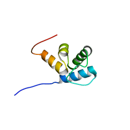
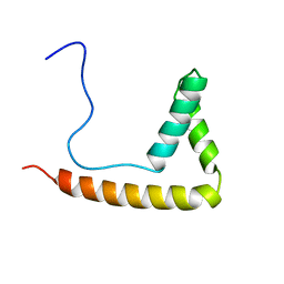
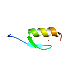
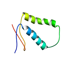
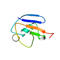
 | | Solution NMR Structure of De novo designed protein, Northeast Structural Genomics Consortium (NESG) Target OR459 | | 分子名称: | De novo designed protein OR459 | | 著者 | Pulavarti, S.V.S.R.K, Kipnis, Y, Sukumaran, D, Maglaqui, M, Janjua, H, Mao, L, Xiao, R, Kornhaber, G, Baker, D, Montelione, G.T, Szyperski, T, Northeast Structural Genomics Consortium (NESG) | | 登録日 | 2014-07-02 | | 公開日 | 2014-09-17 | | 最終更新日 | 2024-05-15 | | 実験手法 | SOLUTION NMR | | 主引用文献 | Solution NMR Structure of De novo designed protein, Northeast Structural Genomics Consortium (NESG) Target OR459
To be Published
|
|
2MRB
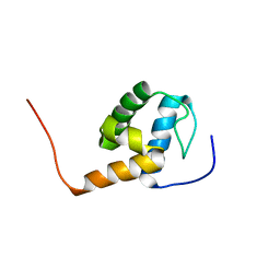
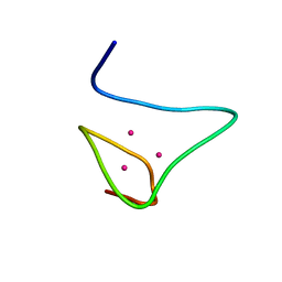 | | THREE-DIMENSIONAL STRUCTURE OF RABBIT LIVER CD-7 METALLOTHIONEIN-2A IN AQUEOUS SOLUTION DETERMINED BY NUCLEAR MAGNETIC RESONANCE | | 分子名称: | CADMIUM ION, CD7 METALLOTHIONEIN-2A | | 著者 | Braun, W, Arseniev, A, Schultze, P, Woergoetter, E, Wagner, G, Vasak, M, Kaegi, J.H.R, Wuthrich, K. | | 登録日 | 1990-05-14 | | 公開日 | 1991-04-15 | | 最終更新日 | 2024-05-22 | | 実験手法 | SOLUTION NMR | | 主引用文献 | Three-dimensional structure of rabbit liver [Cd7]metallothionein-2a in aqueous solution determined by nuclear magnetic resonance.
J.Mol.Biol., 201, 1988
|
|
2MRC
 | |
2MRD
 | |
2MRE
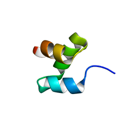
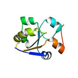
 | | NMR structure of the Rad18-UBZ/ubiquitin complex | | 分子名称: | E3 ubiquitin-protein ligase RAD18, Polyubiquitin-C, ZINC ION | | 著者 | Rizzo, A.A, Salerno, P.E, Bezsonova, I, Korzhnev, D.M. | | 登録日 | 2014-07-03 | | 公開日 | 2014-10-22 | | 最終更新日 | 2024-05-01 | | 実験手法 | SOLUTION NMR | | 主引用文献 | NMR Structure of the Human Rad18 Zinc Finger in Complex with Ubiquitin Defines a Class of UBZ Domains in Proteins Linked to the DNA Damage Response.
Biochemistry, 53, 2014
|
|
2MRF
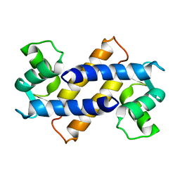
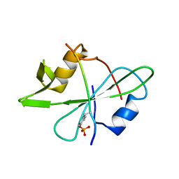
 | | NMR structure of the ubiquitin-binding zinc finger (UBZ) domain from human Rad18 | | 分子名称: | E3 ubiquitin-protein ligase RAD18, ZINC ION | | 著者 | Rizzo, A.A, Salerno, P.E, Bezsonova, I, Korzhnev, D.M. | | 登録日 | 2014-07-03 | | 公開日 | 2014-10-01 | | 最終更新日 | 2024-05-01 | | 実験手法 | SOLUTION NMR | | 主引用文献 | NMR Structure of the Human Rad18 Zinc Finger in Complex with Ubiquitin Defines a Class of UBZ Domains in Proteins Linked to the DNA Damage Response.
Biochemistry, 53, 2014
|
|
2MRI
 | |
2MRJ
 | |
2MRK
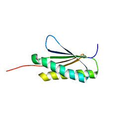
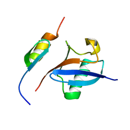
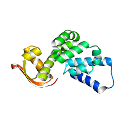
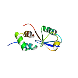
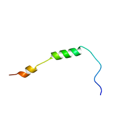
 | | Fyn SH2 domain in complex with the natural inhibitory phosphotyrosine peptide | | 分子名称: | C-terminal Tyrosine-protein kinase Fyn, Tyrosine-protein kinase Fyn | | 著者 | Huculeci, R, Buts, L, Lenaerts, A.J, van Nuland, N. | | 登録日 | 2014-07-09 | | 公開日 | 2015-07-15 | | 最終更新日 | 2016-12-14 | | 実験手法 | SOLUTION NMR | | 主引用文献 | Dynamically Coupled Residues within the SH2 Domain of FYN Are Key to Unlocking Its Activity.
Structure, 24, 2016
|
|
2MRL
 | |
2MRM
 | |
2MRN
 | |
2MRO
 | |
2MRP
 | |
2MRT
 | | CONFORMATION OF CD-7 METALLOTHIONEIN-2 FROM RAT LIVER IN AQUEOUS SOLUTION DETERMINED BY NUCLEAR MAGNETIC RESONANCE SPECTROSCOPY | | 分子名称: | CADMIUM ION, CD7 METALLOTHIONEIN-2 | | 著者 | Braun, W, Schultze, P, Woergoetter, E, Wagner, G, Vasak, M, Kaegi, J.H.R, Wuthrich, K. | | 登録日 | 1990-05-14 | | 公開日 | 1991-04-15 | | 最終更新日 | 2024-05-22 | | 実験手法 | SOLUTION NMR | | 主引用文献 | Conformation of [Cd7]-metallothionein-2 from rat liver in aqueous solution determined by nuclear magnetic resonance spectroscopy.
J.Mol.Biol., 203, 1988
|
|
2MRU
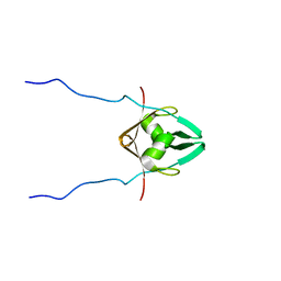
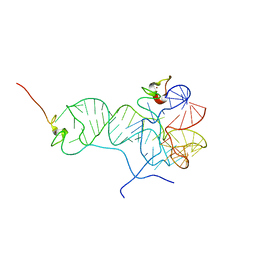
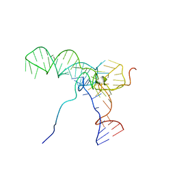
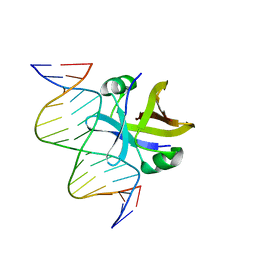 | | Structure of truncated EcMazE-DNA complex | | 分子名称: | Antitoxin MazE, DNA (5'-D(*CP*GP*TP*GP*AP*TP*AP*TP*AP*TP*AP*GP*TP*GP*C)-3'), DNA (5'-D(P*GP*CP*AP*CP*TP*AP*TP*AP*TP*AP*TP*CP*AP*CP*G)-3') | | 著者 | Zorzini, V, Buts, L, Loris, R, van Nuland, N. | | 登録日 | 2014-07-15 | | 公開日 | 2015-02-04 | | 最終更新日 | 2024-05-15 | | 実験手法 | SOLUTION NMR | | 主引用文献 | Escherichia coli antitoxin MazE as transcription factor: insights into MazE-DNA binding.
Nucleic Acids Res., 43, 2015
|
|
2MRW
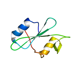
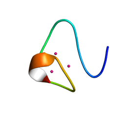
 | | Solution Structure of MciZ from Bacillus subtilis | | 分子名称: | Cell division factor | | 著者 | Castellen, P, Sforca, M.L, Zeri, A.C.M, Gueiros-Filho, F.J. | | 登録日 | 2014-07-16 | | 公開日 | 2015-03-25 | | 最終更新日 | 2024-05-01 | | 実験手法 | SOLUTION NMR | | 主引用文献 | FtsZ filament capping by MciZ, a developmental regulator of bacterial division.
Proc.Natl.Acad.Sci.USA, 112, 2015
|
|
2MRY
 | |
2MRZ
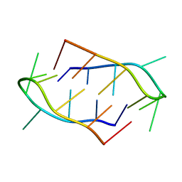 | | Dimeric structure of the Human A-box | | 分子名称: | DNA (5'-D(P*TP*CP*CP*TP*TP*TP*TP*CP*CP*A)-3') | | 著者 | Garavis, M, Escaja, N, Gabelica, V, Villasante, A, Gonzalez, C. | | 登録日 | 2014-07-18 | | 公開日 | 2015-06-03 | | 最終更新日 | 2024-05-01 | | 実験手法 | SOLUTION NMR | | 主引用文献 | Centromeric Alpha-Satellite DNA Adopts Dimeric i-Motif Structures Capped by AT Hoogsteen Base Pairs.
Chemistry, 21, 2015
|
|
2MS0
 | |
2MS1
 | |
2MS2
 | |


