5O1Y
 
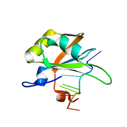
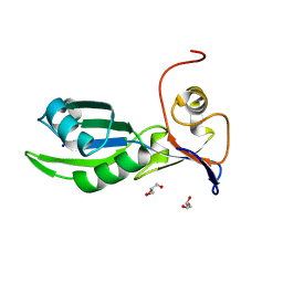
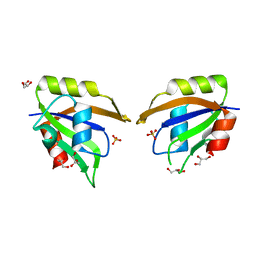
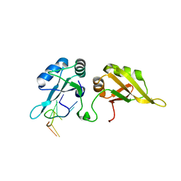
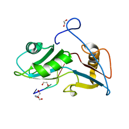 | | Structure of Nrd1 RNA binding domain in complex with RNA (GUAA) | | 分子名称: | 1,2-ETHANEDIOL, 2-AMINO-2-HYDROXYMETHYL-PROPANE-1,3-DIOL, Protein NRD1, ... | | 著者 | Franco-Echevarria, E, Perez-Canadillas, J.M, Gonzalez, B. | | 登録日 | 2017-05-19 | | 公開日 | 2017-08-02 | | 最終更新日 | 2024-01-17 | | 実験手法 | X-RAY DIFFRACTION (2.45 Å) | | 主引用文献 | The structure of transcription termination factor Nrd1 reveals an original mode for GUAA recognition.
Nucleic Acids Res., 45, 2017
|
|
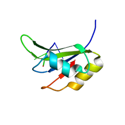
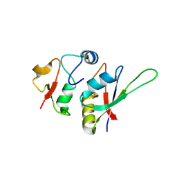
5O1X
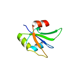
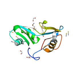 | | Structure of Nrd1 RNA binding domain | | 分子名称: | 1,2-ETHANEDIOL, Protein NRD1, THIOCYANATE ION | | 著者 | Franco-Echevarria, E, Perez-Canadillas, J.M, Gonzalez, B. | | 登録日 | 2017-05-19 | | 公開日 | 2017-08-02 | | 最終更新日 | 2024-01-17 | | 実験手法 | X-RAY DIFFRACTION (1.6 Å) | | 主引用文献 | The structure of transcription termination factor Nrd1 reveals an original mode for GUAA recognition.
Nucleic Acids Res., 45, 2017
|
|
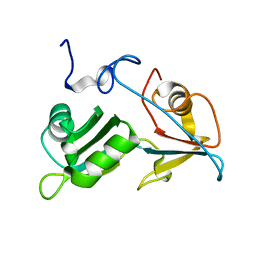
5O1T
 | |
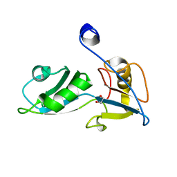
5O1W
 | |
5MPG
 | |
5O3J
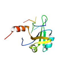
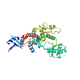
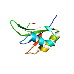
 | | Crystal structure of TIA-1 RRM2 in complex with RNA | | 分子名称: | Nucleolysin TIA-1 isoform p40, RNA (5'-R(P*UP*UP*C)-3') | | 著者 | Sonntag, M, Jagtap, P.K.A, Hennig, J, Sattler, M. | | 登録日 | 2017-05-24 | | 公開日 | 2017-07-05 | | 最終更新日 | 2024-01-17 | | 実験手法 | X-RAY DIFFRACTION (2.97 Å) | | 主引用文献 | Segmental, Domain-Selective Perdeuteration and Small-Angle Neutron Scattering for Structural Analysis of Multi-Domain Proteins.
Angew. Chem. Int. Ed. Engl., 56, 2017
|
|
5XJC
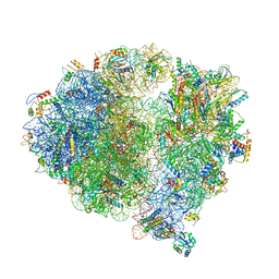
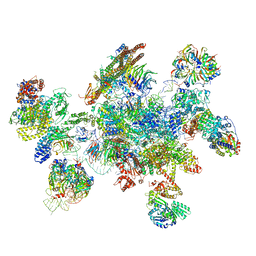
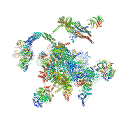
 | | Cryo-EM structure of the human spliceosome just prior to exon ligation at 3.6 angstrom | | 分子名称: | 116 kDa U5 small nuclear ribonucleoprotein component, ADENOSINE-5'-DIPHOSPHATE, ADENOSINE-5'-TRIPHOSPHATE, ... | | 著者 | Zhang, X, Yan, C, Hang, J, Finci, I.L, Lei, J, Shi, Y. | | 登録日 | 2017-04-30 | | 公開日 | 2017-07-05 | | 最終更新日 | 2020-10-14 | | 実験手法 | ELECTRON MICROSCOPY (3.6 Å) | | 主引用文献 | An Atomic Structure of the Human Spliceosome
Cell, 169, 2017
|
|
5MPL
 | |
5O2V
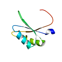 | | NMR structure of TIA-1 RRM1 domain | | 分子名称: | Nucleolysin TIA-1 isoform p40 | | 著者 | Jagtap, P.K.A. | | 登録日 | 2017-05-22 | | 公開日 | 2017-06-28 | | 最終更新日 | 2024-06-19 | | 実験手法 | SOLUTION NMR | | 主引用文献 | Segmental, Domain-Selective Perdeuteration and Small-Angle Neutron Scattering for Structural Analysis of Multi-Domain Proteins.
Angew. Chem. Int. Ed. Engl., 56, 2017
|
|
5X8P
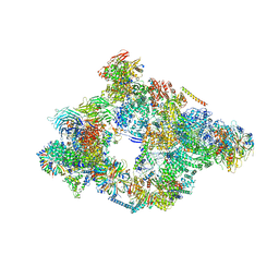
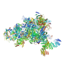
 | | Structure of the 70S chloroplast ribosome from spinach | | 分子名称: | 16S rRNA, 23S rRNA, 30S ribosomal protein S1, ... | | 著者 | Ahmed, T, Shi, J, Bhushan, S. | | 登録日 | 2017-03-03 | | 公開日 | 2017-06-14 | | 最終更新日 | 2024-03-27 | | 実験手法 | ELECTRON MICROSCOPY (3.4 Å) | | 主引用文献 | Unique localization of the plastid-specific ribosomal proteins in the chloroplast ribosome small subunit provides mechanistic insights into the chloroplastic translation
Nucleic Acids Res., 45, 2017
|
|
5X8R
 | | Structure of the 30S small subunit of chloroplast ribosome from spinach | | 分子名称: | 16S rRNA, 30S ribosomal protein S1, chloroplastic, ... | | 著者 | Ahmed, T, Shi, J, Bhushan, S. | | 登録日 | 2017-03-03 | | 公開日 | 2017-06-07 | | 最終更新日 | 2024-03-27 | | 実験手法 | ELECTRON MICROSCOPY (3.7 Å) | | 主引用文献 | Unique localization of the plastid-specific ribosomal proteins in the chloroplast ribosome small subunit provides mechanistic insights into the chloroplastic translation
Nucleic Acids Res., 45, 2017
|
|
5WU6
 | |
5NRL
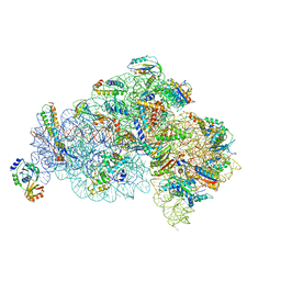
 | | Structure of a pre-catalytic spliceosome. | | 分子名称: | 13 kDa ribonucleoprotein-associated protein, 23 kDa U4/U6.U5 small nuclear ribonucleoprotein component, 66 kDa U4/U6.U5 small nuclear ribonucleoprotein component, ... | | 著者 | Plaschka, C, Lin, P.-C, Nagai, K. | | 登録日 | 2017-04-24 | | 公開日 | 2017-05-31 | | 最終更新日 | 2023-05-24 | | 実験手法 | ELECTRON MICROSCOPY (7.2 Å) | | 主引用文献 | Structure of a pre-catalytic spliceosome.
Nature, 546, 2017
|
|
5GVQ
 | | Solution structure of the first RRM domain of human spliceosomal protein SF3b49 | | 分子名称: | Splicing factor 3B subunit 4 | | 著者 | Kuwasako, K, Nameki, N, Tsuda, K, Takahashi, M, Sato, A, Tochio, N, Inoue, M, Terada, T, Kigawa, T, Kobayashi, N, Shirouzu, M, Ito, T, Sakamoto, T, Wakamatsu, K, Guntert, P, Takahashi, S, Yokoyama, S, Muto, Y, RIKEN Structural Genomics/Proteomics Initiative (RSGI) | | 登録日 | 2016-09-06 | | 公開日 | 2017-04-12 | | 最終更新日 | 2024-05-01 | | 実験手法 | SOLUTION NMR | | 主引用文献 | Solution structure of the first RNA recognition motif domain of human spliceosomal protein SF3b49 and its mode of interaction with a SF3b145 fragment.
Protein Sci., 26, 2017
|
|
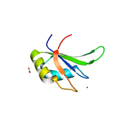
5MDU
 | | Structure of the RNA recognition motif (RRM) of Seb1 from S. pombe. | | 分子名称: | CHLORIDE ION, GLYCEROL, Rpb7-binding protein seb1, ... | | 著者 | Wittmann, S, Renner, M, El Omari, K, Adams, O, Vasiljeva, L, Grimes, J. | | 登録日 | 2016-11-13 | | 公開日 | 2017-04-12 | | 最終更新日 | 2024-05-08 | | 実験手法 | X-RAY DIFFRACTION (1.02 Å) | | 主引用文献 | The conserved protein Seb1 drives transcription termination by binding RNA polymerase II and nascent RNA.
Nat Commun, 8, 2017
|
|
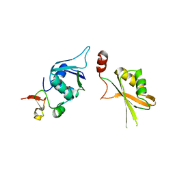
5LSL
 | | Crystal structure of yeast Hsh49p in complex with Cus1p binding domain. | | 分子名称: | Cold sensitive U2 snRNA suppressor 1, Protein HSH49 | | 著者 | van Roon, A.M, Obayashi, E, Sposito, B, Oubridge, C, Nagai, K. | | 登録日 | 2016-09-02 | | 公開日 | 2017-04-12 | | 最終更新日 | 2024-01-17 | | 実験手法 | X-RAY DIFFRACTION (1.65 Å) | | 主引用文献 | Crystal structure of U2 snRNP SF3b components: Hsh49p in complex with Cus1p-binding domain.
RNA, 23, 2017
|
|
5TBX
 | | hnRNP A18 RNA Recognition Motif | | 分子名称: | ACETATE ION, Cold-inducible RNA-binding protein, NICKEL (II) ION | | 著者 | Coburn, K.M, Melville, Z, Aligholizadeh, E, Roth, B.M, Varney, K.M, Weber, D.J. | | 登録日 | 2016-09-13 | | 公開日 | 2017-04-12 | | 最終更新日 | 2024-04-03 | | 実験手法 | X-RAY DIFFRACTION (1.767 Å) | | 主引用文献 | Crystal structure of the human heterogeneous ribonucleoprotein A18 RNA-recognition motif.
Acta Crystallogr F Struct Biol Commun, 73, 2017
|
|
5LSB
 | | Crystal structure of yeast Hsh49p in complex with Cus1p binding domain. | | 分子名称: | Cold sensitive U2 snRNA suppressor 1, Protein HSH49 | | 著者 | van Roon, A.M, Obayashi, E, Sposito, B, Oubridge, C, Nagai, K. | | 登録日 | 2016-08-24 | | 公開日 | 2017-04-12 | | 最終更新日 | 2024-01-17 | | 実験手法 | X-RAY DIFFRACTION (2.7 Å) | | 主引用文献 | Crystal structure of U2 snRNP SF3b components: Hsh49p in complex with Cus1p-binding domain.
RNA, 23, 2017
|
|
5MQF
 | | Cryo-EM structure of a human spliceosome activated for step 2 of splicing (C* complex) | | 分子名称: | 116 kDa U5 small nuclear ribonucleoprotein component, ATP-dependent RNA helicase DHX8, Cell division cycle 5-like protein, ... | | 著者 | Bertram, K, Hartmuth, K, Kastner, B. | | 登録日 | 2016-12-20 | | 公開日 | 2017-03-22 | | 最終更新日 | 2018-11-21 | | 実験手法 | ELECTRON MICROSCOPY (5.9 Å) | | 主引用文献 | Cryo-EM structure of a human spliceosome activated for step 2 of splicing.
Nature, 542, 2017
|
|
5UZG
 | |
5UZM
 | |
5ITH
 | |
5HO4
 | | Crystal structure of hnRNPA2B1 in complex with 10-mer RNA | | 分子名称: | Heterogeneous nuclear ribonucleoproteins A2/B1, RNA (5'-R(*AP*AP*GP*GP*AP*CP*UP*AP*GP*C)-3') | | 著者 | Wu, B.X, Su, S.C, Gan, J.H, Ma, J.B. | | 登録日 | 2016-01-19 | | 公開日 | 2017-02-08 | | 最終更新日 | 2024-03-20 | | 実験手法 | X-RAY DIFFRACTION (1.85 Å) | | 主引用文献 | Molecular basis for the specific and multivariant recognitions of RNA substrates by human hnRNP A2/B1.
Nat Commun, 9, 2018
|
|
5WSG
 | | Cryo-EM structure of the Catalytic Step II spliceosome (C* complex) at 4.0 angstrom resolution | | 分子名称: | 3'-exon-intron, 3'-intron-lariat, 5'-exon, ... | | 著者 | Yan, C, Wan, R, Bai, R, Huang, G, Shi, Y. | | 登録日 | 2016-12-07 | | 公開日 | 2017-01-25 | | 最終更新日 | 2024-03-20 | | 実験手法 | ELECTRON MICROSCOPY (4 Å) | | 主引用文献 | Structure of a yeast step II catalytically activated spliceosome
Science, 355, 2017
|
|
5MPS
 | | Structure of a spliceosome remodeled for exon ligation | | 分子名称: | GUANOSINE-5'-TRIPHOSPHATE, INOSITOL HEXAKISPHOSPHATE, MAGNESIUM ION, ... | | 著者 | Fica, S.M, Oubridge, C, Galej, W.P, Wilkinson, M.E, Newman, A.J, Bai, X.-C, Nagai, K. | | 登録日 | 2016-12-18 | | 公開日 | 2017-01-18 | | 最終更新日 | 2020-10-07 | | 実験手法 | ELECTRON MICROSCOPY (3.85 Å) | | 主引用文献 | Structure of a spliceosome remodelled for exon ligation.
Nature, 542, 2017
|
|


