5O2L
 
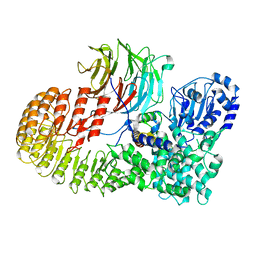
 | | Myosin VI motor domain in the Pre-Transition State | | 分子名称: | ADENOSINE-5'-DIPHOSPHATE, BERYLLIUM TRIFLUORIDE ION, GLYCEROL, ... | | 著者 | Isabet, T, Benisty, H, Houdusse, A. | | 登録日 | 2017-05-22 | | 公開日 | 2018-05-23 | | 最終更新日 | 2024-01-17 | | 実験手法 | X-RAY DIFFRACTION (2.2 Å) | | 主引用文献 | An intermediate along the recovery stroke of myosin VI revealed by X-ray crystallography and molecular dynamics.
Proc. Natl. Acad. Sci. U.S.A., 115, 2018
|
|
8AAU
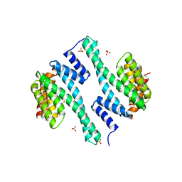
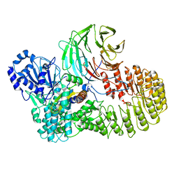
 | | LIM Domain Kinase 1 (LIMK1) bound to LIMKi3 | | 分子名称: | 1,2-ETHANEDIOL, LIM domain kinase 1, MAGNESIUM ION, ... | | 著者 | Mathea, S, Salah, E, Hanke, T, Knapp, S. | | 登録日 | 2022-07-03 | | 公開日 | 2022-08-10 | | 最終更新日 | 2024-02-07 | | 実験手法 | X-RAY DIFFRACTION (1.74 Å) | | 主引用文献 | Development and Characterization of Type I, Type II, and Type III LIM-Kinase Chemical Probes.
J.Med.Chem., 65, 2022
|
|
2ATX
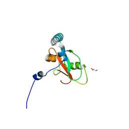
 | | Crystal Structure of the TC10 GppNHp complex | | 分子名称: | MAGNESIUM ION, PHOSPHOAMINOPHOSPHONIC ACID-GUANYLATE ESTER, small GTP binding protein TC10 | | 著者 | Hemsath, L, Dvorsky, R, Fiegen, D, Carlier, M.F, Ahmadian, M.R. | | 登録日 | 2005-08-26 | | 公開日 | 2005-09-13 | | 最終更新日 | 2024-04-03 | | 実験手法 | X-RAY DIFFRACTION (2.65 Å) | | 主引用文献 | An electrostatic steering mechanism of Cdc42 recognition by Wiskott-Aldrich syndrome proteins
Mol.Cell, 20, 2005
|
|
2LSR
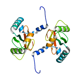
 | | Solution structure of harmonin N terminal domain in complex with a exon68 encoded peptide of cadherin23 | | 分子名称: | Harmonin, peptide from Cadherin-23 | | 著者 | Pan, L, Wu, L, Zhang, C, Zhang, M. | | 登録日 | 2012-05-04 | | 公開日 | 2012-08-15 | | 最終更新日 | 2024-05-15 | | 実験手法 | SOLUTION NMR | | 主引用文献 | Large protein assemblies formed by multivalent interactions between cadherin23 and harmonin suggest a stable anchorage structure at the tip link of stereocilia.
J.Biol.Chem., 287, 2012
|
|
4QWO
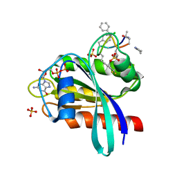
 | | 1.52 Angstrom Crystal Structure of A42R Profilin-like Protein from Monkeypox Virus Zaire-96-I-16 | | 分子名称: | 1,2-ETHANEDIOL, 3,6,9,12,15,18,21-HEPTAOXATRICOSANE-1,23-DIOL, CHLORIDE ION, ... | | 著者 | Minasov, G, Shuvalova, L, Dubrovska, I, Flores, K, Grimshaw, S, Kwon, K, Anderson, W.F, Center for Structural Genomics of Infectious Diseases (CSGID) | | 登録日 | 2014-07-16 | | 公開日 | 2014-08-06 | | 最終更新日 | 2022-10-12 | | 実験手法 | X-RAY DIFFRACTION (1.52 Å) | | 主引用文献 | Structure of the Monkeypox virus profilin-like protein A42R reveals potential functional differences from cellular profilins.
Acta Crystallogr.,Sect.F, 78, 2022
|
|
5XC1
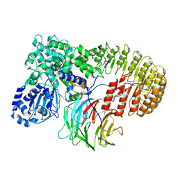
 | | Crystal structure of the complex of an aromatic mutant (W6A) of an alkali thermostable GH10 Xylanase from Bacillus sp. NG-27 with S-1,2-Propanediol | | 分子名称: | Beta-xylanase, MAGNESIUM ION, S-1,2-PROPANEDIOL, ... | | 著者 | Bansia, H, Mahanta, P, Ramakumar, S. | | 登録日 | 2017-03-21 | | 公開日 | 2018-03-28 | | 最終更新日 | 2023-11-22 | | 実験手法 | X-RAY DIFFRACTION (2.26 Å) | | 主引用文献 | Small Glycols Discover Cryptic Pockets on Proteins for Fragment-Based Approaches.
J.Chem.Inf.Model., 2021
|
|
5XC0
 | |
7UAK
 | |
5EFD
 | | Crystal structure of a surface pocket creating mutant (W6A) of an alkali thermostable GH10 xylanase from Bacillus sp. NG-27 | | 分子名称: | 1,2-ETHANEDIOL, Beta-xylanase, CHLORIDE ION, ... | | 著者 | Mahanta, P, Bhardwaj, A, Reddy, V.S, Ramakumar, S. | | 登録日 | 2015-10-23 | | 公開日 | 2016-10-26 | | 最終更新日 | 2024-03-20 | | 実験手法 | X-RAY DIFFRACTION (1.674 Å) | | 主引用文献 | Small Glycols Discover Cryptic Pockets on Proteins for Fragment-Based Approaches.
J.Chem.Inf.Model., 2021
|
|
7UZE
 | | Erythrocyte ankyrin-1 complex class 2 local refinement of AQP1 (C4 symmetry applied) | | 分子名称: | Aquaporin-1, CHOLESTEROL | | 著者 | Vallese, F, Kim, K, Yen, L.Y, Johnston, J.D, Noble, A.J, Cali, T, Clarke, O.B. | | 登録日 | 2022-05-09 | | 公開日 | 2022-07-20 | | 最終更新日 | 2022-07-27 | | 実験手法 | ELECTRON MICROSCOPY (2.4 Å) | | 主引用文献 | Architecture of the human erythrocyte ankyrin-1 complex.
Nat.Struct.Mol.Biol., 29, 2022
|
|
2V6H
 | | Crystal structure of the C1 domain of cardiac myosin binding protein-C | | 分子名称: | MYOSIN-BINDING PROTEIN C, CARDIAC-TYPE | | 著者 | Govata, L, Carpenter, L, Da Fonseca, P.C.A, Helliwell, J.R, Rizkallah, P.J, Flashman, E, Chayen, N.E, Redwood, C, Squire, J.M. | | 登録日 | 2007-07-18 | | 公開日 | 2008-07-22 | | 最終更新日 | 2024-05-08 | | 実験手法 | X-RAY DIFFRACTION (1.55 Å) | | 主引用文献 | Crystal structure of the C1 domain of cardiac myosin binding protein-C: implications for hypertrophic cardiomyopathy.
J. Mol. Biol., 378, 2008
|
|
4F7R
 | |
8H93
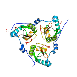
 | | Structure of dimeric mouse SCMC core complex | | 分子名称: | NACHT, LRR and PYD domains-containing protein 5, Oocyte-expressed protein homolog, ... | | 著者 | Chi, P, Ou, G, Han, Z, Li, J, Deng, D. | | 登録日 | 2022-10-24 | | 公開日 | 2024-01-10 | | 最終更新日 | 2024-01-31 | | 実験手法 | ELECTRON MICROSCOPY (3.01 Å) | | 主引用文献 | Structural basis of the subcortical maternal complex and its implications in reproductive disorders.
Nat.Struct.Mol.Biol., 31, 2024
|
|
8H94
 | | Structure of mouse SCMC bound with KH domain of FILIA | | 分子名称: | NACHT, LRR and PYD domains-containing protein 5, Oocyte-expressed protein homolog, ... | | 著者 | Chi, P, Ou, G, Han, Z, Li, J, Deng, D. | | 登録日 | 2022-10-24 | | 公開日 | 2024-01-10 | | 最終更新日 | 2024-01-31 | | 実験手法 | ELECTRON MICROSCOPY (2.9 Å) | | 主引用文献 | Structural basis of the subcortical maternal complex and its implications in reproductive disorders.
Nat.Struct.Mol.Biol., 31, 2024
|
|
8H96
 | | Structure of mouse SCMC core complex | | 分子名称: | NACHT, LRR and PYD domains-containing protein 5, Oocyte-expressed protein homolog, ... | | 著者 | Chi, P, Ou, G, Li, J, Han, Z, Deng, D. | | 登録日 | 2022-10-24 | | 公開日 | 2024-01-10 | | 最終更新日 | 2024-01-31 | | 実験手法 | ELECTRON MICROSCOPY (2.78 Å) | | 主引用文献 | Structural basis of the subcortical maternal complex and its implications in reproductive disorders.
Nat.Struct.Mol.Biol., 31, 2024
|
|
8H95
 | | Structure of mouse SCMC bound with full-length FILIA | | 分子名称: | NACHT, LRR and PYD domains-containing protein 5, Oocyte-expressed protein homolog, ... | | 著者 | Chi, P, Ou, G, Han, Z, Li, L, Deng, D. | | 登録日 | 2022-10-24 | | 公開日 | 2024-01-10 | | 最終更新日 | 2024-01-31 | | 実験手法 | ELECTRON MICROSCOPY (3.38 Å) | | 主引用文献 | Structural basis of the subcortical maternal complex and its implications in reproductive disorders.
Nat.Struct.Mol.Biol., 31, 2024
|
|
7BRQ
 | |
7BRT
 | | Crystal structure of Sec62 LIR fused to GABARAP | | 分子名称: | Translocation protein SEC62,Gamma-aminobutyric acid receptor-associated protein | | 著者 | Yamasaki, A, Noda, N.N. | | 登録日 | 2020-03-30 | | 公開日 | 2020-07-08 | | 最終更新日 | 2023-11-29 | | 実験手法 | X-RAY DIFFRACTION (1.999 Å) | | 主引用文献 | Super-assembly of ER-phagy receptor Atg40 induces local ER remodeling at contacts with forming autophagosomal membranes.
Nat Commun, 11, 2020
|
|
7BRU
 | |
8JJS
 | | Human K-Ras G12D (GDP-bound) in complex with cyclic peptide inhibitor AP10343 | | 分子名称: | GUANOSINE-5'-DIPHOSPHATE, Isoform 2B of GTPase KRas, MAA-ILE-SAR-SAR-7T2-SAR-IAE-LEU-MEA-MLE-7TK, ... | | 著者 | Irie, M, Fukami, T.A, Tanada, M, Ohta, A, Torizawa, T. | | 登録日 | 2023-05-31 | | 公開日 | 2023-07-26 | | 最終更新日 | 2023-11-15 | | 実験手法 | X-RAY DIFFRACTION (1.534 Å) | | 主引用文献 | Development of Orally Bioavailable Peptides Targeting an Intracellular Protein: From a Hit to a Clinical KRAS Inhibitor.
J.Am.Chem.Soc., 145, 2023
|
|
2ETK
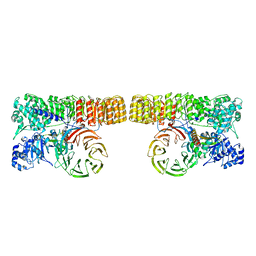
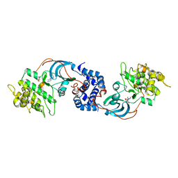 | | Crystal Structure of ROCK 1 bound to hydroxyfasudil | | 分子名称: | 1-(1-HYDROXY-5-ISOQUINOLINESULFONYL)HOMOPIPERAZINE, Rho-associated protein kinase 1 | | 著者 | Jacobs, M. | | 登録日 | 2005-10-27 | | 公開日 | 2005-11-08 | | 最終更新日 | 2023-08-23 | | 実験手法 | X-RAY DIFFRACTION (2.96 Å) | | 主引用文献 | The Structure of Dimeric ROCK I Reveals the Mechanism for Ligand Selectivity.
J.Biol.Chem., 281, 2006
|
|
1MK7
 | | CRYSTAL STRUCTURE OF AN INTEGRIN BETA3-TALIN CHIMERA | | 分子名称: | Integrin Beta3, TALIN | | 著者 | Garcia-Alvarez, B, De Pereda, J.M, Calderwood, D.A, Ulmer, T.S, Critchley, D, Campbell, I.D, Ginsberg, M.H, Liddington, R.C. | | 登録日 | 2002-08-28 | | 公開日 | 2003-01-28 | | 最終更新日 | 2011-07-13 | | 実験手法 | X-RAY DIFFRACTION (2.2 Å) | | 主引用文献 | Structural Determinants of Integrin Recognition by Talin
Mol.Cell, 11, 2003
|
|
1MIX
 | | Crystal structure of a FERM domain of Talin | | 分子名称: | Talin | | 著者 | Garcia-Alvarez, B, de Pereda, J.M, Calderwood, D.A, Ulmer, T.S, Critchley, D, Campbell, I.D, Ginsberg, M.H, Liddington, R.C. | | 登録日 | 2002-08-23 | | 公開日 | 2003-01-28 | | 最終更新日 | 2024-02-14 | | 実験手法 | X-RAY DIFFRACTION (1.75 Å) | | 主引用文献 | Structural determinants of integrin recognition by talin
Mol.Cell, 11, 2003
|
|
1TJF
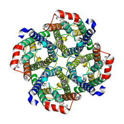
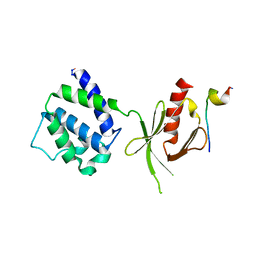
 | | The crystal structure of the N-terminal domain of CAP indicates variable oligomerisation | | 分子名称: | Adenylyl cyclase-associated protein, SULFATE ION | | 著者 | Mohd Yusof, A, Hu, N.J, Wlodawer, A, Hofmann, A. | | 登録日 | 2004-06-04 | | 公開日 | 2005-02-01 | | 最終更新日 | 2023-08-23 | | 実験手法 | X-RAY DIFFRACTION (2.21 Å) | | 主引用文献 | Structural evidence for variable oligomerization of the N-terminal domain of cyclase-associated protein (CAP).
Proteins, 58, 2005
|
|
7MQ9
 | | Cryo-EM structure of the human SSU processome, state pre-A1* | | 分子名称: | 18S rRNA, 40S ribosomal protein S11, 40S ribosomal protein S12, ... | | 著者 | Vanden Broeck, A, Singh, S, Klinge, S. | | 登録日 | 2021-05-05 | | 公開日 | 2021-09-22 | | 最終更新日 | 2024-05-29 | | 実験手法 | ELECTRON MICROSCOPY (3.87 Å) | | 主引用文献 | Nucleolar maturation of the human small subunit processome.
Science, 373, 2021
|
|


