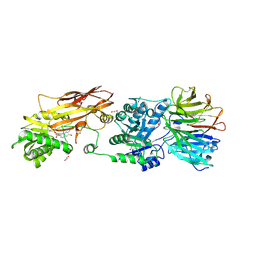
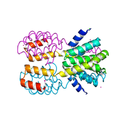
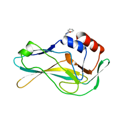
7KID
 
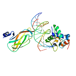
 | | PRMT5:MEP50 Complexed with 5,5-Bicyclic Inhibitor Compound 72 | | 分子名称: | (1S,2R,3aR,4S,6aR)-4-[(2-amino-3,5-difluoroquinolin-7-yl)methyl]-2-(4-amino-5-fluoro-7H-pyrrolo[2,3-d]pyrimidin-7-yl)hexahydropentalene-1,6a(1H)-diol, 1,2-ETHANEDIOL, Methylosome protein 50, ... | | 著者 | Palte, R.L. | | 登録日 | 2020-10-23 | | 公開日 | 2021-04-21 | | 最終更新日 | 2023-10-18 | | 実験手法 | X-RAY DIFFRACTION (2.5 Å) | | 主引用文献 | The Discovery of Two Novel Classes of 5,5-Bicyclic Nucleoside-Derived PRMT5 Inhibitors for the Treatment of Cancer.
J.Med.Chem., 64, 2021
|
|
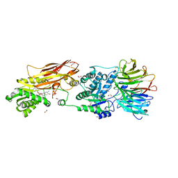
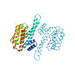
7KIC
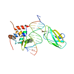
 | | PRMT5:MEP50 Complexed with 5,5-Bicyclic Inhibitor Compound 34 | | 分子名称: | (2R,3R,3aS,6S,6aR)-6-[(2-amino-3-bromoquinolin-7-yl)oxy]-2-(4-methyl-7H-pyrrolo[2,3-d]pyrimidin-7-yl)hexahydro-3aH-cyclopenta[b]furan-3,3a-diol, 1,2-ETHANEDIOL, Methylosome protein 50, ... | | 著者 | Palte, R.L. | | 登録日 | 2020-10-23 | | 公開日 | 2021-04-21 | | 最終更新日 | 2023-10-18 | | 実験手法 | X-RAY DIFFRACTION (2.43 Å) | | 主引用文献 | The Discovery of Two Novel Classes of 5,5-Bicyclic Nucleoside-Derived PRMT5 Inhibitors for the Treatment of Cancer.
J.Med.Chem., 64, 2021
|
|
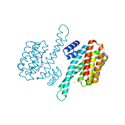
7KIB
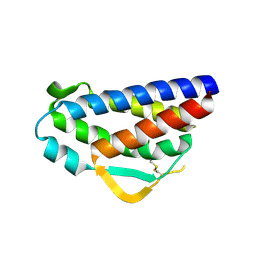
 | | PRMT5:MEP50 Complexed with 5,5-Bicyclic Inhibitor Compound 4 | | 分子名称: | (2R,3R,3aS,6S,6aR)-6-[(2-amino-3-bromoquinolin-7-yl)oxy]-2-(4-amino-7H-pyrrolo[2,3-d]pyrimidin-7-yl)hexahydro-3aH-cyclopenta[b]furan-3,3a-diol, 1,2-ETHANEDIOL, CHLORIDE ION, ... | | 著者 | Palte, R.L, Hayes, R.P. | | 登録日 | 2020-10-23 | | 公開日 | 2021-04-21 | | 最終更新日 | 2023-10-18 | | 実験手法 | X-RAY DIFFRACTION (2.52 Å) | | 主引用文献 | The Discovery of Two Novel Classes of 5,5-Bicyclic Nucleoside-Derived PRMT5 Inhibitors for the Treatment of Cancer.
J.Med.Chem., 64, 2021
|
|
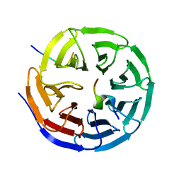
5MJ3
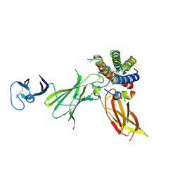
 | | INTERLEUKIN-23 COMPLEX WITH AN ANTAGONISTIC ALPHABODY, CRYSTAL FORM 1 | | 分子名称: | ALPHABODY MA12, Interleukin-12 subunit beta, Interleukin-23 subunit alpha, ... | | 著者 | Desmet, J, Verstraete, K, Bloch, Y, Lorent, E, Wen, Y, Devreese, B, Vandenbroucke, K, Loverix, S, Hettmann, T, Deroo, S, Somers, K, Hendrikx, P, Lasters, I, Savvides, S.N. | | 登録日 | 2016-11-29 | | 公開日 | 2017-01-11 | | 最終更新日 | 2024-01-17 | | 実験手法 | X-RAY DIFFRACTION (1.74 Å) | | 主引用文献 | Structural Basis Of Il-23 Antagonism By An Alphabody Protein Scaffold.
Nat Commun, 5, 2014
|
|
3QYW
 | | Crystal structure of ERK2 in complex with an inhibitor | | 分子名称: | 6-(3-bromophenyl)-7H-purin-2-amine, DIMETHYL SULFOXIDE, Mitogen-activated protein kinase 1, ... | | 著者 | Gelin, M, Pochet, S, Hoh, F, Pirochi, M, Guichou, J.-F, Ferrer, J.-L, Labesse, G. | | 登録日 | 2011-03-04 | | 公開日 | 2011-08-24 | | 最終更新日 | 2017-11-08 | | 実験手法 | X-RAY DIFFRACTION (1.5 Å) | | 主引用文献 | In-plate protein crystallization, in situ ligand soaking and X-ray diffraction.
Acta Crystallogr.,Sect.D, 67, 2011
|
|
2H24
 | | Crystal structure of human IL-10 | | 分子名称: | Interleukin-10 | | 著者 | Yoon, S.I, Walter, M.R. | | 登録日 | 2006-05-18 | | 公開日 | 2006-10-17 | | 最終更新日 | 2023-08-30 | | 実験手法 | X-RAY DIFFRACTION (2 Å) | | 主引用文献 | Conformational changes mediate interleukin-10 receptor 2 (IL-10R2) binding to IL-10 and assembly of the signaling complex.
J.Biol.Chem., 281, 2006
|
|
1JLU
 | | Crystal Structure of the Catalytic Subunit of cAMP-dependent Protein Kinase Complexed with a Phosphorylated Substrate Peptide and Detergent | | 分子名称: | AMP-DEPENDENT PROTEIN KINASE, ALPHA-CATALYTIC SUBUNIT, CAMP-DEPENDENT PROTEIN KINASE INHIBITOR, ... | | 著者 | Madhusudan, Trafny, E.A, Xuong, N.-H, Adams, J.A, Ten Eyck, L.F, Taylor, S.S, Sowadski, J.M. | | 登録日 | 2001-07-16 | | 公開日 | 2001-08-01 | | 最終更新日 | 2023-08-16 | | 実験手法 | X-RAY DIFFRACTION (2.25 Å) | | 主引用文献 | cAMP-dependent protein kinase: crystallographic insights into substrate recognition and phosphotransfer.
Protein Sci., 3, 1994
|
|
3QYZ
 | | Crystal structure of ERK2 in complex with an inhibitor | | 分子名称: | 5'-azido-8-bromo-5'-deoxyadenosine, BETA-MERCAPTOETHANOL, DIMETHYL SULFOXIDE, ... | | 著者 | Gelin, M, Pochet, S, Hoh, F, Pirochi, M, Guichou, J.-F, Ferrer, J.-L, Labesse, G. | | 登録日 | 2011-03-04 | | 公開日 | 2011-08-24 | | 最終更新日 | 2017-11-08 | | 実験手法 | X-RAY DIFFRACTION (1.46 Å) | | 主引用文献 | In-plate protein crystallization, in situ ligand soaking and X-ray diffraction.
Acta Crystallogr.,Sect.D, 67, 2011
|
|
4NAC
 | |
4FU3
 | | CID of human RPRD1B | | 分子名称: | CHLORIDE ION, Regulation of nuclear pre-mRNA domain-containing protein 1B, UNKNOWN ATOM OR ION | | 著者 | Ni, Z, Xu, C, Tempel, W, El Bakkouri, M, Loppnau, P, Guo, X, Bountra, C, Arrowsmith, C.H, Edwards, A.M, Min, J, Greenblatt, J.F, Structural Genomics Consortium (SGC) | | 登録日 | 2012-06-28 | | 公開日 | 2012-08-29 | | 最終更新日 | 2024-04-03 | | 実験手法 | X-RAY DIFFRACTION (1.9 Å) | | 主引用文献 | CID of human RPRD1B
TO BE PUBLISHED
|
|
8C40
 | |
8C3Z
 | |
8C1Y
 | |
5QS6
 | | PanDDA analysis group deposition -- Crystal Structure of human Brachyury G177D variant in complex with Z44592329 | | 分子名称: | N-phenyl-N'-pyridin-3-ylurea, T-box transcription factor T | | 著者 | Newman, J.A, Gavard, A.E, Sherestha, L, Fernandez-Cid, A, Burgess-Brown, N.A, von Delft, F, Arrowsmith, C.H, Edwards, A, Bountra, C, Gileadi, O. | | 登録日 | 2019-06-19 | | 公開日 | 2019-08-21 | | 最終更新日 | 2024-03-06 | | 実験手法 | X-RAY DIFFRACTION (1.67 Å) | | 主引用文献 | PanDDA analysis group deposition
To Be Published
|
|
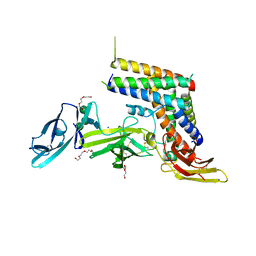
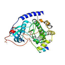
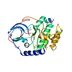
3P4F
 | | Structural and biochemical insights into MLL1 core complex assembly and regulation. | | 分子名称: | Histone-lysine N-methyltransferase MLL, Retinoblastoma-binding protein 5, WD repeat-containing protein 5 | | 著者 | Avdic, V, Zhang, P, Lanouette, S, Groulx, A, Tremblay, V, Brunzelle, J.B, Couture, J.-F. | | 登録日 | 2010-10-06 | | 公開日 | 2010-12-08 | | 最終更新日 | 2023-09-06 | | 実験手法 | X-RAY DIFFRACTION (2.35 Å) | | 主引用文献 | Structural and biochemical insights into MLL1 core complex assembly.
Structure, 19, 2011
|
|
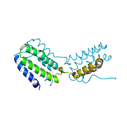
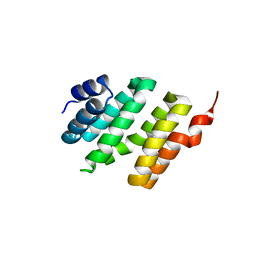
2C6A
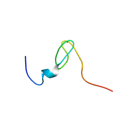 | | Solution structure of the C4 zinc-finger domain of HDM2 | | 分子名称: | UBIQUITIN-PROTEIN LIGASE E3 MDM2, ZINC ION | | 著者 | Yu, G.W, Allen, M.D, Andreeva, A, Fersht, A.R, Bycroft, M. | | 登録日 | 2005-11-08 | | 公開日 | 2006-01-04 | | 最終更新日 | 2024-05-15 | | 実験手法 | SOLUTION NMR | | 主引用文献 | Solution Structure of the C4 Zinc Finger Domain of Hdm2.
Protein Sci., 15, 2006
|
|
1CC0
 | | CRYSTAL STRUCTURE OF THE RHOA.GDP-RHOGDI COMPLEX | | 分子名称: | GUANOSINE-5'-DIPHOSPHATE, MAGNESIUM ION, rho GDP dissociation inhibitor alpha, ... | | 著者 | Longenecker, K.L, Read, P, Derewenda, U, Dauter, Z, Garrard, S, Walker, L, Somlyo, A.V, Somlyo, A.P, Nakamoto, R.K, Derewenda, Z.S. | | 登録日 | 1999-03-03 | | 公開日 | 2000-01-07 | | 最終更新日 | 2023-12-27 | | 実験手法 | X-RAY DIFFRACTION (5 Å) | | 主引用文献 | How RhoGDI binds Rho.
Acta Crystallogr.,Sect.D, 55, 1999
|
|
7UJX
 | | Structure of cAMP-dependent protein kinase using a MD-MX procedure, produced using 2.4 Angstrom data | | 分子名称: | ADENOSINE-5'-DIPHOSPHATE, MAGNESIUM ION, PHOSPHATE ION, ... | | 著者 | Wych, D.C, Aoto, P.C, Wall, M.E. | | 登録日 | 2022-03-31 | | 公開日 | 2022-12-07 | | 最終更新日 | 2023-10-25 | | 実験手法 | X-RAY DIFFRACTION (2.4 Å) | | 主引用文献 | Molecular-dynamics simulation methods for macromolecular crystallography.
Acta Crystallogr D Struct Biol, 79, 2023
|
|
7V0G
 | | Structure of cAMP-dependent protein kinase using a MD-MX procedure, produced using 1.63 Angstrom data | | 分子名称: | ADENOSINE-5'-DIPHOSPHATE, MAGNESIUM ION, PHOSPHATE ION, ... | | 著者 | Wych, D.C, Aoto, P.C, Wall, M.E. | | 登録日 | 2022-05-10 | | 公開日 | 2022-12-07 | | 最終更新日 | 2023-10-25 | | 実験手法 | X-RAY DIFFRACTION (1.63 Å) | | 主引用文献 | Molecular-dynamics simulation methods for macromolecular crystallography.
Acta Crystallogr D Struct Biol, 79, 2023
|
|
4L0Y
 | |
4L18
 | |
3QB1
 | | Interleukin-2 mutant D10 | | 分子名称: | Interleukin-2 | | 著者 | Levin, A.M, Bates, D.L, Ring, A.M, Lin, J.T, Su, L, Krieg, C, Bowman, G.R, Novick, P, Pande, V.S, Khort, H.E, Boyman, O, Fathman, C.G, Garcia, K.C. | | 登録日 | 2011-01-12 | | 公開日 | 2012-04-11 | | 最終更新日 | 2023-09-13 | | 実験手法 | X-RAY DIFFRACTION (3.1 Å) | | 主引用文献 | Exploiting a natural conformational switch to engineer an interleukin-2 'superkine'
Nature, 484, 2012
|
|
6UIB
 | | Crystal structure of IL23 bound to peptide 23-652 | | 分子名称: | 2-acetamido-2-deoxy-beta-D-glucopyranose-(1-4)-2-acetamido-2-deoxy-beta-D-glucopyranose, Interleukin-12 subunit beta, Interleukin-23 subunit alpha, ... | | 著者 | Durbin, J.D, Wang, J, Afshar, S. | | 登録日 | 2019-09-30 | | 公開日 | 2020-07-15 | | 最終更新日 | 2023-10-11 | | 実験手法 | X-RAY DIFFRACTION (2.74 Å) | | 主引用文献 | Integration of phage and yeast display platforms: A reliable and cost effective approach for binning of peptides as displayed on-phage.
Plos One, 15, 2020
|
|
2C6B
 | | Solution structure of the C4 zinc-finger domain of HDM2 | | 分子名称: | UBIQUITIN-PROTEIN LIGASE E3 MDM2, ZINC ION | | 著者 | Yu, G.W, Allen, M.D, Andreeva, A, Fersht, A.R, Bycroft, M. | | 登録日 | 2005-11-08 | | 公開日 | 2006-01-04 | | 最終更新日 | 2024-05-15 | | 実験手法 | SOLUTION NMR | | 主引用文献 | Solution Structure of the C4 Zinc Finger Domain of Hdm2.
Protein Sci., 15, 2006
|
|
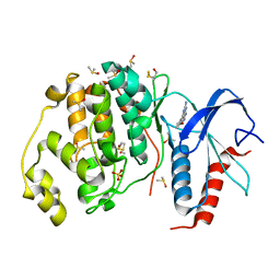
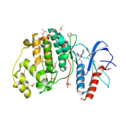
3QWW
 | | Crystal structure of histone lysine methyltransferase SmyD2 in complex with the methyltransferase inhibitor sinefungin | | 分子名称: | SET and MYND domain-containing protein 2, SINEFUNGIN, ZINC ION | | 著者 | Jiang, Y, Sirinupong, N, Brunzelle, J, Yang, Z. | | 登録日 | 2011-02-28 | | 公開日 | 2011-07-06 | | 最終更新日 | 2024-04-03 | | 実験手法 | X-RAY DIFFRACTION (1.8 Å) | | 主引用文献 | Crystal structures of histone and p53 methyltransferase SmyD2 reveal a conformational flexibility of the autoinhibitory C-terminal domain.
Plos One, 6, 2011
|
|


