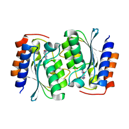
7DC9
 
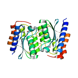 | |
7DCA
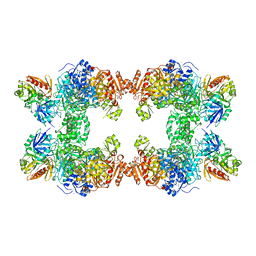
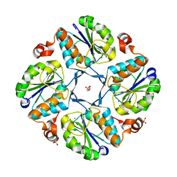
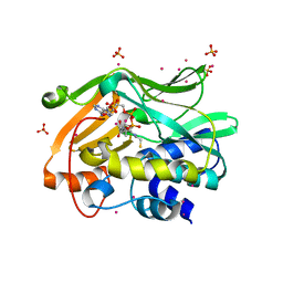
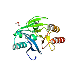
 | | The structure of the Arabidopsis thaliana guanosine deaminase bound by xanthosine | | 分子名称: | 2,3-dihydroxanthosine, Guanosine deaminase, ZINC ION | | 著者 | Xie, W, Jia, Q, Zeng, H. | | 登録日 | 2020-10-23 | | 公開日 | 2021-09-29 | | 最終更新日 | 2023-11-29 | | 実験手法 | X-RAY DIFFRACTION (2.1 Å) | | 主引用文献 | The C-terminal loop of Arabidopsis thaliana guanosine deaminase is essential to catalysis.
Chem.Commun.(Camb.), 57, 2021
|
|
7DCB
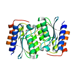 | |
7DCW
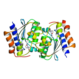 | |
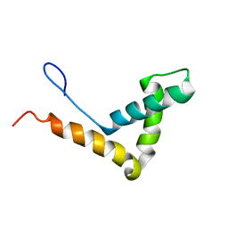
1IE1
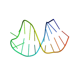
 | | NMR Solution Structure of an In Vitro Selected RNA which is Sequence Specifically Recognized by Hamster Nucleolin RBD12. | | 分子名称: | 5'-R(*GP*GP*CP*CP*GP*AP*AP*AP*UP*CP*CP*CP*GP*AP*AP*GP*UP*AP*GP*GP*CP*C)-3' | | 著者 | Bouvet, P, Allain, F.H.-T, Finger, L.D, Dieckmann, T, Feigon, J. | | 登録日 | 2001-04-05 | | 公開日 | 2001-06-20 | | 最終更新日 | 2024-05-01 | | 実験手法 | SOLUTION NMR | | 主引用文献 | Recognition of pre-formed and flexible elements of an RNA stem-loop by nucleolin.
J.Mol.Biol., 309, 2001
|
|
1J3X
 | |
1I4K
 | | CRYSTAL STRUCTURE OF AN SM-LIKE PROTEIN (AF-SM1) FROM ARCHAEOGLOBUS FULGIDUS AT 2.5A RESOLUTION | | 分子名称: | CITRIC ACID, PUTATIVE SNRNP SM-LIKE PROTEIN | | 著者 | Toro, I, Thore, S, Mayer, C, Basquin, J, Seraphin, B, Suck, D. | | 登録日 | 2001-02-22 | | 公開日 | 2001-08-22 | | 最終更新日 | 2024-04-03 | | 実験手法 | X-RAY DIFFRACTION (2.5 Å) | | 主引用文献 | RNA binding in an Sm core domain: X-ray structure and functional analysis of an archaeal Sm protein complex.
EMBO J., 20, 2001
|
|
1GH0
 | | CRYSTAL STRUCTURE OF C-PHYCOCYANIN FROM SPIRULINA PLATENSIS | | 分子名称: | C-PHYCOCYANIN ALPHA SUBUNIT, C-PHYCOCYANIN BETA SUBUNIT, PHYCOCYANOBILIN | | 著者 | Liang, D.-C, Chang, W.-R, Wang, X.-Q. | | 登録日 | 2000-10-29 | | 公開日 | 2001-06-06 | | 最終更新日 | 2023-12-27 | | 実験手法 | X-RAY DIFFRACTION (2.2 Å) | | 主引用文献 | Structure of C-phycocyanin from Spirulina platensis at 2.2 A resolution: a novel monoclinic crystal form for phycobiliproteins in phycobilisomes.
Acta Crystallogr.,Sect.D, 57, 2001
|
|
1G8K
 | | CRYSTAL STRUCTURE ANALYSIS OF ARSENITE OXIDASE FROM ALCALIGENES FAECALIS | | 分子名称: | 1,2-ETHANEDIOL, 2-AMINO-5,6-DIMERCAPTO-7-METHYL-3,7,8A,9-TETRAHYDRO-8-OXA-1,3,9,10-TETRAAZA-ANTHRACEN-4-ONE GUANOSINE DINUCLEOTIDE, ARSENITE OXIDASE, ... | | 著者 | Ellis, P.J, Conrads, T, Hille, R, Kuhn, P. | | 登録日 | 2000-11-17 | | 公開日 | 2000-12-13 | | 最終更新日 | 2019-11-20 | | 実験手法 | X-RAY DIFFRACTION (1.64 Å) | | 主引用文献 | Crystal structure of the 100 kDa arsenite oxidase from Alcaligenes faecalis in two crystal forms at 1.64 A and 2.03 A.
Structure, 9, 2001
|
|
1GD3
 | | refined solution structure of human cystatin A | | 分子名称: | CYSTATIN A | | 著者 | Shimba, N, Kariya, E, Tate, S, Kaji, H, Kainosho, M. | | 登録日 | 2000-09-08 | | 公開日 | 2001-09-08 | | 最終更新日 | 2023-12-27 | | 実験手法 | SOLUTION NMR | | 主引用文献 | Structural comparison between wild-type and P25S human cystatin A by NMR spectroscopy. Does this mutation affect the a-helix
conformation ?
J.STRUCT.FUNCT.GENOM., 1, 2000
|
|
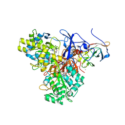
1JDB
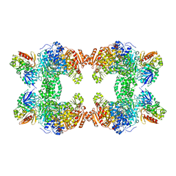
 | | CARBAMOYL PHOSPHATE SYNTHETASE FROM ESCHERICHIA COLI | | 分子名称: | ADENOSINE-5'-DIPHOSPHATE, CARBAMOYL PHOSPHATE SYNTHETASE, CHLORIDE ION, ... | | 著者 | Thoden, J.B, Holden, H.M, Wesenberg, G, Raushel, F.M, Rayment, I. | | 登録日 | 1997-03-25 | | 公開日 | 1998-06-17 | | 最終更新日 | 2024-04-03 | | 実験手法 | X-RAY DIFFRACTION (2.1 Å) | | 主引用文献 | The structure of carbamoyl phosphate synthetase determined to 2.1 A resolution.
Acta Crystallogr.,Sect.D, 55, 1999
|
|
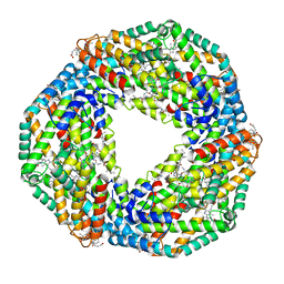
1IRU
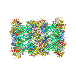 | | Crystal Structure of the mammalian 20S proteasome at 2.75 A resolution | | 分子名称: | 20S proteasome, MAGNESIUM ION | | 著者 | Unno, M, Mizushima, T, Morimoto, Y, Tomisugi, Y, Tanaka, K, Yasuoka, N, Tsukihara, T. | | 登録日 | 2001-10-24 | | 公開日 | 2002-05-22 | | 最終更新日 | 2023-12-27 | | 実験手法 | X-RAY DIFFRACTION (2.75 Å) | | 主引用文献 | The structure of the mammalian 20S proteasome at 2.75 A resolution.
Structure, 10, 2002
|
|
1JQ4
 | | [2Fe-2S] Domain of Methane Monooxygenase Reductase from Methylococcus capsulatus (Bath) | | 分子名称: | FE2/S2 (INORGANIC) CLUSTER, METHANE MONOOXYGENASE COMPONENT C | | 著者 | Mueller, J, Lugovskoy, A.A, Wagner, G, Lippard, S.J. | | 登録日 | 2001-08-03 | | 公開日 | 2002-01-09 | | 最終更新日 | 2024-05-22 | | 実験手法 | SOLUTION NMR | | 主引用文献 | NMR structure of the [2Fe-2S] ferredoxin domain from soluble methane monooxygenase reductase and interaction with its hydroxylase.
Biochemistry, 41, 2002
|
|
1J3D
 | |
1K1E
 | |
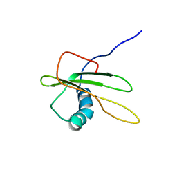
1K6G
 | | Solution Structure of Conserved AGNN Tetraloops: Insights into Rnt1p RNA Processing | | 分子名称: | RNA (5'-R(P*GP*GP*CP*GP*UP*CP*AP*UP*GP*AP*GP*UP*CP*CP*AP*UP*GP*GP*CP*GP*CP*C)-3') | | 著者 | Lebars, I, Lamontagne, B, Yoshizawa, S, Abou Elela, S, Fourmy, D. | | 登録日 | 2001-10-16 | | 公開日 | 2001-12-19 | | 最終更新日 | 2024-05-22 | | 実験手法 | SOLUTION NMR | | 主引用文献 | Solution structure of conserved AGNN tetraloops: insights into Rnt1p RNA processing.
EMBO J., 20, 2001
|
|
1KEE
 | | Inactivation of the Amidotransferase Activity of Carbamoyl Phosphate Synthetase by the Antibiotic Acivicin | | 分子名称: | ADENOSINE-5'-DIPHOSPHATE, CHLORIDE ION, Carbamoyl-phosphate synthetase large chain, ... | | 著者 | Miles, B.W, Thoden, J.B, Holden, H.M, Raushel, F.M. | | 登録日 | 2001-11-15 | | 公開日 | 2001-12-21 | | 最終更新日 | 2023-08-16 | | 実験手法 | X-RAY DIFFRACTION (2.1 Å) | | 主引用文献 | Inactivation of the amidotransferase activity of carbamoyl phosphate synthetase by the antibiotic acivicin.
J.Biol.Chem., 277, 2002
|
|
1KIU
 | |
2PFR
 | | Human N-acetyltransferase 2 | | 分子名称: | Arylamine N-acetyltransferase 2, COENZYME A, SULFATE ION, ... | | 著者 | Tempel, W, Wu, H, Dombrovski, L, Loppnau, P, Weigelt, J, Sundstrom, M, Arrowsmith, C.H, Edwards, A.M, Grant, D.M, Bochkarev, A, Plotnikov, A.N, Structural Genomics Consortium (SGC) | | 登録日 | 2007-04-05 | | 公開日 | 2007-05-15 | | 最終更新日 | 2023-08-30 | | 実験手法 | X-RAY DIFFRACTION (1.92 Å) | | 主引用文献 | The Crystal Structure of Human N-acetyltransferase 2 in complex with CoA
to be published
|
|
2PKH
 | | Structural Genomics, the crystal structure of the C-terminal domain of histidine utilization repressor from Pseudomonas syringae pv. tomato str. DC3000 | | 分子名称: | 1,2-ETHANEDIOL, Histidine utilization repressor | | 著者 | Tan, K, Zhou, M, Clancy, S, Joachimiak, A, Midwest Center for Structural Genomics (MCSG) | | 登録日 | 2007-04-17 | | 公開日 | 2007-05-15 | | 最終更新日 | 2011-07-13 | | 実験手法 | X-RAY DIFFRACTION (1.95 Å) | | 主引用文献 | The crystal structure of the C-terminal domain of histidine utilization repressor from Pseudomonas syringae pv. tomato str. DC3000.
To be Published
|
|
2PLX
 | |
3SQG
 | | Crystal structure of a methyl-coenzyme M reductase purified from Black Sea mats | | 分子名称: | (17[2]S)-17[2]-methylthio-coenzyme F43, 1-THIOETHANESULFONIC ACID, CALCIUM ION, ... | | 著者 | Shima, S, Krueger, M, Weinert, T, Demmer, U, Thauer, R.K, Ermler, U. | | 登録日 | 2011-07-05 | | 公開日 | 2011-11-30 | | 最終更新日 | 2017-11-08 | | 実験手法 | X-RAY DIFFRACTION (2.1 Å) | | 主引用文献 | Structure of a methyl-coenzyme M reductase from Black Sea mats that oxidize methane anaerobically.
Nature, 481, 2011
|
|
3SYQ
 | | Crystal structure of the G protein-gated inward rectifier K+ channel GIRK2 (Kir3.2) R201A mutant in complex with PIP2 | | 分子名称: | G protein-activated inward rectifier potassium channel 2, POTASSIUM ION, [(2R)-2-octanoyloxy-3-[oxidanyl-[(1R,2R,3S,4R,5R,6S)-2,3,6-tris(oxidanyl)-4,5-diphosphonooxy-cyclohexyl]oxy-phosphoryl]oxy-propyl] octanoate | | 著者 | Whorton, M.R, MacKinnon, R. | | 登録日 | 2011-07-18 | | 公開日 | 2011-10-12 | | 最終更新日 | 2023-09-13 | | 実験手法 | X-RAY DIFFRACTION (3.44 Å) | | 主引用文献 | Crystal Structure of the Mammalian GIRK2 K(+) Channel and Gating Regulation by G Proteins, PIP(2), and Sodium.
Cell(Cambridge,Mass.), 147, 2011
|
|
3AML
 | | Structure of the Starch Branching Enzyme I (BEI) from Oryza sativa L | | 分子名称: | 4-(2-HYDROXYETHYL)-1-PIPERAZINE ETHANESULFONIC ACID, ACETATE ION, BETA-MERCAPTOETHANOL, ... | | 著者 | Kakuta, Y, Chaen, K, Noguchi, J, Vu, N, Kimura, M. | | 登録日 | 2010-08-20 | | 公開日 | 2011-09-28 | | 最終更新日 | 2023-11-01 | | 実験手法 | X-RAY DIFFRACTION (1.7 Å) | | 主引用文献 | Crystal structure of the branching enzyme I (BEI) from Oryza sativa L with implications for catalysis and substrate binding.
Glycobiology, 21, 2011
|
|
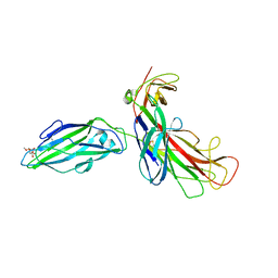
1KO3
 | | VIM-2, a Zn-beta-lactamase from Pseudomonas aeruginosa with Cys221 reduced | | 分子名称: | ACETATE ION, CHLORIDE ION, HYDROXIDE ION, ... | | 著者 | Garcia-Saez, I, Docquier, J.-D, Rossolini, G.M, Dideberg, O. | | 登録日 | 2001-12-20 | | 公開日 | 2003-09-02 | | 最終更新日 | 2023-10-25 | | 実験手法 | X-RAY DIFFRACTION (1.91 Å) | | 主引用文献 | The three-dimensional structure of VIM-2, a Zn-beta-lactamase from Pseudomonas aeruginosa in its reduced and oxidised form
J.Mol.Biol., 375, 2008
|
|


