2DX2
 
 | |
2DX3
 | |
2DX4
 | |
2DX5
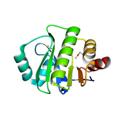
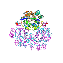
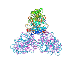
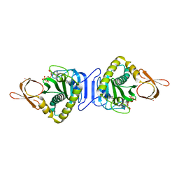
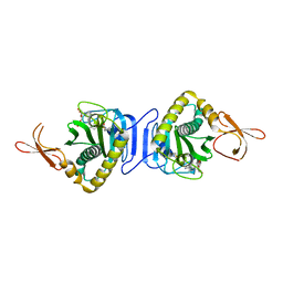
 | | The complex structure between the mouse EAP45-GLUE domain and ubiquitin | | 分子名称: | Ubiquitin, Vacuolar protein sorting protein 36 | | 著者 | Hirano, S, Suzuki, N, Slagsvold, T, Kawasaki, M, Trambaiolo, D, Kato, R, Stenmark, H, Wakatsuki, S. | | 登録日 | 2006-08-24 | | 公開日 | 2006-10-10 | | 最終更新日 | 2024-03-13 | | 実験手法 | X-RAY DIFFRACTION (3.35 Å) | | 主引用文献 | Structural basis of ubiquitin recognition by mammalian Eap45 GLUE domain
Nat.Struct.Mol.Biol., 13, 2006
|
|
2DX6
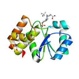
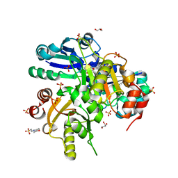
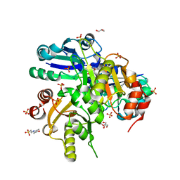
 | | Crystal structure of conserved hypothetical protein, TTHA0132 from Thermus thermophilus HB8 | | 分子名称: | ACETATE ION, Hypothetical protein TTHA0132, ISOPROPYL ALCOHOL | | 著者 | Kishishita, S, Murayama, K, Terada, T, Shirouzu, M, Kuramitsu, S, Yokoyama, S, RIKEN Structural Genomics/Proteomics Initiative (RSGI) | | 登録日 | 2006-08-24 | | 公開日 | 2007-02-24 | | 最終更新日 | 2011-07-13 | | 実験手法 | X-RAY DIFFRACTION (1.78 Å) | | 主引用文献 | Crystal structure of conserved hypothetical protein, TTHA0132 from Thermus thermophilus HB8
To be Published
|
|
2DX7
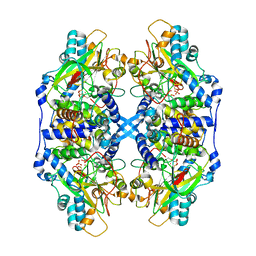
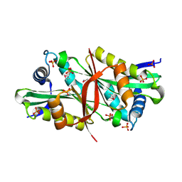
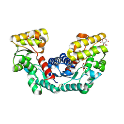 | | Crystal structure of Pyrococcus horikoshii OT3 aspartate racemase complex with citric acid | | 分子名称: | CITRIC ACID, aspartate racemase | | 著者 | Ohtaki, A, Arakawa, T, Iizuka, R, Odaka, M, Yohda, M. | | 登録日 | 2006-08-24 | | 公開日 | 2007-08-28 | | 最終更新日 | 2023-10-25 | | 実験手法 | X-RAY DIFFRACTION (2 Å) | | 主引用文献 | Structure of aspartate racemase complexed with a dual substrate analogue, citric acid, and implications for the reaction mechanism.
Proteins, 70, 2008
|
|
2DX8
 | |
2DXA
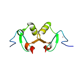
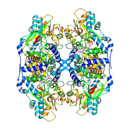
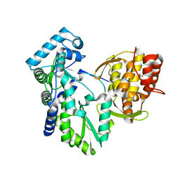
 | | Crystal structure of trans editing enzyme ProX from E.coli | | 分子名称: | CHLORIDE ION, Protein ybaK | | 著者 | Kato-Murayama, M, Murayama, K, Terada, T, Shirouzu, M, Yokoyama, S, RIKEN Structural Genomics/Proteomics Initiative (RSGI) | | 登録日 | 2006-08-25 | | 公開日 | 2007-02-25 | | 最終更新日 | 2023-11-15 | | 実験手法 | X-RAY DIFFRACTION (1.58 Å) | | 主引用文献 | Crystal structure of trans editing enzyme ProX from E.coli
To be Published
|
|
2DXB
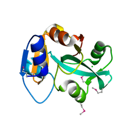
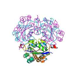
 | | Recombinant thiocyanate hydrolase comprising partially-modified cobalt centers | | 分子名称: | COBALT (III) ION, PHOSPHATE ION, Thiocyanate hydrolase subunit alpha, ... | | 著者 | Arakawa, T, Kawano, Y, Katayama, Y, Yohda, M, Odaka, M. | | 登録日 | 2006-08-25 | | 公開日 | 2007-08-28 | | 最終更新日 | 2023-11-15 | | 実験手法 | X-RAY DIFFRACTION (2.25 Å) | | 主引用文献 | Structural Basis for Catalytic Activation of Thiocyanate Hydrolase Involving Metal-Ligated Cysteine Modification
J.Am.Chem.Soc., 131, 2009
|
|
2DXC
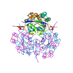
 | | Recombinant thiocyanate hydrolase, fully-matured form | | 分子名称: | COBALT (III) ION, L(+)-TARTARIC ACID, Thiocyanate hydrolase subunit alpha, ... | | 著者 | Arakawa, T, Kawano, Y, Katayama, Y, Yohda, M, Odaka, M. | | 登録日 | 2006-08-25 | | 公開日 | 2007-08-28 | | 最終更新日 | 2023-11-15 | | 実験手法 | X-RAY DIFFRACTION (1.9 Å) | | 主引用文献 | Structural Basis for Catalytic Activation of Thiocyanate Hydrolase Involving Metal-Ligated Cysteine Modification
J.Am.Chem.Soc., 131, 2009
|
|
2DXD
 | | Crystal structure of nucleoside diphosphate kinase in complex with ATP analog | | 分子名称: | ADENOSINE MONOPHOSPHATE, CHLORIDE ION, Nucleoside diphosphate kinase | | 著者 | Kato-Murayama, M, Murayama, K, Terada, T, Shirouzu, M, Yokoyama, S, RIKEN Structural Genomics/Proteomics Initiative (RSGI) | | 登録日 | 2006-08-25 | | 公開日 | 2007-02-25 | | 最終更新日 | 2023-10-25 | | 実験手法 | X-RAY DIFFRACTION (1.77 Å) | | 主引用文献 | Crystal structure of nucleoside diphosphate kinase in complex with ATP analog
To be Published
|
|
2DXE
 | | Crystal structure of nucleoside diphosphate kinase in complex with GDP | | 分子名称: | CHLORIDE ION, GUANOSINE-5'-DIPHOSPHATE, MAGNESIUM ION, ... | | 著者 | Kato-Murayama, M, Murayama, K, Terada, T, Shirouzu, M, Yokoyama, S, RIKEN Structural Genomics/Proteomics Initiative (RSGI) | | 登録日 | 2006-08-25 | | 公開日 | 2007-02-25 | | 最終更新日 | 2023-10-25 | | 実験手法 | X-RAY DIFFRACTION (1.7 Å) | | 主引用文献 | Crystal structure of nucleoside diphosphate kinase in complex with GDP
To be Published
|
|
2DXF
 | | Crystal structure of nucleoside diphosphate kinase in complex with GTP analog | | 分子名称: | CHLORIDE ION, Nucleoside diphosphate kinase, PHOSPHOAMINOPHOSPHONIC ACID-GUANYLATE ESTER | | 著者 | Kato-Murayama, M, Murayama, K, Terada, T, Shirouzu, M, Yokoyama, S, RIKEN Structural Genomics/Proteomics Initiative (RSGI) | | 登録日 | 2006-08-25 | | 公開日 | 2007-02-25 | | 最終更新日 | 2023-10-25 | | 実験手法 | X-RAY DIFFRACTION (1.7 Å) | | 主引用文献 | Crystal structure of nucleoside diphosphate kinase in complex with GTP analog
To be Published
|
|
2DXI
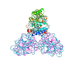
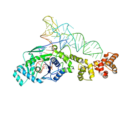 | | 2.2 A crystal structure of glutamyl-tRNA synthetase from Thermus thermophilus complexed with tRNA(Glu), ATP, and L-glutamol | | 分子名称: | (4S)-4-AMINO-5-HYDROXYPENTANOIC ACID, ADENOSINE-5'-TRIPHOSPHATE, MAGNESIUM ION, ... | | 著者 | Sekine, S, Yokoyama, S, RIKEN Structural Genomics/Proteomics Initiative (RSGI) | | 登録日 | 2006-08-28 | | 公開日 | 2006-10-31 | | 最終更新日 | 2023-11-15 | | 実験手法 | X-RAY DIFFRACTION (2.2 Å) | | 主引用文献 | Structural bases of transfer RNA-dependent amino acid recognition and activation by glutamyl-tRNA synthetase
Structure, 14, 2006
|
|
2DXL
 | |
2DXM
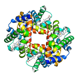 | | Neutron Structure Analysis of Deoxy Human Hemoglobin | | 分子名称: | Hemoglobin subunit alpha, Hemoglobin subunit beta, PROTOPORPHYRIN IX CONTAINING FE | | 著者 | Morimoto, Y. | | 登録日 | 2006-08-28 | | 公開日 | 2007-12-04 | | 最終更新日 | 2024-04-03 | | 実験手法 | NEUTRON DIFFRACTION (2.1 Å) | | 主引用文献 | Protonation states of buried histidine residues in human deoxyhemoglobin revealed by neutron crystallography.
J.Am.Chem.Soc., 129, 2007
|
|
2DXN
 | |
2DXP
 | |
2DXQ
 | | Putative acetyltransferase from Agrobacterium tumefaciens str. C58 | | 分子名称: | Acetyltransferase, PHOSPHATE ION | | 著者 | Binkowski, T.A, Xu, X, Edwards, A, Savchenko, A, Joachimiak, A, Midwest Center for Structural Genomics (MCSG) | | 登録日 | 2006-08-30 | | 公開日 | 2006-09-27 | | 最終更新日 | 2011-07-13 | | 実験手法 | X-RAY DIFFRACTION (1.8 Å) | | 主引用文献 | Putative acetyltransferase from Agrobacterium tumefaciens str. C58
To be published
|
|
2DXR
 | | Crystal structure of the complex formed between C-terminal half of bovine lactoferrin and sorbitol at 2.85 A resolution | | 分子名称: | 2-acetamido-2-deoxy-beta-D-glucopyranose-(1-4)-2-acetamido-2-deoxy-beta-D-glucopyranose, CARBONATE ION, FE (III) ION, ... | | 著者 | Mir, R, Prem kumar, R, Sinha, M, Singh, N, Sharma, S, Bhushan, A, Kaur, P, Singh, T.P. | | 登録日 | 2006-08-30 | | 公開日 | 2006-09-12 | | 最終更新日 | 2023-10-25 | | 実験手法 | X-RAY DIFFRACTION (2.85 Å) | | 主引用文献 | Crystal structure of the complex formed between C-terminal half of bovine lactoferrin and sorbitol at 2.85 A resolution
To be Published
|
|
2DXS
 | | Crystal structure of HCV NS5B RNA polymerase complexed with a tetracyclic inhibitor | | 分子名称: | Genome polyprotein, N-[(13-CYCLOHEXYL-6,7-DIHYDROINDOLO[1,2-D][1,4]BENZOXAZEPIN-10-YL)CARBONYL]-2-METHYL-L-ALANINE | | 著者 | Adachi, T, Tsuruha, J, Doi, S, Murase, K, Ikegashira, K, Watanabe, S, Uehara, K, Orita, T, Nomura, A, Kamada, M. | | 登録日 | 2006-08-30 | | 公開日 | 2006-12-26 | | 最終更新日 | 2023-10-25 | | 実験手法 | X-RAY DIFFRACTION (2.2 Å) | | 主引用文献 | Discovery of Conformationally Constrained Tetracyclic Compounds as Potent Hepatitis C Virus NS5B RNA Polymerase Inhibitors
J.Med.Chem., 49, 2006
|
|
2DXT
 | |
2DXU
 | | Crystal Structure Of Biotin Protein Ligase From Pyrococcus Horikoshii Complexed with Biotinyl-5'-AMP, Mutation R48A | | 分子名称: | BIOTINYL-5-AMP, biotin--[acetyl-CoA-carboxylase] ligase | | 著者 | Bagautdinov, B, Taketa, M, Matsuura, Y, Kunishima, N, RIKEN Structural Genomics/Proteomics Initiative (RSGI) | | 登録日 | 2006-08-30 | | 公開日 | 2007-03-01 | | 最終更新日 | 2023-10-25 | | 実験手法 | X-RAY DIFFRACTION (1.28 Å) | | 主引用文献 | Protein biotinylation visualized by a complex structure of biotin protein ligase with a substrate
J.Biol.Chem., 283, 2008
|
|
2DXV
 | |
2DXW
 | |


