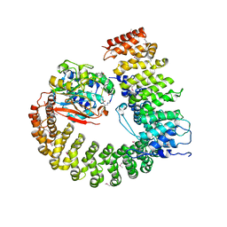
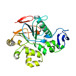
2IAE
 
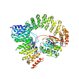 | | Crystal structure of a protein phosphatase 2A (PP2A) holoenzyme. | | 分子名称: | MANGANESE (II) ION, Serine/threonine-protein phosphatase 2A 56 kDa regulatory subunit gamma isoform, Serine/threonine-protein phosphatase 2A 65 kDa regulatory subunit A alpha isoform, ... | | 著者 | Cho, U.S, Xu, W. | | 登録日 | 2006-09-07 | | 公開日 | 2006-12-26 | | 最終更新日 | 2023-11-15 | | 実験手法 | X-RAY DIFFRACTION (3.5 Å) | | 主引用文献 | Crystal structure of a protein phosphatase 2A heterotrimeric holoenzyme.
Nature, 445, 2007
|
|
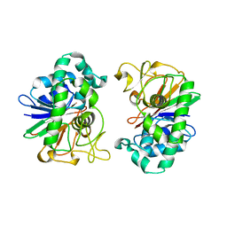
2HY1
 | | Crystal structure of Rv0805 | | 分子名称: | FE (III) ION, MANGANESE (II) ION, PHOSPHATE ION, ... | | 著者 | Shenoy, A.R, Capuder, M, Draskovic, P, Lamba, D, Visweswariah, S.S, Podobnik, M. | | 登録日 | 2006-08-04 | | 公開日 | 2006-12-26 | | 最終更新日 | 2024-02-14 | | 実験手法 | X-RAY DIFFRACTION (1.932 Å) | | 主引用文献 | Structural and Biochemical Analysis of the Rv0805 Cyclic Nucleotide Phosphodiesterase from Mycobacterium tuberculosis.
J.Mol.Biol., 365, 2007
|
|
2HYP
 | | Crystal structure of Rv0805 D66A mutant | | 分子名称: | BROMIDE ION, CACODYLATE ION, FE (III) ION, ... | | 著者 | Shenoy, A.R, Capuder, M, Draskovic, P, Lamba, D, Visweswariah, S.S, Podobnik, M. | | 登録日 | 2006-08-07 | | 公開日 | 2006-12-26 | | 最終更新日 | 2024-02-21 | | 実験手法 | X-RAY DIFFRACTION (2.05 Å) | | 主引用文献 | Structural and Biochemical Analysis of the Rv0805 Cyclic Nucleotide Phosphodiesterase from Mycobacterium tuberculosis.
J.Mol.Biol., 365, 2007
|
|
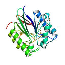
2NXF
 | | Crystal Structure of a dimetal phosphatase from Danio rerio LOC 393393 | | 分子名称: | ETHANOL, PHOSPHATE ION, Putative dimetal phosphatase, ... | | 著者 | Bitto, E, Wesenberg, G.E, Phillips Jr, G.N, McCoy, J.G, Bingman, C.A, Center for Eukaryotic Structural Genomics (CESG) | | 登録日 | 2006-11-17 | | 公開日 | 2006-12-12 | | 最終更新日 | 2023-12-27 | | 実験手法 | X-RAY DIFFRACTION (1.7 Å) | | 主引用文献 | Crystal Structure of a dimetal phosphatase from Danio rerio LOC 393393
To be Published
|
|
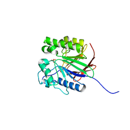
2NYM
 | | Crystal Structure of Protein Phosphatase 2A (PP2A) with C-terminus truncated catalytic subunit | | 分子名称: | MANGANESE (II) ION, Protein phosphatase 2, Serine/threonine-protein phosphatase 2A 56 kDa regulatory subunit gamma isoform, ... | | 著者 | Chen, Y, Xing, Y, Xu, Y, Chao, Y, Lin, Z, Jeffrey, P.D, Shi, Y. | | 登録日 | 2006-11-21 | | 公開日 | 2006-12-12 | | 最終更新日 | 2023-11-15 | | 実験手法 | X-RAY DIFFRACTION (3.6 Å) | | 主引用文献 | Structure of the Protein Phosphatase 2A Holoenzyme.
Cell(Cambridge,Mass.), 127, 2006
|
|
2NYL
 | | Crystal structure of Protein Phosphatase 2A (PP2A) holoenzyme with the catalytic subunit carboxyl terminus truncated | | 分子名称: | MANGANESE (II) ION, Protein phosphatase 2, regulatory subunit A (PR 65), ... | | 著者 | Xing, Y, Xu, Y, Chen, Y, Chao, Y, Lin, Z, Shi, Y. | | 登録日 | 2006-11-20 | | 公開日 | 2006-12-12 | | 最終更新日 | 2023-11-15 | | 実験手法 | X-RAY DIFFRACTION (3.8 Å) | | 主引用文献 | Structure of the Protein Phosphatase 2A Holoenzyme.
Cell(Cambridge,Mass.), 127, 2006
|
|
2NPP
 | | Structure of the Protein Phosphatase 2A Holoenzyme | | 分子名称: | MANGANESE (II) ION, Protein Phosphatase 2, regulatory subunit A (PR 65), ... | | 著者 | Xu, Y, Chen, Y, Xing, Y, Chao, Y, Shi, Y. | | 登録日 | 2006-10-28 | | 公開日 | 2006-12-12 | | 最終更新日 | 2023-11-15 | | 実験手法 | X-RAY DIFFRACTION (3.3 Å) | | 主引用文献 | Structure of the protein phosphatase 2A holoenzyme
Cell(Cambridge,Mass.), 127, 2006
|
|
2DFJ
 | |
2IE3
 | | Structure of the Protein Phosphatase 2A Core Enzyme Bound to Tumor-inducing Toxins | | 分子名称: | MANGANESE (II) ION, Protein Phosphatase 2, regulatory subunit A (PR 65), ... | | 著者 | Xing, Y, Xu, Y, Chen, Y, Jeffrey, P.D, Chao, Y, Shi, Y. | | 登録日 | 2006-09-17 | | 公開日 | 2006-11-07 | | 最終更新日 | 2023-11-15 | | 実験手法 | X-RAY DIFFRACTION (2.8 Å) | | 主引用文献 | Structure of Protein Phosphatase 2A Core Enzyme Bound to Tumor-Inducing Toxins
Cell(Cambridge,Mass.), 127, 2006
|
|
2IE4
 | | Structure of the Protein Phosphatase 2A Core Enzyme Bound to okadaic acid | | 分子名称: | MANGANESE (II) ION, OKADAIC ACID, Protein Phosphatase 2, ... | | 著者 | Xing, Y, Xu, Y, Chen, Y, Jeffrey, P.D, Chao, Y, Shi, Y. | | 登録日 | 2006-09-17 | | 公開日 | 2006-11-07 | | 最終更新日 | 2024-02-21 | | 実験手法 | X-RAY DIFFRACTION (2.6 Å) | | 主引用文献 | Structure of Protein Phosphatase 2A Core Enzyme Bound to Tumor-Inducing Toxins
Cell(Cambridge,Mass.), 127, 2006
|
|
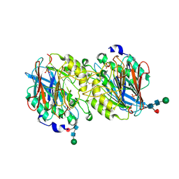
2BDX
 | | X-ray Crystal Structure of dihydromicrocystin-LA bound to Protein Phosphatase-1 | | 分子名称: | DIHYDROMICROCYSTIN-LA, MANGANESE (II) ION, Serine/threonine protein phosphatase PP1-gamma catalytic subunit | | 著者 | Maynes, J.T, Luu, H.A, Cherney, M.M, Andersen, R.J, Williams, D, Holmes, C.F, James, M.N. | | 登録日 | 2005-10-21 | | 公開日 | 2006-01-17 | | 最終更新日 | 2023-11-15 | | 実験手法 | X-RAY DIFFRACTION (2.3 Å) | | 主引用文献 | Crystal Structures of Protein Phosphatase-1 Bound to Motuporin and Dihydromicrocystin-LA: Elucidation of the Mechanism of Enzyme Inhibition by Cyanobacterial Toxins.
J.Mol.Biol., 356, 2006
|
|
2BCD
 | | X-ray crystal structure of Protein Phosphatase-1 with the marine toxin motuporin bound | | 分子名称: | BETA-MERCAPTOETHANOL, MANGANESE (II) ION, MOTUPORIN, ... | | 著者 | Maynes, J.T, Luu, H.A, Cherney, M.M, Andersen, R.J, Williams, D, Holmes, C.F, James, M.N. | | 登録日 | 2005-10-19 | | 公開日 | 2006-01-17 | | 最終更新日 | 2023-11-15 | | 実験手法 | X-RAY DIFFRACTION (2.1 Å) | | 主引用文献 | Crystal Structures of Protein Phosphatase-1 Bound to Motuporin and Dihydromicrocystin-LA: Elucidation of the Mechanism of Enzyme Inhibition by Cyanobacterial Toxins.
J.Mol.Biol., 356, 2006
|
|
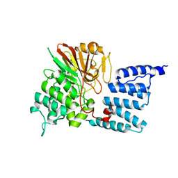
2BQ8
 | | Crystal structure of human purple acid phosphatase with an inhibitory conformation of the repression loop | | 分子名称: | FE (II) ION, SULFATE ION, TARTRATE-RESISTANT ACID PHOSPHATASE TYPE 5, ... | | 著者 | Straeter, N, Jasper, B, Krebs, B. | | 登録日 | 2005-04-27 | | 公開日 | 2005-10-24 | | 最終更新日 | 2023-12-13 | | 実験手法 | X-RAY DIFFRACTION (2.2 Å) | | 主引用文献 | Crystal Structures of Recombinant Human Purple Acid Phosphatase with and without an Inhibitory Conformation of the Repression Loop.
J.Mol.Biol., 351, 2005
|
|
2AHD
 | |
1WAR
 | | Recombinant Human Purple Acid Phosphatase expressed in Pichia Pastoris | | 分子名称: | 2-acetamido-2-deoxy-beta-D-glucopyranose, FE (III) ION, HUMAN PURPLE ACID PHOSPHATASE, ... | | 著者 | Duff, A.P, Langley, D.B, Han, R, Averill, B.A, Freeman, H.C, Guss, J.M. | | 登録日 | 2004-10-28 | | 公開日 | 2005-07-06 | | 最終更新日 | 2023-12-13 | | 実験手法 | X-RAY DIFFRACTION (2.22 Å) | | 主引用文献 | Crystal Structures of Recombinant Human Purple Acid Phosphatase with and without an Inhibitory Conformation of the Repression Loop.
J.Mol.Biol., 351, 2005
|
|
1V73
 | | Crystal Structure of Cold-Active Protein-Tyrosine Phosphatase of a Psychrophile Shewanella SP. | | 分子名称: | ACETIC ACID, CALCIUM ION, psychrophilic phosphatase I | | 著者 | Tsuruta, H, Mikami, B, Aizono, Y. | | 登録日 | 2003-12-09 | | 公開日 | 2005-03-01 | | 最終更新日 | 2023-12-27 | | 実験手法 | X-RAY DIFFRACTION (1.82 Å) | | 主引用文献 | Crystal Structure of Cold-Active Protein-Tyrosine Phosphatase from a Psychrophile, Shewanella sp
J.Biochem.(Tokyo), 137, 2005
|
|
1WAO
 | | PP5 structure | | 分子名称: | MANGANESE (II) ION, SERINE/THREONINE PROTEIN PHOSPHATASE 5 | | 著者 | Barford, D. | | 登録日 | 2004-10-27 | | 公開日 | 2005-02-22 | | 最終更新日 | 2024-05-08 | | 実験手法 | X-RAY DIFFRACTION (2.9 Å) | | 主引用文献 | Molecular Basis for Tpr Domain-Mediated Regulation of Protein Phosphatase 5
Embo J., 24, 2005
|
|
1XZW
 | | Sweet potato purple acid phosphatase/phosphate complex | | 分子名称: | 2-acetamido-2-deoxy-beta-D-glucopyranose, 2-acetamido-2-deoxy-beta-D-glucopyranose-(1-4)-2-acetamido-2-deoxy-beta-D-glucopyranose, FE (III) ION, ... | | 著者 | Schenk, G, Carrington, L.E, Gahan, L.R, Hamilton, S.E, de Jersey, J, Guddat, L.W. | | 登録日 | 2004-11-12 | | 公開日 | 2004-12-14 | | 最終更新日 | 2023-10-25 | | 実験手法 | X-RAY DIFFRACTION (2.5 Å) | | 主引用文献 | Phosphate forms an unusual tripodal complex with the Fe-Mn center of sweet potato purple acid phosphatase
Proc.Natl.Acad.Sci.USA, 102, 2005
|
|
1T71
 | | Crystal structure of a novel phosphatase Mycoplasma pneumoniaefrom | | 分子名称: | FE (III) ION, phosphatase | | 著者 | Shin, D.H, Jancarik, J, Kim, R, Yokota, H, Kim, S.-H, Berkeley Structural Genomics Center (BSGC) | | 登録日 | 2004-05-07 | | 公開日 | 2004-12-07 | | 最終更新日 | 2024-02-14 | | 実験手法 | X-RAY DIFFRACTION (2.1 Å) | | 主引用文献 | Crystal structure of a novel phosphatase from Mycoplasma pneumoniae
To be Published
|
|
1T70
 | | Crystal structure of a novel phosphatase from Deinococcus radiodurans | | 分子名称: | Phosphatase | | 著者 | Shin, D.H, Wang, W, Kim, R, Yokota, H, Kim, S.H, Berkeley Structural Genomics Center (BSGC) | | 登録日 | 2004-05-07 | | 公開日 | 2004-12-07 | | 最終更新日 | 2024-02-14 | | 実験手法 | X-RAY DIFFRACTION (2.3 Å) | | 主引用文献 | Structural and enzymatic characterization of DR1281: A calcineurin-like phosphoesterase from Deinococcus radiodurans.
Proteins, 70, 2008
|
|
1XM7
 | |
1S95
 | | Structure of serine/threonine protein phosphatase 5 | | 分子名称: | (4S)-2-METHYL-2,4-PENTANEDIOL, MANGANESE (II) ION, PHOSPHATE ION, ... | | 著者 | Swingle, M.R, Honkanen, R.E, Ciszak, E.M. | | 登録日 | 2004-02-03 | | 公開日 | 2004-08-24 | | 最終更新日 | 2023-08-23 | | 実験手法 | X-RAY DIFFRACTION (1.6 Å) | | 主引用文献 | Structural basis for the catalytic activity of human serine/threonine protein phosphatase-5.
J.Biol.Chem., 279, 2004
|
|
1U32
 | | Crystal structure of a Protein Phosphatase-1: Calcineurin Hybrid Bound to Okadaic Acid | | 分子名称: | BETA-MERCAPTOETHANOL, MANGANESE (II) ION, OKADAIC ACID, ... | | 著者 | Maynes, J.T, Perreault, K.R, Cherney, M.M, Luu, H.A, James, M.N.G, Holmes, C.F.B. | | 登録日 | 2004-07-20 | | 公開日 | 2004-08-17 | | 最終更新日 | 2023-10-25 | | 実験手法 | X-RAY DIFFRACTION (2 Å) | | 主引用文献 | Crystal Structure and Mutagenesis of a Protein Phosphatase-1:Calcineurin Hybrid Elucidate the Role of the {beta}12-{beta}13 Loop in Inhibitor Binding
J.Biol.Chem., 279, 2004
|
|
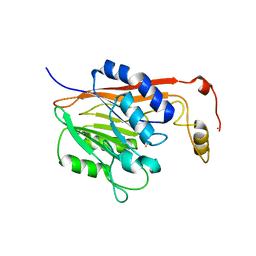
1SU1
 | | Structural and biochemical characterization of Yfce, a phosphoesterase from E. coli | | 分子名称: | Hypothetical protein yfcE, SULFATE ION, ZINC ION | | 著者 | Miller, D.J, Shuvalova, L, Evdokimova, E, Savchenko, A, Yakunin, A, Anderson, W.F, Midwest Center for Structural Genomics (MCSG) | | 登録日 | 2004-03-25 | | 公開日 | 2004-08-17 | | 最終更新日 | 2024-02-14 | | 実験手法 | X-RAY DIFFRACTION (2.25 Å) | | 主引用文献 | Structural and biochemical characterization of a novel Mn2+-dependent phosphodiesterase encoded by the yfcE gene.
Protein Sci., 16, 2007
|
|
1S3M
 | | Structural and Functional Characterization of a Novel Archaeal Phosphodiesterase | | 分子名称: | Hypothetical protein MJ0936, NICKEL (II) ION | | 著者 | Chen, S, Busso, D, Yakunin, A.F, Kuznetsova, E, Proudfoot, M, Jancrick, J, Kim, R, Kim, S.-H, Berkeley Structural Genomics Center (BSGC) | | 登録日 | 2004-01-13 | | 公開日 | 2004-08-10 | | 最終更新日 | 2024-02-14 | | 実験手法 | X-RAY DIFFRACTION (2.5 Å) | | 主引用文献 | Structural and functional characterization of a novel phosphodiesterase from Methanococcus jannaschii
J.Biol.Chem., 279, 2004
|
|


