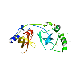
1Z3M
 
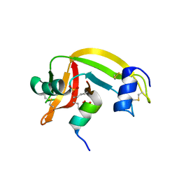 | | Crystal structure of mutant Ribonuclease S (F8Nva) | | 分子名称: | Ribonuclease pancreatic, S-Peptide, S-protein, ... | | 著者 | Das, M, Vasudeva Rao, B, Ghosh, S, Varadarajan, R. | | 登録日 | 2005-03-14 | | 公開日 | 2005-03-29 | | 最終更新日 | 2023-11-15 | | 実験手法 | X-RAY DIFFRACTION (2 Å) | | 主引用文献 | Attempts to delineate the relative contributions of changes in hydrophobicity and packing to changes in stability of ribonuclease S mutants.
Biochemistry, 44, 2005
|
|
1Z3Z
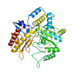 | | The crystal structure of a DGD mutant: Q52A | | 分子名称: | 2-(N-MORPHOLINO)-ETHANESULFONIC ACID, POTASSIUM ION, PYRIDOXAL-5'-PHOSPHATE, ... | | 著者 | Fogle, E.J, Liu, W, Toney, M.D. | | 登録日 | 2005-03-14 | | 公開日 | 2006-01-03 | | 最終更新日 | 2021-10-20 | | 実験手法 | X-RAY DIFFRACTION (2.9 Å) | | 主引用文献 | Role of q52 in catalysis of decarboxylation and transamination in dialkylglycine decarboxylase.
Biochemistry, 44, 2005
|
|
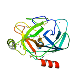
1Z5H
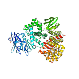 | | Crystal structures of the Tricorn interacting Factor F3 from Thermoplasma acidophilum | | 分子名称: | SULFATE ION, Tricorn protease interacting factor F3, ZINC ION | | 著者 | Kyrieleis, O.J.P, Goettig, P, Kiefersauer, R, Huber, R, Brandstetter, H. | | 登録日 | 2005-03-18 | | 公開日 | 2005-06-28 | | 最終更新日 | 2024-03-13 | | 実験手法 | X-RAY DIFFRACTION (2.3 Å) | | 主引用文献 | Crystal Structures of the Tricorn Interacting Factor F3 from Thermoplasma acidophilum, a Zinc Aminopeptidase in Three Different Conformations
J.MOL.BIOL., 349, 2005
|
|
3DOE
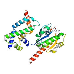 | | Complex of ARL2 and BART, Crystal Form 1 | | 分子名称: | ADP-ribosylation factor-like protein 2, ADP-ribosylation factor-like protein 2-binding protein, GUANOSINE-5'-TRIPHOSPHATE, ... | | 著者 | Zhang, T, Li, S, Ding, J. | | 登録日 | 2008-07-04 | | 公開日 | 2009-03-03 | | 最終更新日 | 2023-11-01 | | 実験手法 | X-RAY DIFFRACTION (2.25 Å) | | 主引用文献 | Crystal structure of the ARL2-GTP-BART complex reveals a novel recognition and binding mode of small GTPase with effector
Structure, 17, 2009
|
|
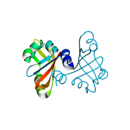
2P0K
 | | Crystal structure of SCMH1 | | 分子名称: | CHLORIDE ION, PHOSPHATE ION, Polycomb protein SCMH1 | | 著者 | Herzanych, N, Senisterra, G, Liu, Y, Crombet, L, Loppnau, P, Kozieradzki, I, Vedadi, M, Weigelt, J, Sundstrom, M, Arrowsmith, C.H, Edwards, A.M, Bochkarev, A, Min, J, Structural Genomics Consortium (SGC) | | 登録日 | 2007-02-28 | | 公開日 | 2007-03-27 | | 最終更新日 | 2023-08-30 | | 実験手法 | X-RAY DIFFRACTION (1.75 Å) | | 主引用文献 | Structure of SCMH1
To be Published
|
|
7K5J
 | | Structure of an E1-E2-ubiquitin thioester mimetic | | 分子名称: | ADENOSINE MONOPHOSPHATE, Ubiquitin, Ubiquitin-activating enzyme E1 1, ... | | 著者 | Yuan, L, Lv, Z, Olsen, S.K. | | 登録日 | 2020-09-16 | | 公開日 | 2021-04-28 | | 最終更新日 | 2023-10-18 | | 実験手法 | X-RAY DIFFRACTION (3.42 Å) | | 主引用文献 | Crystal structures of an E1-E2-ubiquitin thioester mimetic reveal molecular mechanisms of transthioesterification.
Nat Commun, 12, 2021
|
|
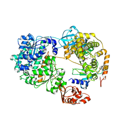
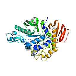
1ZAH
 | | Fructose-1,6-bisphosphate aldolase from rabbit muscle | | 分子名称: | Fructose-bisphosphate aldolase A | | 著者 | St-Jean, M, Lafrance-Vanasse, J, Liotard, B, Sygusch, J. | | 登録日 | 2005-04-06 | | 公開日 | 2005-05-10 | | 最終更新日 | 2023-08-23 | | 実験手法 | X-RAY DIFFRACTION (1.8 Å) | | 主引用文献 | High Resolution Reaction Intermediates of Rabbit Muscle Fructose-1,6-bisphosphate Aldolase: substrate cleavage and induced fit.
J.Biol.Chem., 280, 2005
|
|
2OWH
 | | Structure of an early-microsecond photolyzed state of CO-bjFixLH | | 分子名称: | PROTOPORPHYRIN IX CONTAINING FE, Sensor protein fixL | | 著者 | Key, J, Srajer, V, Pahl, R, Moffat, K. | | 登録日 | 2007-02-16 | | 公開日 | 2007-06-19 | | 最終更新日 | 2024-02-21 | | 実験手法 | X-RAY DIFFRACTION (2.5 Å) | | 主引用文献 | Time-resolved crystallographic studies of the heme domain of the oxygen sensor FixL: structural dynamics of ligand rebinding and their relation to signal transduction.
Biochemistry, 46, 2007
|
|
1ZFX
 | |
3RZY
 | |
1ZJB
 | | Crystal structure of the trehalulose synthase MutB from Pseudomonas mesoacidophila MX-45 (monoclinic form) | | 分子名称: | 2-AMINO-2-HYDROXYMETHYL-PROPANE-1,3-DIOL, CALCIUM ION, Trehalulose synthase | | 著者 | Ravaud, S, Robert, X, Haser, R, Aghajari, N. | | 登録日 | 2005-04-28 | | 公開日 | 2006-10-17 | | 最終更新日 | 2023-08-23 | | 実験手法 | X-RAY DIFFRACTION (1.8 Å) | | 主引用文献 | Expression, purification, crystallization and preliminary X-ray crystallographic studies of the trehalulose synthase MutB from Pseudomonas mesoacidophila MX-45.
Acta Crystallogr.,Sect.F, 61, 2005
|
|
1G36
 | | TRYPSIN INHIBITOR COMPLEX | | 分子名称: | 4-{[1-METHYL-5-(2-METHYL-BENZOIMIDAZOL-1-YLMETHYL)-1H-BENZOIMIDAZOL-2-YLMETHYL]-AMINO}-BENZAMIDINE, CALCIUM ION, SULFATE ION, ... | | 著者 | Nar, H. | | 登録日 | 2000-10-23 | | 公開日 | 2001-10-23 | | 最終更新日 | 2011-07-13 | | 実験手法 | X-RAY DIFFRACTION (1.9 Å) | | 主引用文献 | Structural basis for inhibition promiscuity of dual specific thrombin and factor Xa blood coagulation inhibitors.
Structure, 9, 2001
|
|
3S7O
 | | Crystal Structure of the Infrared Fluorescent D207H variant of Deinococcus Bacteriophytochrome chromophore binding domain at 1.24 angstrom resolution | | 分子名称: | 3-[2-[(Z)-[3-(2-carboxyethyl)-5-[(Z)-(4-ethenyl-3-methyl-5-oxidanylidene-pyrrol-2-ylidene)methyl]-4-methyl-pyrrol-1-ium -2-ylidene]methyl]-5-[(Z)-[(3E)-3-ethylidene-4-methyl-5-oxidanylidene-pyrrolidin-2-ylidene]methyl]-4-methyl-1H-pyrrol-3- yl]propanoic acid, Bacteriophytochrome, GLYCEROL | | 著者 | Forest, K.T, Auldridge, M.E, Satyshur, K.A, Anstrom, D.M. | | 登録日 | 2011-05-26 | | 公開日 | 2011-12-21 | | 最終更新日 | 2023-09-13 | | 実験手法 | X-RAY DIFFRACTION (1.24 Å) | | 主引用文献 | Structure-guided engineering enhances a phytochrome-based infrared fluorescent protein.
J.Biol.Chem., 287, 2012
|
|
2AYY
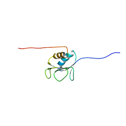 | | Solution structure of the E.coli RcsC C-terminus (residues 700-816) containing linker region | | 分子名称: | Sensor kinase protein rcsC | | 著者 | Rogov, V.V, Rogova, N.Y, Bernhard, F, Koglin, A, Lohr, F, Dotsch, V. | | 登録日 | 2005-09-09 | | 公開日 | 2006-09-26 | | 最終更新日 | 2024-05-29 | | 実験手法 | SOLUTION NMR | | 主引用文献 | A New Structural Domain in the Escherichia coli RcsC Hybrid Sensor Kinase Connects Histidine Kinase and Phosphoreceiver Domains
J.Mol.Biol., 364, 2006
|
|
3S7N
 | |
3B33
 | |
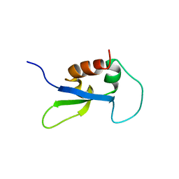
2E6Z
 | | Solution structure of the second KOW motif of human transcription elongation factor SPT5 | | 分子名称: | Transcription elongation factor SPT5 | | 著者 | Tanabe, W, Suzuki, S, Muto, Y, Inoue, M, Kigawa, T, Terada, T, Shirouzu, M, Yokoyama, S, RIKEN Structural Genomics/Proteomics Initiative (RSGI) | | 登録日 | 2007-01-05 | | 公開日 | 2007-07-10 | | 最終更新日 | 2024-05-29 | | 実験手法 | SOLUTION NMR | | 主引用文献 | Solution structure of the second KOW motif of human transcription elongation factor SPT5
To be Published
|
|
3EWK
 | |
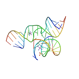
1Y90
 | | HIV-1 Dis(Mal) Duplex Mn-Soaked | | 分子名称: | 5'-R(*CP*(5BU)P*UP*GP*CP*UP*GP*AP*GP*GP*UP*GP*CP*AP*CP*AP*CP*AP*GP*CP*AP*AP*G)-3', MANGANESE (II) ION | | 著者 | Ennifar, E, Walter, P, Dumas, P. | | 登録日 | 2004-12-14 | | 公開日 | 2004-12-21 | | 最終更新日 | 2024-02-14 | | 実験手法 | X-RAY DIFFRACTION (3.08 Å) | | 主引用文献 | A crystallographic study of the binding of 13 metal ions to two related RNA duplexes
Nucleic Acids Res., 31, 2003
|
|
2P4B
 | | Crystal structure of E.coli RseB | | 分子名称: | Sigma-E factor regulatory protein rseB, octyl beta-D-glucopyranoside | | 著者 | Kim, D.Y, Kim, K.K. | | 登録日 | 2007-03-12 | | 公開日 | 2007-05-22 | | 最終更新日 | 2024-03-13 | | 実験手法 | X-RAY DIFFRACTION (2.4 Å) | | 主引用文献 | Crystal structure of RseB and a model of its binding mode to RseA
Proc.Natl.Acad.Sci.Usa, 104, 2007
|
|
1Y6T
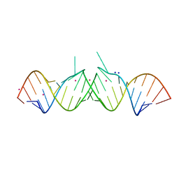 | | HIV-1 Dis(Mal) Duplex Co Hexamine-Soaked | | 分子名称: | 5'-R(*CP*UP*UP*GP*CP*UP*GP*AP*GP*GP*UP*GP*CP*AP*CP*AP*CP*AP*GP*CP*AP*AP*G)-3', COBALT (III) ION, SODIUM ION | | 著者 | Ennifar, E, Walter, P, Dumas, P. | | 登録日 | 2004-12-07 | | 公開日 | 2004-12-21 | | 最終更新日 | 2024-02-14 | | 実験手法 | X-RAY DIFFRACTION (2.6 Å) | | 主引用文献 | A crystallographic study of the binding of 13 metal ions to two related RNA duplexes
Nucleic Acids Res., 31, 2003
|
|
2DY7
 | |
1Y99
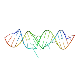 | | HIV-1 subtype A DIS RNA duplex | | 分子名称: | 5'-R(*CP*UP*UP*GP*CP*UP*GP*AP*GP*GP*UP*GP*CP*AP*CP*AP*CP*AP*GP*CP*AP*AP*G)-3', MAGNESIUM ION | | 著者 | Ennifar, E, Yusupov, M, Walter, P, Marquet, R, Ehresmann, B, Ehresmann, C, Dumas, P. | | 登録日 | 2004-12-15 | | 公開日 | 2004-12-21 | | 最終更新日 | 2023-08-23 | | 実験手法 | X-RAY DIFFRACTION (2.4 Å) | | 主引用文献 | The crystal structure of the dimerization initiation site of genomic HIV-1 RNA reveals an extended duplex with two adenine bulges
Structure Fold.Des., 7, 1999
|
|
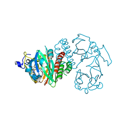
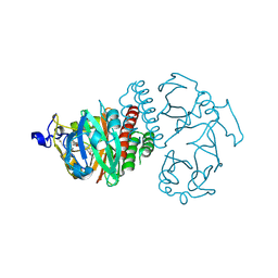
3BES
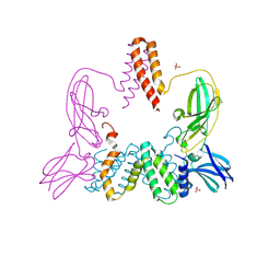 | | Structure of a Poxvirus ifngbp/ifng Complex | | 分子名称: | 2-acetamido-2-deoxy-beta-D-glucopyranose, Interferon gamma, Interferon-gamma binding protein C4R, ... | | 著者 | Nuara, A.A, Walter, M.R. | | 登録日 | 2007-11-20 | | 公開日 | 2008-02-12 | | 最終更新日 | 2020-07-29 | | 実験手法 | X-RAY DIFFRACTION (2.2 Å) | | 主引用文献 | Structure and mechanism of IFN-gamma antagonism by an orthopoxvirus IFN-gamma-binding protein.
Proc.Natl.Acad.Sci.Usa, 105, 2008
|
|
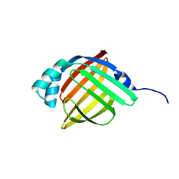
2EFI
 | | Solution structure of the chromo domain of Mortality factor 4-like protein 1 from human | | 分子名称: | Mortality factor 4-like protein 1 | | 著者 | Li, H, Sato, M, Tochio, N, Tomizawa, T, Koshiba, S, Harada, T, Watanabe, S, Kigawa, T, Yokoyama, S, RIKEN Structural Genomics/Proteomics Initiative (RSGI) | | 登録日 | 2007-02-22 | | 公開日 | 2007-08-28 | | 最終更新日 | 2024-05-29 | | 実験手法 | SOLUTION NMR | | 主引用文献 | Solution structure of the chromo domain of Mortality factor 4-like protein 1 from human
To be Published
|
|


