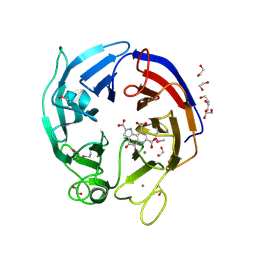
3DGD
 
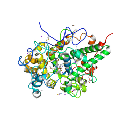
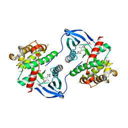
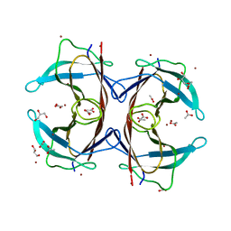 | | Crystal structure of the F87M/L110M mutant of human transthyretin at pH 4.6 | | 分子名称: | ACETATE ION, GLYCEROL, Transthyretin, ... | | 著者 | Palmieri, L.C, Freire, J.B.B, Foguel, D, Lima, L.M.T.R. | | 登録日 | 2008-06-13 | | 公開日 | 2008-07-22 | | 最終更新日 | 2023-08-30 | | 実験手法 | X-RAY DIFFRACTION (1.383 Å) | | 主引用文献 | Novel Zn2+-binding sites in human transthyretin: implications for amyloidogenesis and retinol-binding protein recognition.
J.Biol.Chem., 285, 2010
|
|
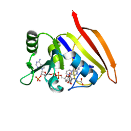
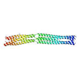
9B68
 | | GluA2 flip Q in complex with TARPgamma2 at pH8, class1, structure of NTD | | 分子名称: | 2-acetamido-2-deoxy-beta-D-glucopyranose, 2-acetamido-2-deoxy-beta-D-glucopyranose-(1-4)-2-acetamido-2-deoxy-beta-D-glucopyranose, Isoform Flip of Glutamate receptor 2, ... | | 著者 | Nakagawa, T, Greger, I.H. | | 登録日 | 2024-03-23 | | 公開日 | 2024-07-31 | | 最終更新日 | 2024-10-30 | | 実験手法 | ELECTRON MICROSCOPY (3.6 Å) | | 主引用文献 | Proton-triggered rearrangement of the AMPA receptor N-terminal domains impacts receptor kinetics and synaptic localization.
Nat.Struct.Mol.Biol., 31, 2024
|
|
4W7W
 | |
4W89
 | | Crystal structure of XEG5A, a GH5 xyloglucan-specific endo-beta-1,4-glucanase from metagenomic library, in complex with cellotriose | | 分子名称: | MAGNESIUM ION, Xyloglucan-specific endo-beta-1,4-glucanase, beta-D-glucopyranose-(1-4)-beta-D-glucopyranose-(1-4)-alpha-D-glucopyranose | | 著者 | Santos, C.R, Cordeiro, R.L, Wong, D.W.S, Murakami, M.T. | | 登録日 | 2014-08-22 | | 公開日 | 2015-03-11 | | 最終更新日 | 2023-12-27 | | 実験手法 | X-RAY DIFFRACTION (2.4 Å) | | 主引用文献 | Structural Basis for Xyloglucan Specificity and alpha-d-Xylp(1 6)-d-Glcp Recognition at the -1 Subsite within the GH5 Family.
Biochemistry, 54, 2015
|
|
9M0I
 | | Structure of the intermediate of lactoperoxidase formed with thiocynate and hydrogen peroxidase at 1.99 A resolution. | | 分子名称: | 2-acetamido-2-deoxy-beta-D-glucopyranose, 2-acetamido-2-deoxy-beta-D-glucopyranose-(1-4)-2-acetamido-2-deoxy-beta-D-glucopyranose, CALCIUM ION, ... | | 著者 | Viswanathan, V, Maurya, A, Sirohi, H.V, Kushwaha, G.S, Sinha, M, Kaur, P, Sharma, S, Singh, T.P. | | 登録日 | 2025-02-24 | | 公開日 | 2025-03-26 | | 実験手法 | X-RAY DIFFRACTION (1.995 Å) | | 主引用文献 | Structure of the intermediate of lactoperoxidase formed with thiocynate and hydrogen peroxidase at 1.99 A resolution.
To Be Published
|
|
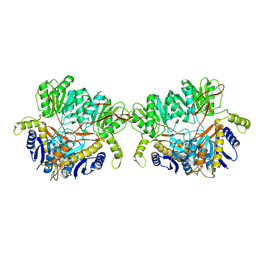
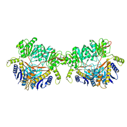
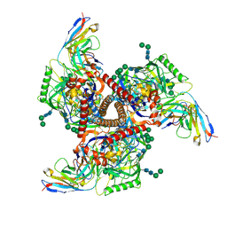
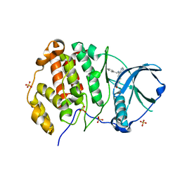
3DFR
 | | CRYSTAL STRUCTURES OF ESCHERICHIA COLI AND LACTOBACILLUS CASEI DIHYDROFOLATE REDUCTASE REFINED AT 1.7 ANGSTROMS RESOLUTION. I. GENERAL FEATURES AND BINDING OF METHOTREXATE | | 分子名称: | DIHYDROFOLATE REDUCTASE, METHOTREXATE, NADPH DIHYDRO-NICOTINAMIDE-ADENINE-DINUCLEOTIDE PHOSPHATE | | 著者 | Filman, D.J, Matthews, D.A, Bolin, J.T, Kraut, J. | | 登録日 | 1982-06-25 | | 公開日 | 1982-10-21 | | 最終更新日 | 2024-02-21 | | 実験手法 | X-RAY DIFFRACTION (1.7 Å) | | 主引用文献 | Crystal structures of Escherichia coli and Lactobacillus casei dihydrofolate reductase refined at 1.7 A resolution. I. General features and binding of methotrexate.
J.Biol.Chem., 257, 1982
|
|
8ACX
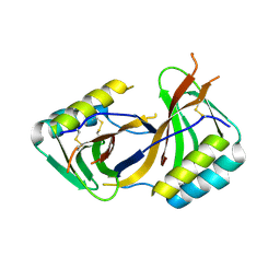 | |
9B69
 | | GluA2 flip Q in complex with TARPgamma2 at pH8, class12, structure of NTD | | 分子名称: | 2-acetamido-2-deoxy-beta-D-glucopyranose, 2-acetamido-2-deoxy-beta-D-glucopyranose-(1-4)-2-acetamido-2-deoxy-beta-D-glucopyranose, Isoform Flip of Glutamate receptor 2, ... | | 著者 | Nakagawa, T, Greger, I.H. | | 登録日 | 2024-03-23 | | 公開日 | 2024-07-31 | | 最終更新日 | 2024-10-30 | | 実験手法 | ELECTRON MICROSCOPY (3.69 Å) | | 主引用文献 | Proton-triggered rearrangement of the AMPA receptor N-terminal domains impacts receptor kinetics and synaptic localization.
Nat.Struct.Mol.Biol., 31, 2024
|
|
3DAS
 | | Structure of the PQQ-bound form of Aldose Sugar Dehydrogenase (Adh) from Streptomyces coelicolor | | 分子名称: | 1,2-ETHANEDIOL, CALCIUM ION, PYRROLOQUINOLINE QUINONE, ... | | 著者 | Southall, S.M, Doel, J.J, Oubrie, A, Richardson, D.J. | | 登録日 | 2008-05-30 | | 公開日 | 2009-06-02 | | 最終更新日 | 2024-10-30 | | 実験手法 | X-RAY DIFFRACTION (1.6 Å) | | 主引用文献 | Structural and Enzymatic Characterization of a Thermostable PQQ-dependent Soluble Aldose Sugar Dehydrogenase
To be Published
|
|
6TPM
 | | Crystal structure of AmpC from E.coli with Relebactam (MK-7655) | | 分子名称: | (2S,5R)-1-formyl-N-(piperidin-4-yl)-5-[(sulfooxy)amino]piperidine-2-carboxamide, 2-(N-MORPHOLINO)-ETHANESULFONIC ACID, Beta-lactamase, ... | | 著者 | Lang, P.A, Leissing, T.M, Schofield, C.J, Brem, J. | | 登録日 | 2019-12-13 | | 公開日 | 2020-11-25 | | 最終更新日 | 2024-11-13 | | 実験手法 | X-RAY DIFFRACTION (1.72 Å) | | 主引用文献 | Structural Investigations of the Inhibition of Escherichia coli AmpC beta-Lactamase by Diazabicyclooctanes.
Antimicrob.Agents Chemother., 65, 2021
|
|
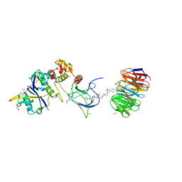
9BCG
 | | Myeloid cell leukemia-1 (Mcl-1) complexed with compound | | 分子名称: | 7-[(4R,5S,6P)-7-chloro-10-[3-(4-chloro-3,5-dimethylphenoxy)propyl]-4-methyl-1-oxo-6-(1,3,5-trimethyl-1H-pyrazol-4-yl)-3,4-dihydropyrazino[1,2-a]indol-2(1H)-yl]-4,5-dimethoxy-1-methyl-1H-indole-2-carboxylic acid, Maltose/maltodextrin-binding periplasmic protein,Induced myeloid leukemia cell differentiation protein Mcl-1, alpha-D-glucopyranose-(1-4)-alpha-D-glucopyranose | | 著者 | Zhao, B, Fesik, S.W. | | 登録日 | 2024-04-09 | | 公開日 | 2024-08-07 | | 最終更新日 | 2024-09-11 | | 実験手法 | X-RAY DIFFRACTION (1.898 Å) | | 主引用文献 | Discovery of a Myeloid Cell Leukemia 1 (Mcl-1) Inhibitor That Demonstrates Potent In Vivo Activities in Mouse Models of Hematological and Solid Tumors.
J.Med.Chem., 67, 2024
|
|
8B5T
 | |
9BGE
 | | Cryo-EM structure of mAb8-24 bound to 426c.WITO.TM.SOSIP | | 分子名称: | 2-acetamido-2-deoxy-beta-D-glucopyranose, 2-acetamido-2-deoxy-beta-D-glucopyranose-(1-4)-2-acetamido-2-deoxy-beta-D-glucopyranose, 426c.WITO.TM.SOSIP, ... | | 著者 | Hurlburt, N.K, Pancera, M. | | 登録日 | 2024-04-18 | | 公開日 | 2024-09-04 | | 最終更新日 | 2024-11-06 | | 実験手法 | ELECTRON MICROSCOPY (4.2 Å) | | 主引用文献 | Short CDRL1 in intermediate VRC01-like mAbs is not sufficient to overcome key glycan barriers on HIV-1 Env.
J.Virol., 98, 2024
|
|
4W79
 | | Crystal Structure of Human Protein N-terminal Glutamine Amidohydrolase | | 分子名称: | 1,2-ETHANEDIOL, CARBONATE ION, Protein N-terminal glutamine amidohydrolase, ... | | 著者 | Bitto, E, Bingman, C.A, McCoy, J.G, Wesenberg, G.E, Phillips Jr, G.N, Center for Eukaryotic Structural Genomics (CESG) | | 登録日 | 2014-08-21 | | 公開日 | 2014-09-17 | | 最終更新日 | 2024-10-23 | | 実験手法 | X-RAY DIFFRACTION (1.5 Å) | | 主引用文献 | Crystal structure of human protein N-terminal glutamine amidohydrolase, an initial component of the N-end rule pathway.
Plos One, 9, 2014
|
|
8BB2
 | | Structure of human WDR5 and pVHL:ElonginC:ElonginB bound to PROTAC with PEG linker (conformation #2) | | 分子名称: | 1,2-ETHANEDIOL, Elongin-B, Elongin-C, ... | | 著者 | Kraemer, A, Doelle, A, Knapp, S, Structural Genomics Consortium (SGC) | | 登録日 | 2022-10-12 | | 公開日 | 2022-11-09 | | 最終更新日 | 2024-01-31 | | 実験手法 | X-RAY DIFFRACTION (2.05 Å) | | 主引用文献 | Structure of human WDR5 and pVHL:ElonginC:ElonginB bound to PROTAC with PEG linker (conformation #2)
To Be Published
|
|
4W8B
 | | Crystal structure of XEG5B, a GH5 xyloglucan-specific beta-1,4-glucanase from ruminal metagenomic library, in complex with XXLG | | 分子名称: | Exo-xyloglucanase, GLYCEROL, SULFATE ION, ... | | 著者 | Santos, C.R, Cordeiro, R.L, Wong, D.W.S, Murakami, M.T. | | 登録日 | 2014-08-22 | | 公開日 | 2015-03-11 | | 最終更新日 | 2023-12-27 | | 実験手法 | X-RAY DIFFRACTION (1.15 Å) | | 主引用文献 | Structural Basis for Xyloglucan Specificity and alpha-d-Xylp(1 6)-d-Glcp Recognition at the -1 Subsite within the GH5 Family.
Biochemistry, 54, 2015
|
|
8BB3
 | | Structure of human WDR5 and pVHL:ElonginC:ElonginB bound to PROTAC with PEG linker (conformation #1) | | 分子名称: | 1,2-ETHANEDIOL, Elongin-B, Elongin-C, ... | | 著者 | Kraemer, A, Doelle, A, Knapp, S, Structural Genomics Consortium (SGC) | | 登録日 | 2022-10-12 | | 公開日 | 2022-11-09 | | 最終更新日 | 2024-01-31 | | 実験手法 | X-RAY DIFFRACTION (1.8 Å) | | 主引用文献 | Structure of human WDR5 and pVHL:ElonginC:ElonginB bound to PROTAC with PEG linker (conformation #1)
To Be Published
|
|
8BEM
 | |
8BB5
 | | Structure of human WDR5 and pVHL:ElonginC:ElonginB bound to PROTAC with Aryl linker | | 分子名称: | 1,2-ETHANEDIOL, Elongin-B, Elongin-C, ... | | 著者 | Kraemer, A, Doelle, A, Knapp, S, Structural Genomics Consortium (SGC) | | 登録日 | 2022-10-12 | | 公開日 | 2022-11-09 | | 最終更新日 | 2024-01-31 | | 実験手法 | X-RAY DIFFRACTION (2.2 Å) | | 主引用文献 | Tracking the PROTAC degradation pathway in living cells highlights the importance of ternary complex measurement for PROTAC optimization.
Cell Chem Biol, 30, 2023
|
|
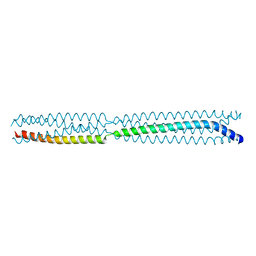
8B8B
 | | Multimerization domain of Munia virus 1 phosphoprotein | | 分子名称: | Munia Bornavirus 1 phosphoprotein, NITRATE ION | | 著者 | Chenavier, F, Tarbouriech, N, Bourhis, J.M, Tomonaga, K, Horie, M, Crepin, T. | | 登録日 | 2022-10-04 | | 公開日 | 2022-11-23 | | 最終更新日 | 2024-05-01 | | 実験手法 | X-RAY DIFFRACTION (2.15 Å) | | 主引用文献 | Borna Disease Virus 1 Phosphoprotein Forms a Tetramer and Interacts with Host Factors Involved in DNA Double-Strand Break Repair and mRNA Processing.
Viruses, 14, 2022
|
|
8B8A
 | | Multimerization domain of borna disease virus 1 phosphoprotein | | 分子名称: | Phosphoprotein | | 著者 | Tarbouriech, N, Legrand, P, Bourhis, J.M, Chenavier, F, Freslon, L, Kawasaki, J, Horie, M, Tomonaga, K, Bachiri, K, Coyaud, E, Gonzalez-Dunia, D, Ruigrok, R.W.H, Crepin, T. | | 登録日 | 2022-10-04 | | 公開日 | 2022-11-23 | | 最終更新日 | 2024-01-31 | | 実験手法 | X-RAY DIFFRACTION (2.75 Å) | | 主引用文献 | Borna Disease Virus 1 Phosphoprotein Forms a Tetramer and Interacts with Host Factors Involved in DNA Double-Strand Break Repair and mRNA Processing.
Viruses, 14, 2022
|
|
8BGC
 | | Crystal structure of human Casein Kinase II subunit alpha (CK2a1) in complex with compound 2 (AA-CS-9-003) | | 分子名称: | 5-[(phenylmethyl)amino]pyrimido[4,5-c]quinoline-8-carboxylic acid, Casein kinase II subunit alpha, SULFATE ION | | 著者 | Kraemer, A, Axtman, A.D, Knapp, S, Structural Genomics Consortium (SGC) | | 登録日 | 2022-10-27 | | 公開日 | 2022-11-23 | | 最終更新日 | 2024-01-31 | | 実験手法 | X-RAY DIFFRACTION (2.8 Å) | | 主引用文献 | Discovery of a Potent and Selective Naphthyridine-Based Chemical Probe for Casein Kinase 2.
Acs Med.Chem.Lett., 14, 2023
|
|
5D29
 | | X-ray structure of human glutamate carboxypeptidase II (GCPII) in complex with a hydroxamate inhibitor JHU241 | | 分子名称: | 2-acetamido-2-deoxy-beta-D-glucopyranose, 2-acetamido-2-deoxy-beta-D-glucopyranose-(1-4)-2-acetamido-2-deoxy-beta-D-glucopyranose, 4-[(2~{S})-2-carboxy-5-(oxidanylamino)-5-oxidanylidene-pentyl]benzoic acid, ... | | 著者 | Barinka, C, Novakova, Z, Pavlicek, J. | | 登録日 | 2015-08-05 | | 公開日 | 2016-04-27 | | 最終更新日 | 2024-10-16 | | 実験手法 | X-RAY DIFFRACTION (1.8 Å) | | 主引用文献 | Unprecedented Binding Mode of Hydroxamate-Based Inhibitors of Glutamate Carboxypeptidase II: Structural Characterization and Biological Activity.
J.Med.Chem., 59, 2016
|
|
8BCJ
 | |
6TOY
 | |


