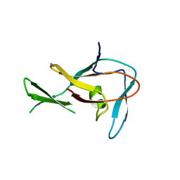
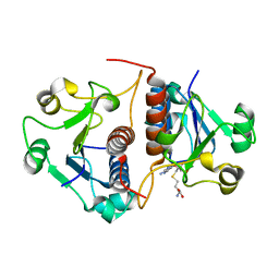
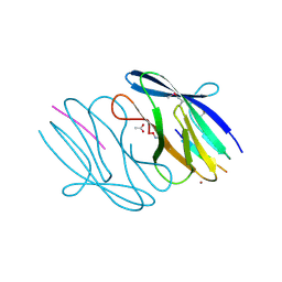
3N23
 
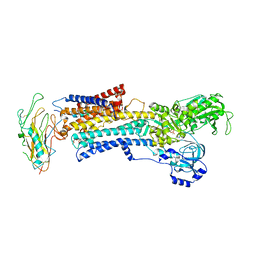 | | Crystal structure of the high affinity complex between ouabain and the E2P form of the sodium-potassium pump | | 分子名称: | MAGNESIUM ION, Na+/K+ ATPase gamma subunit transcript variant a, OUABAIN, ... | | 著者 | Yatime, L, Laursen, M, Morth, J.P, Esmann, M, Nissen, P, Fedosova, N.U. | | 登録日 | 2010-05-17 | | 公開日 | 2011-01-19 | | 最終更新日 | 2025-03-26 | | 実験手法 | X-RAY DIFFRACTION (4.6 Å) | | 主引用文献 | Structural insights into the high affinity binding of cardiotonic steroids to the Na+,K+-ATPase.
J.Struct.Biol., 174, 2011
|
|
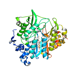
3E8U
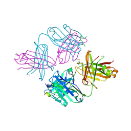 | | Crystal structure and thermodynamic analysis of diagnostic Fab 106.3 complexed with BNP 5-13 (C10A) reveal basis of selective molecular recognition | | 分子名称: | BNP peptide epitope, Fab 106.3 heavy chain, Fab 106.3 light chain | | 著者 | Longenecker, K.L, Ruan, Q, Fry, E.H, Saldana, S.S, Brophy, S.E, Richardson, P.L, Tetin, S.Y. | | 登録日 | 2008-08-20 | | 公開日 | 2009-07-07 | | 最終更新日 | 2024-11-20 | | 実験手法 | X-RAY DIFFRACTION (2.1 Å) | | 主引用文献 | Crystal structure and thermodynamic analysis of diagnostic mAb 106.3 complexed with BNP 5-13 (C10A).
Proteins, 76, 2009
|
|
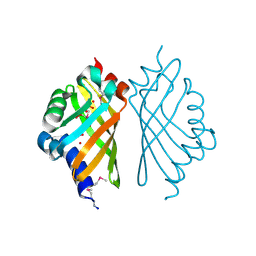
3E95
 | | Crystal Structure of the Plasmodium Falciparum ubiquitin conjugating enzyme complex, PfUBC13-PfUev1a | | 分子名称: | UNKNOWN ATOM OR ION, Ubiquitin carrier protein, Ubiquitin-conjugating enzyme E2 | | 著者 | Wernimont, A.K, Lam, A, Ali, A, Brokx, S, Lin, Y.H, Zhao, Y, Lew, J, Ravichandran, M, Wasney, G, Vedadi, M, Kozieradzki, I, Schapira, M, Bochkarev, A, Wilkstrom, M, BOuntra, C, Arrowsmith, C.H, Edwards, A.M, Hui, R, Qiu, W, Brand, V.B, Structural Genomics Consortium (SGC) | | 登録日 | 2008-08-21 | | 公開日 | 2008-09-30 | | 最終更新日 | 2024-10-30 | | 実験手法 | X-RAY DIFFRACTION (2.5 Å) | | 主引用文献 | Crystal Structure of the Plasmodium Falciparum ubiquitin conjugating enzyme complex, PfUBC13-PfUev1a
TO BE PUBLISHED
|
|
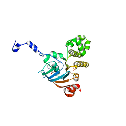
3E9G
 | | Crystal structure long-form (residue1-124) of Eaf3 chromo domain | | 分子名称: | Chromatin modification-related protein EAF3 | | 著者 | Sun, B, Hong, J, Zhang, P, Lin, D, Ding, J. | | 登録日 | 2008-08-22 | | 公開日 | 2008-11-04 | | 最終更新日 | 2023-11-01 | | 実験手法 | X-RAY DIFFRACTION (2.5 Å) | | 主引用文献 | Molecular Basis of the Interaction of Saccharomyces cerevisiae Eaf3 Chromo Domain with Methylated H3K36
J.Biol.Chem., 283, 2008
|
|
3NF4
 | |
3E9R
 | | Crystal structure of purine nucleoside phosphorylase from Schistosoma mansoni in complex with adenine | | 分子名称: | ACETATE ION, ADENINE, DIMETHYL SULFOXIDE, ... | | 著者 | Pereira, H.M, Rezende, M.M, Oliva, G, Garratt, R.C. | | 登録日 | 2008-08-23 | | 公開日 | 2009-09-01 | | 最終更新日 | 2023-08-30 | | 実験手法 | X-RAY DIFFRACTION (1.85 Å) | | 主引用文献 | Adenosine binding to low-molecular-weight purine nucleoside phosphorylase: the structural basis for recognition based on its complex with the enzyme from Schistosoma mansoni.
Acta Crystallogr.,Sect.D, 66, 2010
|
|
3NFS
 | | Crystal structure the Fab fragment of therapeutic antibody daclizumab | | 分子名称: | Heavy chain of Fab fragment of daclizumab, Light chain of Fab fragment of daclizumab | | 著者 | Yang, H, Wang, J, Du, J, Zhong, C, Guo, Y, Ding, J. | | 登録日 | 2010-06-10 | | 公開日 | 2010-09-15 | | 最終更新日 | 2024-11-06 | | 実験手法 | X-RAY DIFFRACTION (2.6 Å) | | 主引用文献 | Structural basis of immunosuppression by the therapeutic antibody daclizumab
Cell Res., 20, 2010
|
|
3EO8
 | |
3VIV
 | | 1510-N membrane-bound stomatin-specific protease K138A mutant in complex with a substrate peptide | | 分子名称: | 441aa long hypothetical nfeD protein, CHLORIDE ION, GLYCEROL, ... | | 著者 | Yokoyama, H, Matsui, I, Fujii, S. | | 登録日 | 2011-10-12 | | 公開日 | 2012-05-23 | | 最終更新日 | 2023-11-08 | | 実験手法 | X-RAY DIFFRACTION (2.25 Å) | | 主引用文献 | Crystal structure of a membrane stomatin-specific protease in complex with a substrate Peptide
Biochemistry, 51, 2012
|
|
3NGK
 | |
3EP0
 | | Methyltransferase domain of human PR domain-containing protein 12 | | 分子名称: | PR domain zinc finger protein 12 | | 著者 | Amaya, M.F, Zeng, H, Loppnau, P, Bountra, C, Weigelt, J, Arrowsmith, C.H, Edwards, A.M, Botchkarev, A, Min, J, Plotnikov, A.N, Wu, H, Structural Genomics Consortium (SGC) | | 登録日 | 2008-09-29 | | 公開日 | 2008-11-04 | | 最終更新日 | 2023-09-06 | | 実験手法 | X-RAY DIFFRACTION (2.1 Å) | | 主引用文献 | The crystal structure of methyltransferase domain of human PR
Domain-containing protein 12.
To be Published
|
|
3VKR
 | | Assimilatory nitrite reductase (Nii3) - NO2 complex from tobbaco leaf analysed with high X-ray dose | | 分子名称: | CHLORIDE ION, IRON/SULFUR CLUSTER, NITRITE ION, ... | | 著者 | Nakano, S, Takahashi, M, Sakamoto, A, Morikawa, H, Katayanagi, K. | | 登録日 | 2011-11-20 | | 公開日 | 2012-04-25 | | 最終更新日 | 2023-12-27 | | 実験手法 | X-RAY DIFFRACTION (1.6 Å) | | 主引用文献 | The reductive reaction mechanism of tobacco nitrite reductase derived from a combination of crystal structures and ultraviolet-visible microspectroscopy
Proteins, 80, 2012
|
|
3N3A
 | | Ribonucleotide Reductase Dimanganese(II)-NrdF from Escherichia coli in Complex with Reduced NrdI | | 分子名称: | FLAVIN MONONUCLEOTIDE, MANGANESE (II) ION, Protein nrdI, ... | | 著者 | Boal, A.K, Cotruvo Jr, J.A, Stubbe, J, Rosenzweig, A.C. | | 登録日 | 2010-05-19 | | 公開日 | 2010-08-18 | | 最終更新日 | 2023-09-06 | | 実験手法 | X-RAY DIFFRACTION (1.99 Å) | | 主引用文献 | Structural basis for activation of class Ib ribonucleotide reductase.
Science, 329, 2010
|
|
3EPV
 | | X-ray Structure of the Metal-sensor CnrX in both the Apo- and Copper-bound Forms | | 分子名称: | COPPER (II) ION, Nickel and cobalt resistance protein cnrR | | 著者 | Pompidor, G, Maillard, A.P, Girard, E, Gambarelli, S, Kahn, R, Coves, J. | | 登録日 | 2008-09-30 | | 公開日 | 2008-11-25 | | 最終更新日 | 2024-10-30 | | 実験手法 | X-RAY DIFFRACTION (1.742 Å) | | 主引用文献 | X-ray structure of the metal-sensor CnrX in both the apo- and copper-bound forms.
Febs Lett., 2008
|
|
3EBC
 | | Structure of N141A HincII with Cognate DNA | | 分子名称: | 5'-D(*DGP*DCP*DCP*DCP*DGP*DTP*DCP*DGP*DAP*DCP*DCP*DGP*DGP*DC)-3', 5'-D(*DGP*DCP*DCP*DGP*DGP*DTP*DCP*DGP*DAP*DCP*DGP*DGP*DGP*DC)-3', MANGANESE (II) ION, ... | | 著者 | Little, E.J, Babic, A.C, Horton, N.C. | | 登録日 | 2008-08-27 | | 公開日 | 2008-12-23 | | 最終更新日 | 2024-02-21 | | 実験手法 | X-RAY DIFFRACTION (2.55 Å) | | 主引用文献 | Early Interrogation and Recognition of DNA Sequence by Indirect Readout
Structure, 16, 2008
|
|
3N4K
 | | Putative RNA methyltransferase from Yersinia pestis in complex with S-ADENOSYL-L-HOMOCYSTEINE. | | 分子名称: | RNA methyltransferase, S-ADENOSYL-L-HOMOCYSTEINE | | 著者 | Osipiuk, J, Maltseva, N, Peterson, S, Anderson, W.F, Joachimiak, A, Center for Structural Genomics of Infectious Diseases (CSGID) | | 登録日 | 2010-05-21 | | 公開日 | 2010-06-23 | | 最終更新日 | 2023-09-06 | | 実験手法 | X-RAY DIFFRACTION (1.76 Å) | | 主引用文献 | X-ray crystal structure of putative RNA methyltransferase from Yersinia pestis.
To be Published
|
|
3VLZ
 | | Assimilatory nitrite reductase (Nii3) - N226K mutant - SO3 full complex from tobacco leaf | | 分子名称: | CHLORIDE ION, IRON/SULFUR CLUSTER, Nitrite reductase, ... | | 著者 | Nakano, S, Takahashi, M, Sakamoto, A, Morikawa, H, Katayanagi, K. | | 登録日 | 2011-12-05 | | 公開日 | 2012-09-26 | | 最終更新日 | 2023-12-27 | | 実験手法 | X-RAY DIFFRACTION (2.07 Å) | | 主引用文献 | X-ray crystal structure of a mutant assimilatory nitrite reductase that shows sulfite reductase-like activity
Chem.Biodivers., 9, 2012
|
|
3EBT
 | |
3NHB
 | | Nucleotide Binding Domain of Human ABCB6 (ADP bound structure) | | 分子名称: | ADENOSINE-5'-DIPHOSPHATE, ATP-binding cassette sub-family B member 6, mitochondrial, ... | | 著者 | Haffke, M, Menzel, A, Carius, Y, Jahn, D, Heinz, D.W. | | 登録日 | 2010-06-14 | | 公開日 | 2010-08-25 | | 最終更新日 | 2023-09-06 | | 実験手法 | X-RAY DIFFRACTION (2.15 Å) | | 主引用文献 | Structures of the nucleotide-binding domain of the human ABCB6 transporter and its complexes with nucleotides.
Acta Crystallogr.,Sect.D, 66, 2010
|
|
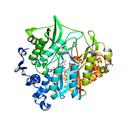
3N55
 | | SO1698 protein, an aspartic peptidase from Shewanella oneidensis. | | 分子名称: | ACETATE ION, BETA-MERCAPTOETHANOL, Peptidase, ... | | 著者 | Osipiuk, J, Mulligan, R, Collart, F, Joachimiak, A, Midwest Center for Structural Genomics (MCSG) | | 登録日 | 2010-05-24 | | 公開日 | 2010-06-02 | | 最終更新日 | 2025-03-26 | | 実験手法 | X-RAY DIFFRACTION (1.57 Å) | | 主引用文献 | Characterization of member of DUF1888 protein family, self-cleaving and self-assembling endopeptidase.
J.Biol.Chem., 287, 2012
|
|
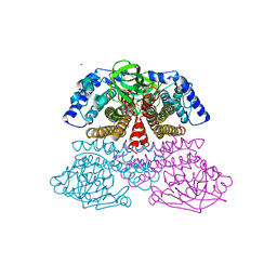
3MXG
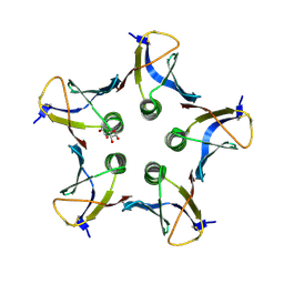 | | Structure of Shiga Toxin type 2 (Stx2) B Pentamer Mutant Q40L | | 分子名称: | D-xylose, Shiga-like toxin 2 subunit B | | 著者 | Kovall, R.A, Vander Wielen, B.D, Friedmann, D.R, Flagler, M.J. | | 登録日 | 2010-05-07 | | 公開日 | 2011-01-12 | | 最終更新日 | 2024-11-20 | | 実験手法 | X-RAY DIFFRACTION (2.49 Å) | | 主引用文献 | Molecular basis of differential B-pentamer stability of Shiga toxins 1 and 2.
Plos One, 5, 2010
|
|
6DEG
 | |
3ED7
 | | Replacement of Val3 in Human Thymidylate Synthase Affects Its Kinetic Properties and Intracellular Stability | | 分子名称: | SULFATE ION, Thymidylate synthase | | 著者 | Huang, X, Gibson, L.M, Bell, B.J, Lovelace, L.L, Pena, M.M, Berger, F.G, Berger, S.H. | | 登録日 | 2008-09-02 | | 公開日 | 2010-03-02 | | 最終更新日 | 2024-02-21 | | 実験手法 | X-RAY DIFFRACTION (1.56 Å) | | 主引用文献 | Replacement of Val3 in human thymidylate synthase affects its kinetic properties and intracellular stability .
Biochemistry, 49, 2010
|
|
6DS1
 | |
6CZE
 | | Crystal structure of Mycobacterium tuberculosis dethiobiotin synthetase in complex with inosine triphosphate (ITP) - promiscuous binding mode with disordered nucleoside | | 分子名称: | ATP-dependent dethiobiotin synthetase BioD, INOSINE 5'-TRIPHOSPHATE, MAGNESIUM ION | | 著者 | Thompson, A.P, Wegener, K.L, Bruning, J.B, Polyak, S.W. | | 登録日 | 2018-04-09 | | 公開日 | 2018-11-21 | | 最終更新日 | 2024-03-13 | | 実験手法 | X-RAY DIFFRACTION (2.3 Å) | | 主引用文献 | Mycobacterium tuberculosis Dethiobiotin Synthetase Facilitates Nucleoside Triphosphate Promiscuity through Alternate Binding Modes
Acs Catalysis, 8(11), 2018
|
|


