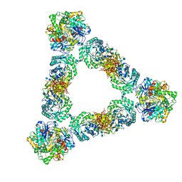
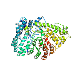
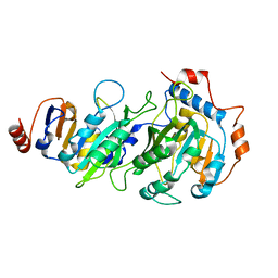
7DXM
 
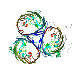
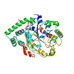 | | Crystal structure of DltD | | 分子名称: | Protein DltD, SULFATE ION | | 著者 | Yan, X.X, Zeng, Q, Tian, L.F. | | 登録日 | 2021-01-19 | | 公開日 | 2021-07-21 | | 最終更新日 | 2023-11-29 | | 実験手法 | X-RAY DIFFRACTION (2.96 Å) | | 主引用文献 | Crystal Structure of an O-acyltransfer Terminal Protein stDltD and Its Implications for dlt Operon-mediated D-alanylation of S. thermophilus.
Prog.Biochem.Biophys., 48, 2022
|
|
8ORB
 | |
7A9M
 | |
3HFB
 | |
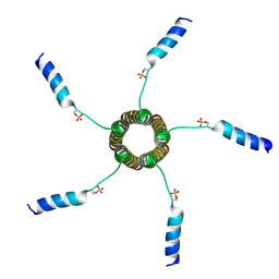
7RZN
 | | Crystal Structure of Ferritin grown by microbatch method in presence of agarose and electric field 2.1KV | | 分子名称: | CADMIUM ION, Ferritin light chain, SULFATE ION | | 著者 | Aditya, S, Priyadharshine, R, Maham, I, Miller, J.D, Stojanoff, V. | | 登録日 | 2021-08-27 | | 公開日 | 2022-11-09 | | 最終更新日 | 2023-10-18 | | 実験手法 | X-RAY DIFFRACTION (1.97 Å) | | 主引用文献 | Crystal Structure of Ferritin grown by microbatch method in presence of agarose and electric field 2.1KV
To Be Published
|
|
7AC9
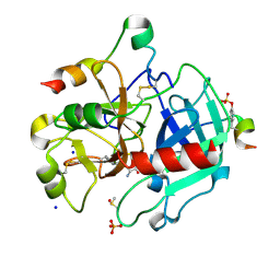 | | Thrombin in complex with D-arginine (j77) | | 分子名称: | 2-acetamido-2-deoxy-beta-D-glucopyranose, D-ARGININE, DIMETHYL SULFOXIDE, ... | | 著者 | Scanlan, W, Heine, A, Klebe, G, Abazi, N. | | 登録日 | 2020-09-10 | | 公開日 | 2021-10-06 | | 最終更新日 | 2024-01-31 | | 実験手法 | X-RAY DIFFRACTION (1.393 Å) | | 主引用文献 | Thrombin in complex with D-arginine (j77)
To be published
|
|
3HE3
 | |
2MJZ
 | |
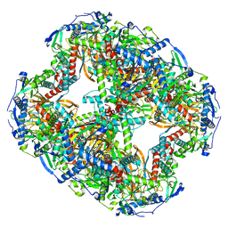
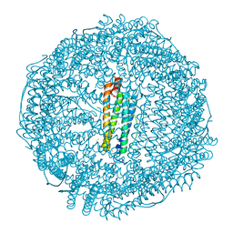
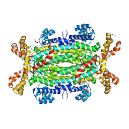
7KDV
 | | Murine core lysosomal multienzyme complex (LMC) composed of acid beta-galactosidase (GLB1) and protective protein cathepsin A (PPCA, CTSA) | | 分子名称: | 2-acetamido-2-deoxy-beta-D-glucopyranose, 2-acetamido-2-deoxy-beta-D-glucopyranose-(1-4)-2-acetamido-2-deoxy-beta-D-glucopyranose, Beta-galactosidase, ... | | 著者 | Gorelik, A, Illes, K, Hasan, S.M.N, Nagar, B, Mazhab-Jafari, M.T. | | 登録日 | 2020-10-09 | | 公開日 | 2021-03-17 | | 最終更新日 | 2021-05-26 | | 実験手法 | ELECTRON MICROSCOPY (4.59 Å) | | 主引用文献 | Structure of the murine lysosomal multienzyme complex core.
Sci Adv, 7, 2021
|
|
2M3B
 | |
7EAL
 | |
8OPB
 | |
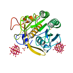
7E7L
 | | The crystal structure of arylacetate decarboxylase from Olsenella scatoligenes. | | 分子名称: | 4-HYDROXYPHENYLACETATE, Hydroxyphenylacetic acid decarboxylase | | 著者 | Lu, Q, Duan, Y, Zhang, Y, Yuchi, Z. | | 登録日 | 2021-02-26 | | 公開日 | 2021-07-28 | | 最終更新日 | 2023-11-29 | | 実験手法 | X-RAY DIFFRACTION (3.53 Å) | | 主引用文献 | The Glycyl Radical Enzyme Arylacetate Decarboxylase from Olsenella scatoligenes
Acs Catalysis, 11, 2021
|
|
7EAO
 | |
8OQ3
 | |
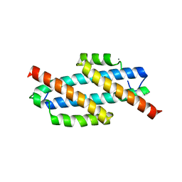
2M2W
 | | Ternary complex of ASFV Pol X with DNA and MgdGTP | | 分子名称: | 2'-DEOXYGUANOSINE-5'-TRIPHOSPHATE, 5'-D(P*GP*GP*CP*GP*AP*AP*GP*CP*CP*GP*GP*GP*TP*GP*CP*GP*AP*AP*GP*CP*AP*CP*(DOC))-3', MAGNESIUM ION, ... | | 著者 | Wu, W, Su, M, Tsai, M. | | 登録日 | 2013-01-03 | | 公開日 | 2014-04-02 | | 最終更新日 | 2024-05-01 | | 実験手法 | SOLUTION NMR | | 主引用文献 | How a low-fidelity DNA polymerase chooses non-Watson-Crick from Watson-Crick incorporation.
J.Am.Chem.Soc., 136, 2014
|
|
7RX8
 | |
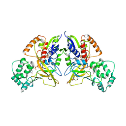
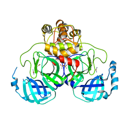
2J91
 | | Crystal structure of Human Adenylosuccinate Lyase in complex with AMP | | 分子名称: | ADENOSINE MONOPHOSPHATE, ADENYLOSUCCINATE LYASE, CHLORIDE ION, ... | | 著者 | Stenmark, P, Moche, M, Arrowsmith, C, Berglund, H, Busam, R, Collins, R, Edwards, A, Ericsson, U.B, Flodin, S, Flores, A, Graslund, S, Hammarstrom, M, Hallberg, B.M, Holmberg Schiavone, L, Hogbom, M, Johansson, I, Karlberg, T, Kosinska, U, Kotenyova, T, Magnusdottir, A, Nilsson, M.E, Nilsson-Ehle, P, Nyman, T, Ogg, D, Persson, C, Sagemark, J, Sundstrom, M, Uppenberg, J, Uppsten, M, Thorsell, A.G, van Den Berg, S, Wallden, K, Weigelt, J, Nordlund, P. | | 登録日 | 2006-11-01 | | 公開日 | 2006-11-07 | | 最終更新日 | 2023-12-13 | | 実験手法 | X-RAY DIFFRACTION (1.8 Å) | | 主引用文献 | Crystal Structure of Human Adenylosuccinate Lyase
To be Published
|
|
3HGW
 | |
2Z9L
 | |
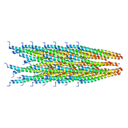
2J1N
 | | osmoporin OmpC | | 分子名称: | CHLORIDE ION, DODECANE, MAGNESIUM ION, ... | | 著者 | Basle, A, Storici, P, Rummel, G, Rosenbusch, J.P, Schirmer, T. | | 登録日 | 2006-08-15 | | 公開日 | 2006-09-06 | | 最終更新日 | 2023-12-13 | | 実験手法 | X-RAY DIFFRACTION (2 Å) | | 主引用文献 | Crystal Structure of Osmoporin Ompc from E. Coli at 2.0 A.
J.Mol.Biol., 362, 2006
|
|
2M0Y
 | |
6ZVP
 | | Atomic model of the EM-based structure of the full-length tyrosine hydroxylase in complex with dopamine (residues 40-497) in which the regulatory domain (residues 40-165) has been included only with the backbone atoms | | 分子名称: | FE (III) ION, L-DOPAMINE, Tyrosine 3-monooxygenase | | 著者 | Bueno-Carrasco, M.T, Cuellar, J, Santiago, C, Valpuesta, J.M, Martinez, A, Flydal, M.I. | | 登録日 | 2020-07-27 | | 公開日 | 2021-11-17 | | 最終更新日 | 2022-02-02 | | 実験手法 | ELECTRON MICROSCOPY (4 Å) | | 主引用文献 | Structural mechanism for tyrosine hydroxylase inhibition by dopamine and reactivation by Ser40 phosphorylation.
Nat Commun, 13, 2022
|
|
7A2G
 | | Full-length structure of the substrate-free tyrosine hydroxylase (apo-TH). | | 分子名称: | FE (III) ION, Tyrosine 3-monooxygenase | | 著者 | Bueno-Carrasco, M.T, Cuellar, J, Santiago, C, Flydal, M.I, Martinez, A, Valpuesta, J.M. | | 登録日 | 2020-08-17 | | 公開日 | 2021-12-01 | | 最終更新日 | 2024-07-10 | | 実験手法 | ELECTRON MICROSCOPY (4.1 Å) | | 主引用文献 | Structural mechanism for tyrosine hydroxylase inhibition by dopamine and reactivation by Ser40 phosphorylation.
Nat Commun, 13, 2022
|
|
7JN3
 | | Cryo-EM structure of Rous sarcoma virus cleaved synaptic complex (CSC) with HIV-1 integrase strand transfer inhibitor MK-2048 | | 分子名称: | (6S)-2-(3-chloro-4-fluorobenzyl)-8-ethyl-10-hydroxy-N,6-dimethyl-1,9-dioxo-1,2,6,7,8,9-hexahydropyrazino[1',2':1,5]pyrrolo[2,3-d]pyridazine-4-carboxamide, DNA (5'-D(*AP*AP*TP*GP*TP*TP*GP*TP*CP*TP*TP*AP*TP*GP*CP*AP*AP*T)-3'), DNA (5'-D(*AP*TP*TP*GP*CP*AP*TP*AP*AP*GP*AP*CP*AP*AP*CP*A)-3'), ... | | 著者 | Pandey, K.K, Bera, S, Shi, K, Aihara, H, Grandgenett, D.P. | | 登録日 | 2020-08-03 | | 公開日 | 2021-03-17 | | 最終更新日 | 2024-05-15 | | 実験手法 | ELECTRON MICROSCOPY (3.21 Å) | | 主引用文献 | Cryo-EM structure of the Rous sarcoma virus octameric cleaved synaptic complex intasome.
Commun Biol, 4, 2021
|
|


