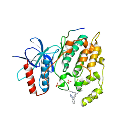
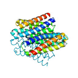
6S9T
 
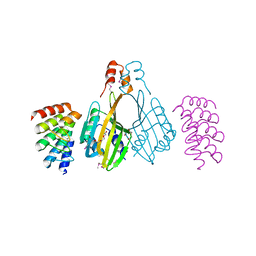 | | Dimerization domain of Xenopus laevis LDB1 in complex with darpin 3 | | 分子名称: | Darpin 3, LIM domain-binding protein 1, TETRAETHYLENE GLYCOL | | 著者 | Renko, M, Schaefer, J.V, Pluckthun, A, Bienz, M. | | 登録日 | 2019-07-15 | | 公開日 | 2019-10-09 | | 最終更新日 | 2019-10-23 | | 実験手法 | X-RAY DIFFRACTION (2.05 Å) | | 主引用文献 | Rotational symmetry of the structured Chip/LDB-SSDP core module of the Wnt enhanceosome.
Proc.Natl.Acad.Sci.USA, 116, 2019
|
|
6P6U
 | |
3WOH
 | |
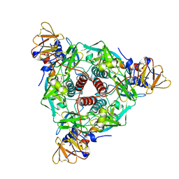
3WW3
 | | X-ray structures of Cellulomonas parahominis L-ribose isomerase with no ligand | | 分子名称: | L-ribose isomerase, MANGANESE (II) ION | | 著者 | Terami, Y, Yoshida, H, Takata, G, Kamitori, S. | | 登録日 | 2014-06-13 | | 公開日 | 2015-04-29 | | 最終更新日 | 2024-05-29 | | 実験手法 | X-RAY DIFFRACTION (1.9 Å) | | 主引用文献 | Essentiality of tetramer formation of Cellulomonas parahominis L-ribose isomerase involved in novel L-ribose metabolic pathway.
Appl.Microbiol.Biotechnol., 99, 2015
|
|
3O2M
 | |
3WW1
 | | X-ray structure of Cellulomonas parahominis L-ribose isomerase with L-ribose | | 分子名称: | L-ribose, L-ribose isomerase, MANGANESE (II) ION, ... | | 著者 | Terami, Y, Yoshida, H, Takata, G, Kamitori, S. | | 登録日 | 2014-06-13 | | 公開日 | 2015-04-29 | | 最終更新日 | 2024-05-29 | | 実験手法 | X-RAY DIFFRACTION (1.95 Å) | | 主引用文献 | Essentiality of tetramer formation of Cellulomonas parahominis L-ribose isomerase involved in novel L-ribose metabolic pathway.
Appl.Microbiol.Biotechnol., 99, 2015
|
|
5JD2
 | | SFX structure of corestreptavidin-selenobiotin complex | | 分子名称: | 5-[(3aS,4S,6aR)-2-oxohexahydro-1H-selenopheno[3,4-d]imidazol-4-yl]pentanoic acid, Streptavidin | | 著者 | DeMirci, H, Hunter, M.S, Boutet, S. | | 登録日 | 2016-04-15 | | 公開日 | 2016-11-16 | | 最終更新日 | 2024-03-06 | | 実験手法 | X-RAY DIFFRACTION (1.9 Å) | | 主引用文献 | Selenium single-wavelength anomalous diffraction de novo phasing using an X-ray-free electron laser.
Nat Commun, 7, 2016
|
|
3WW4
 | | X-ray structures of Cellulomonas parahominis L-ribose isomerase with L-allose | | 分子名称: | L-allose, L-ribose isomerase, MANGANESE (II) ION, ... | | 著者 | Terami, Y, Yoshida, H, Takata, G, Kamitori, S. | | 登録日 | 2014-06-13 | | 公開日 | 2015-04-29 | | 最終更新日 | 2024-05-29 | | 実験手法 | X-RAY DIFFRACTION (1.95 Å) | | 主引用文献 | Essentiality of tetramer formation of Cellulomonas parahominis L-ribose isomerase involved in novel L-ribose metabolic pathway.
Appl.Microbiol.Biotechnol., 99, 2015
|
|
6B6U
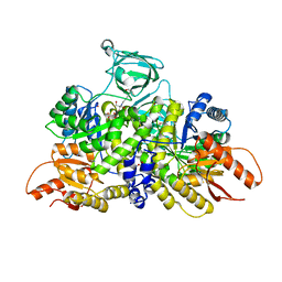 | | Pyruvate Kinase M2 mutant - S437Y | | 分子名称: | 1,2-ETHANEDIOL, 2-[3-(2-HYDROXY-1,1-DIHYDROXYMETHYL-ETHYLAMINO)-PROPYLAMINO]-2-HYDROXYMETHYL-PROPANE-1,3-DIOL, CHLORIDE ION, ... | | 著者 | Srivastava, D, Dey, M. | | 登録日 | 2017-10-03 | | 公開日 | 2017-12-20 | | 最終更新日 | 2023-10-04 | | 実験手法 | X-RAY DIFFRACTION (1.35 Å) | | 主引用文献 | Structural Investigation of a Dimeric Variant of Pyruvate Kinase Muscle Isoform 2.
Biochemistry, 56, 2017
|
|
6XYE
 | |
4EKV
 | | Streptavidin 8-aa-loop H127C mutein with reversible biotin binding | | 分子名称: | BIOTIN, CHLORIDE ION, Streptavidin | | 著者 | Barrette-Ng, I.H, Honetschlaeger, C, Wong, S.L, Ng, K.K.S. | | 登録日 | 2012-04-09 | | 公開日 | 2012-06-13 | | 最終更新日 | 2023-09-13 | | 実験手法 | X-RAY DIFFRACTION (2 Å) | | 主引用文献 | Development of a tetrameric streptavidin mutein with reversible biotin binding capability: engineering a mobile loop as an exit door for biotin.
Plos One, 7, 2012
|
|
5JEI
 | | Crystal structure of the GluA2 LBD in complex with FW | | 分子名称: | 1,2-ETHANEDIOL, 2-(2-METHOXYETHOXY)ETHANOL, 2-AMINO-3-(5-FLUORO-2,4-DIOXO-3,4-DIHYDRO-2H-PYRIMIDIN-1-YL)-PROPIONIC ACID, ... | | 著者 | Eibl, C, Salazar, H, Chebli, M, Plested, A.J.R. | | 登録日 | 2016-04-18 | | 公開日 | 2017-02-22 | | 最終更新日 | 2024-01-10 | | 実験手法 | X-RAY DIFFRACTION (1.229 Å) | | 主引用文献 | Mechanism of partial agonism in AMPA-type glutamate receptors.
Nat Commun, 8, 2017
|
|
6Q58
 | |
1ZNA
 | |
6Q6B
 | | Structure of the copper storage protein, Ccsp, from Streptomyces lividans loaded with 10 copper equivalents | | 分子名称: | COPPER (I) ION, COPPER (II) ION, Cytosolic copper storage protein | | 著者 | Straw, M.L, Hough, M.A, Worrall, J.A.R. | | 登録日 | 2018-12-10 | | 公開日 | 2019-07-10 | | 最終更新日 | 2024-01-24 | | 実験手法 | X-RAY DIFFRACTION (1.9 Å) | | 主引用文献 | A Histidine Residue and a Tetranuclear Cuprous-thiolate Cluster Dominate the Copper Loading Landscape of a Copper Storage Protein from Streptomyces lividans.
Chemistry, 25, 2019
|
|
4N3E
 | | Crystal structure of Hyp-1, a St John's wort PR-10 protein, in complex with 8-anilino-1-naphthalene sulfonate (ANS) | | 分子名称: | 4-(2-HYDROXYETHYL)-1-PIPERAZINE ETHANESULFONIC ACID, 8-ANILINO-1-NAPHTHALENE SULFONATE, Phenolic oxidative coupling protein, ... | | 著者 | Sliwiak, J, Dauter, Z, Mccoy, A.J, Read, R.J, Jaskolski, M. | | 登録日 | 2013-10-07 | | 公開日 | 2014-02-26 | | 最終更新日 | 2023-09-20 | | 実験手法 | X-RAY DIFFRACTION (2.43 Å) | | 主引用文献 | Likelihood-based molecular-replacement solution for a highly pathological crystal with tetartohedral twinning and sevenfold translational noncrystallographic symmetry.
Acta Crystallogr.,Sect.D, 70, 2014
|
|
3FXI
 | | Crystal structure of the human TLR4-human MD-2-E.coli LPS Ra complex | | 分子名称: | 2-acetamido-2-deoxy-beta-D-glucopyranose, 2-acetamido-2-deoxy-beta-D-glucopyranose-(1-4)-2-acetamido-2-deoxy-beta-D-glucopyranose, 3-HYDROXY-TETRADECANOIC ACID, ... | | 著者 | Park, B.S, Song, D.H, Kim, H.M, Lee, J.-O. | | 登録日 | 2009-01-21 | | 公開日 | 2009-03-03 | | 最終更新日 | 2023-11-01 | | 実験手法 | X-RAY DIFFRACTION (3.1 Å) | | 主引用文献 | The structural basis of lipopolysaccharide recognition by the TLR4-MD-2 complex
Nature, 458, 2009
|
|
3WW2
 | | X-ray structures of Cellulomonas parahominis L-ribose isomerase with L-psicose | | 分子名称: | L-psicose, L-ribose isomerase, MANGANESE (II) ION, ... | | 著者 | Terami, Y, Yoshida, H, Takata, G, Kamitori, S. | | 登録日 | 2014-06-13 | | 公開日 | 2015-04-29 | | 最終更新日 | 2024-05-29 | | 実験手法 | X-RAY DIFFRACTION (2 Å) | | 主引用文献 | Essentiality of tetramer formation of Cellulomonas parahominis L-ribose isomerase involved in novel L-ribose metabolic pathway.
Appl.Microbiol.Biotechnol., 99, 2015
|
|
3AIX
 | |
3R52
 | | Structure analysis of a wound-inducible lectin ipomoelin from sweet potato | | 分子名称: | CADMIUM ION, Ipomoelin, methyl alpha-D-glucopyranoside | | 著者 | Liu, K.L, Chang, W.C, Jeng, S.T, Cheng, Y.S. | | 登録日 | 2011-03-18 | | 公開日 | 2012-03-28 | | 最終更新日 | 2023-09-13 | | 実験手法 | X-RAY DIFFRACTION (2.1 Å) | | 主引用文献 | Ipomoelin, a jacalin-related lectin with a compact tetrameric association and versatile carbohydrate binding properties regulated by its N terminus.
Plos One, 7, 2012
|
|
3AIZ
 | |
2QOG
 | | Crotoxin B, the basic PLA2 from Crotalus durissus terrificus. | | 分子名称: | CALCIUM ION, Phospholipase A2 CB1, Phospholipase A2 CB2 | | 著者 | Marchi-Salvador, D.P, Correa, L.C, Fontes, M.R.M. | | 登録日 | 2007-07-20 | | 公開日 | 2008-04-01 | | 最終更新日 | 2011-07-13 | | 実験手法 | X-RAY DIFFRACTION (2.28 Å) | | 主引用文献 | Insights into the role of oligomeric state on the biological activities of crotoxin: crystal structure of a tetrameric phospholipase A2 formed by two isoforms of crotoxin B from Crotalus durissus terrificus venom.
Proteins, 72, 2008
|
|
3GQG
 | | Crystal structure at acidic pH of the ferric form of the Root effect hemoglobin from Trematomus bernacchii. | | 分子名称: | Hemoglobin subunit alpha, Hemoglobin subunit beta, PROTOPORPHYRIN IX CONTAINING FE | | 著者 | Vergara, A, Franzese, M, Merlino, A, Bonomi, G, Mazzarella, L. | | 登録日 | 2009-03-24 | | 公開日 | 2009-10-13 | | 最終更新日 | 2023-09-06 | | 実験手法 | X-RAY DIFFRACTION (1.73 Å) | | 主引用文献 | Correlation between hemichrome stability and the root effect in tetrameric hemoglobins.
Biophys.J., 97, 2009
|
|
3I0R
 | | crystal structure of HIV reverse transcriptase in complex with inhibitor 3 | | 分子名称: | Reverse transcriptase/ribonuclease H, S-{2-[(2-chloro-4-sulfamoylphenyl)amino]-2-oxoethyl} 6-methyl-3,4-dihydroquinoline-1(2H)-carbothioate, p51 RT | | 著者 | Yan, Y, Prasad, S. | | 登録日 | 2009-06-25 | | 公開日 | 2009-08-25 | | 最終更新日 | 2023-09-06 | | 実験手法 | X-RAY DIFFRACTION (2.98 Å) | | 主引用文献 | Substituted tetrahydroquinolines as potent allosteric inhibitors of reverse transcriptase and its key mutants.
Bioorg.Med.Chem.Lett., 19, 2009
|
|
3I0S
 | | crystal structure of HIV reverse transcriptase in complex with inhibitor 7 | | 分子名称: | Reverse transcriptase/ribonuclease H, S-{2-[(2-chloro-4-sulfamoylphenyl)amino]-2-oxoethyl} 6,8-dichloro-3,4-dihydroquinoline-1(2H)-carbothioate, p51 RT | | 著者 | Yan, Y, Prasad, S. | | 登録日 | 2009-06-25 | | 公開日 | 2009-08-25 | | 最終更新日 | 2023-09-06 | | 実験手法 | X-RAY DIFFRACTION (2.7 Å) | | 主引用文献 | Substituted tetrahydroquinolines as potent allosteric inhibitors of reverse transcriptase and its key mutants.
Bioorg.Med.Chem.Lett., 19, 2009
|
|


