5WFX
 
 | |
7BBJ
 | |
5WBQ
 | |
2P6V
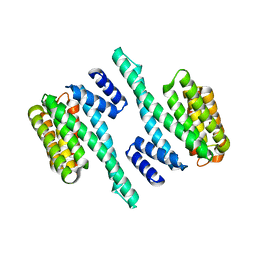
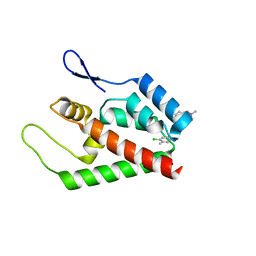
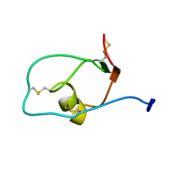
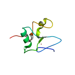
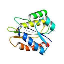
 | | Structure of TAFH domain of the human TAF4 subunit of TFIID | | 分子名称: | SULFATE ION, Transcription initiation factor TFIID subunit 4 | | 著者 | Wang, X, Truckses, D.M, Takada, S, Matsumura, T, Tanese, N, Jacobson, R.H. | | 登録日 | 2007-03-19 | | 公開日 | 2007-05-15 | | 最終更新日 | 2011-07-13 | | 実験手法 | X-RAY DIFFRACTION (2 Å) | | 主引用文献 | Conserved region I of human coactivator TAF4 binds to a short hydrophobic motif present in transcriptional regulators.
Proc.Natl.Acad.Sci.Usa, 104, 2007
|
|
2JPR
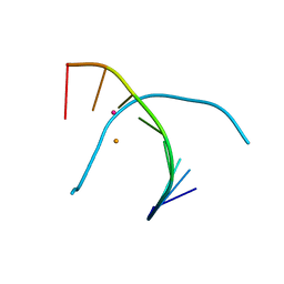
 | | Joint refinement of the HIV-1 CA-NTD in complex with the assembly inhibitor CAP-1 | | 分子名称: | 1-(3-chloro-4-methylphenyl)-3-{2-[({5-[(dimethylamino)methyl]-2-furyl}methyl)thio]ethyl}urea, Gag-Pol polyprotein | | 著者 | Kelly, B.N, Kyere, S, Kinde, I, Tang, C, Howard, B.R, Robinson, H, Sundquist, W.I, Summers, M.F, Hill, C.P. | | 登録日 | 2007-05-22 | | 公開日 | 2007-10-09 | | 最終更新日 | 2024-05-29 | | 実験手法 | SOLUTION NMR | | 主引用文献 | Structure of the Antiviral Assembly Inhibitor CAP-1 Complex with the HIV-1 CA Protein
J.Mol.Biol., 373, 2007
|
|
5WHQ
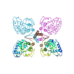
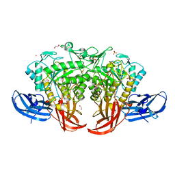
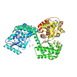
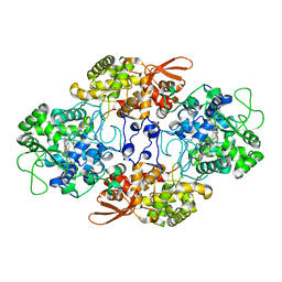 | | Crystal structure of the catalase-peroxidase from Neurospora crassa at 2.9 A | | 分子名称: | Catalase-peroxidase, POTASSIUM ION, PROTOPORPHYRIN IX CONTAINING FE | | 著者 | Diaz-Vilchis, A, Vega-Garcia, V, Rudino-Pinera, E, Hansberg, W. | | 登録日 | 2017-07-18 | | 公開日 | 2018-01-17 | | 最終更新日 | 2023-10-04 | | 実験手法 | X-RAY DIFFRACTION (2.9 Å) | | 主引用文献 | Structure, kinetics, molecular and redox properties of a cytosolic and developmentally regulated fungal catalase-peroxidase.
Arch. Biochem. Biophys., 640, 2018
|
|
2P9C
 | |
5WFV
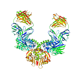
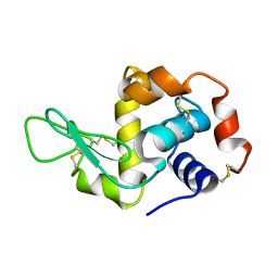
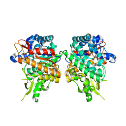
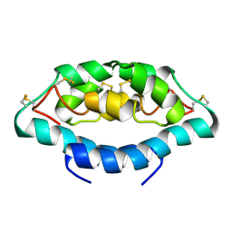
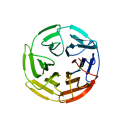 | | Kelch domain of human Keap1 bound to Nrf2 ETGE peptide | | 分子名称: | Kelch-like ECH-associated protein 1, Nrf2 ETGE peptide, SULFATE ION | | 著者 | Carolan, J.P, Lynch, A.J, Allen, K.N. | | 登録日 | 2017-07-12 | | 公開日 | 2018-09-19 | | 最終更新日 | 2023-10-04 | | 実験手法 | X-RAY DIFFRACTION (1.91 Å) | | 主引用文献 | Interaction Energetics and Druggability of the Protein-Protein Interaction between Kelch-like ECH-Associated Protein 1 (KEAP1) and Nuclear Factor Erythroid 2 Like 2 (Nrf2).
Biochemistry, 59, 2020
|
|
2JXD
 | |
2PAH
 | |
5WHO
 | |
5AMY
 | |
5AWP
 | |
5AXG
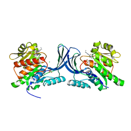
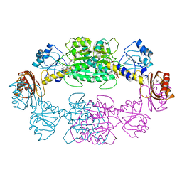
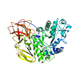
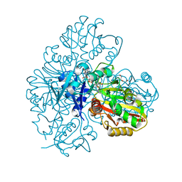
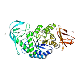
 | | Crystal structure of thermophilic dextranase from Thermoanaerobacter pseudethanolicus | | 分子名称: | 1,2-ETHANEDIOL, 3-CYCLOHEXYL-1-PROPYLSULFONIC ACID, Dextranase, ... | | 著者 | Suzuki, N, Kishine, N, Fujimoto, Z, Sakurai, M, Momma, M, Ko, J.A, Nam, S.H, Kimura, A, Kim, Y.M. | | 登録日 | 2015-07-29 | | 公開日 | 2015-11-11 | | 最終更新日 | 2023-11-08 | | 実験手法 | X-RAY DIFFRACTION (1.85 Å) | | 主引用文献 | Crystal structure of thermophilic dextranase from Thermoanaerobacter pseudethanolicus
J.Biochem., 159, 2016
|
|
7BEW
 | |
7BBS
 | |
2JTG
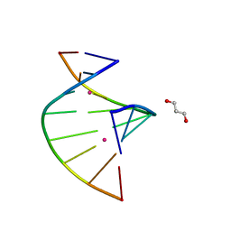
 | | Solution structure of the THAP-zinc finger of THAP1 | | 分子名称: | THAP domain-containing protein 1, ZINC ION | | 著者 | Bessiere, D, Campagne, S, Milon, A, Gervais, V. | | 登録日 | 2007-07-31 | | 公開日 | 2007-12-11 | | 最終更新日 | 2024-05-29 | | 実験手法 | SOLUTION NMR | | 主引用文献 | Structure-function analysis of the THAP zinc finger of THAP1, a large C2CH DNA-binding module linked to Rb/E2F pathways
J.Biol.Chem., 283, 2008
|
|
2JV7
 | |
5WSQ
 | |
2NT1
 | |
5WSS
 | |
5B2V
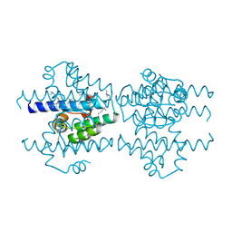
 | | Crystal Structure of P450BM3 with N-perfluorohexanoyl-L-tryptophan | | 分子名称: | (2~{S})-3-(1~{H}-indol-3-yl)-2-[2,2,3,3,4,4,5,5,6,6,6-undecakis(fluoranyl)hexanoylamino]propanoic acid, Bifunctional cytochrome P450/NADPH--P450 reductase, PROTOPORPHYRIN IX CONTAINING FE | | 著者 | Cong, Z, Shoji, O, Kasai, C, Sugimoto, H, Shiro, Y, Watanabe, Y. | | 登録日 | 2016-02-03 | | 公開日 | 2017-02-08 | | 最終更新日 | 2023-11-08 | | 実験手法 | X-RAY DIFFRACTION (2.3 Å) | | 主引用文献 | Crystal Structure of P450BM3 with decoy molecules
to be published
|
|
6TNM
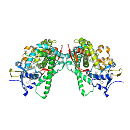
 | | E. coli aerobic trifunctional enzyme subunit-alpha | | 分子名称: | ADENOSINE-5'-TRIPHOSPHATE, Fatty acid oxidation complex subunit alpha, GLYCEROL, ... | | 著者 | Sah-Teli, S.K, Hynonen, M.J, Wierenga, R.K, Venkatesan, R. | | 登録日 | 2019-12-09 | | 公開日 | 2020-03-25 | | 最終更新日 | 2024-01-24 | | 実験手法 | X-RAY DIFFRACTION (2.95 Å) | | 主引用文献 | Insights into the stability and substrate specificity of the E. coli aerobic beta-oxidation trifunctional enzyme complex.
J.Struct.Biol., 210, 2020
|
|
5B3K
 | |
6TP0
 | |


