7NUL
 
 | |
7NUQ
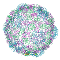
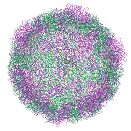
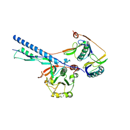
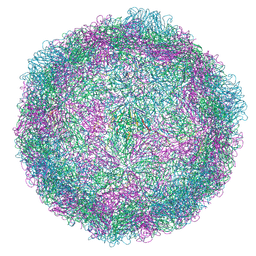 | | Rhinovirus 14 virion-like at pH 6.2 | | 分子名称: | Genome polyprotein, Octanucleotide | | 著者 | Hrebik, D, Plevka, P. | | 登録日 | 2021-03-12 | | 公開日 | 2021-05-19 | | 最終更新日 | 2024-07-10 | | 実験手法 | ELECTRON MICROSCOPY (2.8 Å) | | 主引用文献 | ICAM-1 induced rearrangements of capsid and genome prime rhinovirus 14 for activation and uncoating.
Proc.Natl.Acad.Sci.USA, 118, 2021
|
|
7NSX
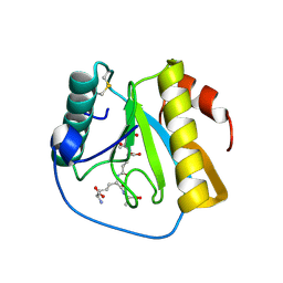
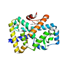
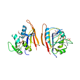
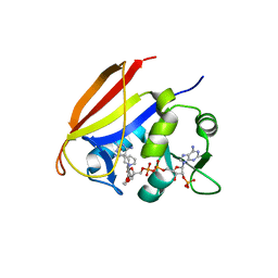
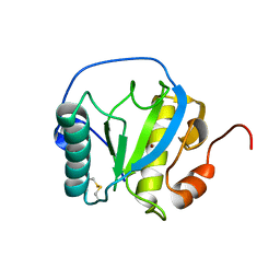 | | Drosophila PGRP-LB wild-type | | 分子名称: | Isoform A of Peptidoglycan-recognition protein LB, ZINC ION | | 著者 | Orlans, J, Aller, P, Da Silva, P. | | 登録日 | 2021-03-08 | | 公開日 | 2021-05-19 | | 最終更新日 | 2024-11-13 | | 実験手法 | X-RAY DIFFRACTION (1.9 Å) | | 主引用文献 | PGRP-LB: An Inside View into the Mechanism of the Amidase Reaction.
Int J Mol Sci, 22, 2021
|
|
7NHL
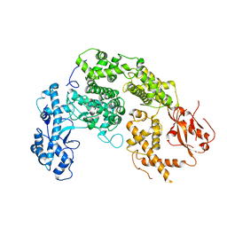
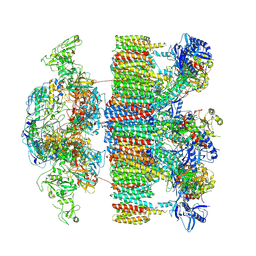
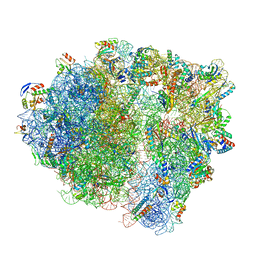 | | VgaA-LC, an antibiotic resistance ABCF, in complex with 70S ribosome from Staphylococcus aureus | | 分子名称: | 16S rRNA, 23S rRNA, 30S ribosomal protein S10, ... | | 著者 | Crowe-McAuliffe, C, Murina, V, Hauryliuk, V, Wilson, D.N. | | 登録日 | 2021-02-10 | | 公開日 | 2021-05-05 | | 最終更新日 | 2024-05-01 | | 実験手法 | ELECTRON MICROSCOPY (3.1 Å) | | 主引用文献 | Structural basis of ABCF-mediated resistance to pleuromutilin, lincosamide, and streptogramin A antibiotics in Gram-positive pathogens.
Nat Commun, 12, 2021
|
|
7O6E
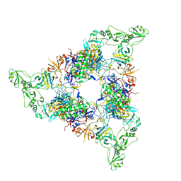
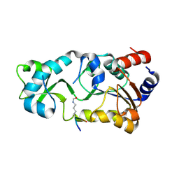
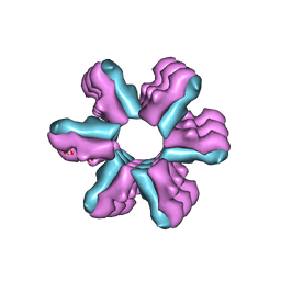
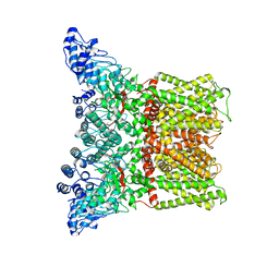
 | | 2.12 A cryo-EM structure of Mycobacterium tuberculosis Ferritin | | 分子名称: | Ferritin BfrB | | 著者 | Gijsbers, A, Zhang, Y, Gao, Y, Peters, P.J, Ravelli, R.B.G. | | 登録日 | 2021-04-10 | | 公開日 | 2021-05-19 | | 最終更新日 | 2024-07-10 | | 実験手法 | ELECTRON MICROSCOPY (2.1 Å) | | 主引用文献 | Mycobacterium tuberculosis ferritin: a suitable workhorse protein for cryo-EM development.
Acta Crystallogr D Struct Biol, 77, 2021
|
|
7NUN
 | |
7NT0
 | | Drosophila PGRP-LB Y78F mutant in complex with tracheal cytotoxin (TCT) | | 分子名称: | GLCNAC(BETA1-4)-MURNAC(1,6-ANHYDRO)-L-ALA-GAMMA-D-GLU-MESO-A2PM-D-ALA, Isoform A of Peptidoglycan-recognition protein LB, ZINC ION | | 著者 | Orlans, J, Aller, P, Da Silva, P. | | 登録日 | 2021-03-08 | | 公開日 | 2021-05-19 | | 最終更新日 | 2024-10-16 | | 実験手法 | X-RAY DIFFRACTION (1.8 Å) | | 主引用文献 | PGRP-LB: An Inside View into the Mechanism of the Amidase Reaction.
Int J Mol Sci, 22, 2021
|
|
7NUR
 | |
7NPS
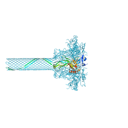
 | | Structure of the periplasmic assembly from the ESX-5 inner membrane complex, C1 model | | 分子名称: | ESX-5 secretion system ATPase EccB5, Mycosin-5 | | 著者 | Fahrenkamp, D, Bunduc, C.M, Wald, J, Ummels, R, Bitter, W, Houben, E.N.G, Marlovits, T.C. | | 登録日 | 2021-02-28 | | 公開日 | 2021-05-26 | | 最終更新日 | 2024-10-23 | | 実験手法 | ELECTRON MICROSCOPY (3.81 Å) | | 主引用文献 | Structure and dynamics of a mycobacterial type VII secretion system.
Nature, 593, 2021
|
|
7NPT
 | | Cytosolic bridge of an intact ESX-5 inner membrane complex | | 分子名称: | ESX-5 secretion system protein EccC5, ESX-5 secretion system protein EccD5 | | 著者 | Fahrenkamp, D, Bunduc, C.M, Wald, J, Ummels, R, Bitter, W, Houben, E.N.G, Marlovits, T.C. | | 登録日 | 2021-02-28 | | 公開日 | 2021-05-26 | | 最終更新日 | 2024-07-10 | | 実験手法 | ELECTRON MICROSCOPY (3.27 Å) | | 主引用文献 | Structure and dynamics of a mycobacterial type VII secretion system.
Nature, 593, 2021
|
|
7NPC
 | | ROR(gamma)t ligand binding domain in complex with allosteric ligand FM156 | | 分子名称: | 4-[[3-[2-chloranyl-6-(trifluoromethyl)phenyl]-5-(1~{H}-pyrrol-3-yl)-1,2-oxazol-4-yl]methoxy]benzoic acid, GLYCEROL, Nuclear receptor ROR-gamma | | 著者 | de Vries, R.M.J.M, Meijer, F.A, Brunsveld, L. | | 登録日 | 2021-02-26 | | 公開日 | 2021-06-02 | | 最終更新日 | 2024-01-31 | | 実験手法 | X-RAY DIFFRACTION (1.47 Å) | | 主引用文献 | Structure-Activity Relationship Studies of Trisubstituted Isoxazoles as Selective Allosteric Ligands for the Retinoic-Acid-Receptor-Related Orphan Receptor gamma t.
J.Med.Chem., 64, 2021
|
|
7NP7
 | | Structure of an intact ESX-5 inner membrane complex, Composite C1 model | | 分子名称: | ESX-5 secretion system ATPase EccB5, ESX-5 secretion system protein EccC5, ESX-5 secretion system protein EccD5, ... | | 著者 | Fahrenkamp, D, Bunduc, C.M, Wald, J, Ummels, R, Bitter, W, Houben, E.N.G, Marlovits, T.C. | | 登録日 | 2021-02-26 | | 公開日 | 2021-06-02 | | 最終更新日 | 2024-10-16 | | 実験手法 | ELECTRON MICROSCOPY (4.03 Å) | | 主引用文献 | Structure and dynamics of a mycobacterial type VII secretion system.
Nature, 593, 2021
|
|
3DOB
 | | Peptide-binding domain of Heat shock 70 kDa protein F44E5.5 from C.elegans. | | 分子名称: | BETA-MERCAPTOETHANOL, Heat shock 70 kDa protein F44E5.5 | | 著者 | Osipiuk, J, Hatzos, C, Gu, M, Zhang, R, Voisine, C, Morimoto, R.I, Joachimiak, A, Midwest Center for Structural Genomics (MCSG) | | 登録日 | 2008-07-03 | | 公開日 | 2008-07-22 | | 最終更新日 | 2023-08-30 | | 実験手法 | X-RAY DIFFRACTION (2.39 Å) | | 主引用文献 | X-ray crystal structure of Peptide-binding domain of Heat shock 70 kDa protein F44E5.5 from C.elegans.
To be Published
|
|
3K35
 | | Crystal Structure of Human SIRT6 | | 分子名称: | ADENOSINE-5-DIPHOSPHORIBOSE, NAD-dependent deacetylase sirtuin-6, SULFATE ION, ... | | 著者 | Pan, P.W, Dong, A, Qiu, W, Loppnau, P, Wang, J, Ravichandran, M, Bochkarev, A, Bountra, C, Weigelt, J, Arrowsmith, C.H, Min, J, Edwards, A.M, Structural Genomics Consortium (SGC) | | 登録日 | 2009-10-01 | | 公開日 | 2009-12-08 | | 最終更新日 | 2023-09-06 | | 実験手法 | X-RAY DIFFRACTION (2 Å) | | 主引用文献 | Structure and biochemical functions of SIRT6.
J.Biol.Chem., 286, 2011
|
|
3JR3
 | | Sir2 bound to acetylated peptide | | 分子名称: | Acetylated Peptide, NAD-dependent deacetylase, ZINC ION | | 著者 | Hawse, W.F, Wolberger, C. | | 登録日 | 2009-09-08 | | 公開日 | 2009-09-29 | | 最終更新日 | 2024-10-30 | | 実験手法 | X-RAY DIFFRACTION (1.5 Å) | | 主引用文献 | Structure-based mechanism of ADP-ribosylation by sirtuins.
J.Biol.Chem., 284, 2009
|
|
3K7T
 | | Crystal structure of apo-form 6-hydroxy-L-nicotine oxidase, crystal form P3121 | | 分子名称: | (1R)-2-{[(S)-(2-aminoethoxy)(hydroxy)phosphoryl]oxy}-1-[(pentadecanoyloxy)methyl]ethyl (12E)-hexadeca-9,12-dienoate, 6-hydroxy-L-nicotine oxidase, FLAVIN-ADENINE DINUCLEOTIDE | | 著者 | Bourenkov, G.P, Kachalova, G.S, Bartunik, H.D. | | 登録日 | 2009-10-13 | | 公開日 | 2010-01-19 | | 最終更新日 | 2023-09-06 | | 実験手法 | X-RAY DIFFRACTION (2.85 Å) | | 主引用文献 | Crystal Structure Analysis of Free and Substrate-Bound 6-Hydroxy-l-Nicotine Oxidase from Arthrobacter nicotinovorans.
J.Mol.Biol., 396, 2010
|
|
3JV2
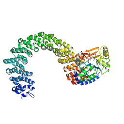
 | | Crystal Structure of B. subtilis SecA with bound peptide | | 分子名称: | ADENOSINE-5'-DIPHOSPHATE, MAGNESIUM ION, Protein translocase subunit SecA, ... | | 著者 | Zimmer, J. | | 登録日 | 2009-09-15 | | 公開日 | 2009-11-24 | | 最終更新日 | 2023-09-06 | | 実験手法 | X-RAY DIFFRACTION (2.5 Å) | | 主引用文献 | Conformational flexibility and peptide interaction of the translocation ATPase SecA.
J.Mol.Biol., 394, 2009
|
|
3J1Z
 | | Inward-Facing Conformation of the Zinc Transporter YiiP revealed by Cryo-electron Microscopy | | 分子名称: | Cation efflux family protein | | 著者 | Coudray, N, Valvo, S, Hu, M, Lasala, R, Kim, C, Vink, M, Zhou, M, Provasi, D, Filizola, M, Tao, J, Fang, J, Penczek, P.A, Ubarretxena-Belandia, I, Stokes, D.L, Transcontinental EM Initiative for Membrane Protein Structure (TEMIMPS) | | 登録日 | 2012-07-24 | | 公開日 | 2012-10-10 | | 最終更新日 | 2024-02-21 | | 実験手法 | ELECTRON MICROSCOPY (13 Å) | | 主引用文献 | Inward-facing conformation of the zinc transporter YiiP revealed by cryoelectron microscopy.
Proc.Natl.Acad.Sci.USA, 110, 2013
|
|
3CIN
 | |
3JVX
 | | Crystal structure of Bacillus anthracis dihydrofolate reductase complexed with NADPH and 2,4-diamino-5-(3-(3,4,5-trimethoxyphenyl)prop-1-ynyl)-6-ethylpyrimidine (UCP120A) | | 分子名称: | 6-ethyl-5-[3-(3,4,5-trimethoxyphenyl)prop-1-yn-1-yl]pyrimidine-2,4-diamine, Dihydrofolate reductase, NADPH DIHYDRO-NICOTINAMIDE-ADENINE-DINUCLEOTIDE PHOSPHATE | | 著者 | Anderson, A.C, Beierlein, J.M, Karri, N.G. | | 登録日 | 2009-09-17 | | 公開日 | 2010-09-15 | | 最終更新日 | 2023-09-06 | | 実験手法 | X-RAY DIFFRACTION (2.25 Å) | | 主引用文献 | Mutational Studies into Trimethoprim Resistance in Bacillus anthracis Dihydrofolate Reductase
To be Published
|
|
3JW5
 | |
3J5R
 | |
3J9C
 | | CryoEM single particle reconstruction of anthrax toxin protective antigen pore at 2.9 Angstrom resolution | | 分子名称: | CALCIUM ION, Protective antigen PA-63 | | 著者 | Jiang, J, Pentelute, B.L, Collier, R.J, Zhou, Z.H. | | 登録日 | 2014-12-25 | | 公開日 | 2015-03-11 | | 最終更新日 | 2024-02-21 | | 実験手法 | ELECTRON MICROSCOPY (2.9 Å) | | 主引用文献 | Atomic structure of anthrax protective antigen pore elucidates toxin translocation.
Nature, 521, 2015
|
|
3K7V
 | | Protein phosphatase 2A core complex bound to dinophysistoxin-1 | | 分子名称: | (2R)-3-[(2S,5R,6R,8S)-8-{(1R,2E)-3-[(2R,4a'R,5R,6'S,8'R,8a'S)-6'-{(1S,3S)-3-[(2S,3R,6R,11R)-3,11-dimethyl-1,7-dioxaspiro[5.5]undec-2-yl]-1-hydroxybutyl}-8'-hydroxy-7'-methylideneoctahydro-3H,3'H-spiro[furan-2,2'-pyrano[3,2-b]pyran]-5-yl]-1-methylprop-2-en-1-yl}-5-hydroxy-10-methyl-1,7-dioxaspiro[5.5]undec-10-en-2-yl]-2-hydroxy-2-methylpropanoic acid, MANGANESE (II) ION, SULFATE ION, ... | | 著者 | Jeffrey, P.D, Huhn, J, Shi, Y. | | 登録日 | 2009-10-13 | | 公開日 | 2009-11-03 | | 最終更新日 | 2023-09-06 | | 実験手法 | X-RAY DIFFRACTION (2.85 Å) | | 主引用文献 | A structural basis for the reduced toxicity of dinophysistoxin-2.
Chem.Res.Toxicol., 22, 2009
|
|
3JAI
 | | Structure of a mammalian ribosomal termination complex with ABCE1, eRF1(AAQ), and the UGA stop codon | | 分子名称: | 18S ribosomal RNA, 28S ribosomal RNA, 5.8S ribosomal RNA, ... | | 著者 | Brown, A, Shao, S, Murray, J, Hegde, R.S, Ramakrishnan, V. | | 登録日 | 2015-06-10 | | 公開日 | 2015-08-12 | | 最終更新日 | 2024-11-27 | | 実験手法 | ELECTRON MICROSCOPY (3.65 Å) | | 主引用文献 | Structural basis for stop codon recognition in eukaryotes.
Nature, 524, 2015
|
|


