5X57
 
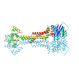
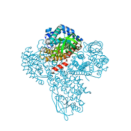
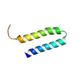
 | | Structure of GAR domain of ACF7 | | 分子名称: | Microtubule-actin cross-linking factor 1, isoforms 1/2/3/5, NICKEL (II) ION | | 著者 | Yang, F, Wang, T, Zhang, Y, Wu, X.Y. | | 登録日 | 2017-02-15 | | 公開日 | 2017-07-05 | | 最終更新日 | 2024-03-27 | | 実験手法 | X-RAY DIFFRACTION (1.45 Å) | | 主引用文献 | ACF7 regulates inflammatory colitis and intestinal wound response by orchestrating tight junction dynamics.
Nat Commun, 8, 2017
|
|
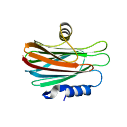
2MOT
 | | Backbone Structure of Actin Depolymerizing Factor (ADF) of Toxoplasma gondii Based on Prot3DNMR Approach | | 分子名称: | Actin depolymerizing factor ADF | | 著者 | Kumar, D, Raikwal, N, Raval, I, Jaiswal, N, Shukla, V, Arora, A. | | 登録日 | 2014-05-05 | | 公開日 | 2015-05-27 | | 最終更新日 | 2024-05-15 | | 実験手法 | SOLUTION NMR | | 主引用文献 | Prot3DNMR: A Simple and Swift Strategy for Backbone Structure Determination of Proteins by NMR
To be Published
|
|
7O7I
 | |
4LHX
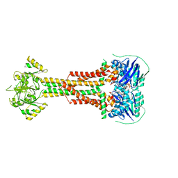
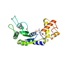
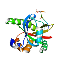
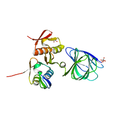
 | | Crystal structure of nucleotide-free Rab8:Rabin8 | | 分子名称: | Rab-3A-interacting protein, Ras-related protein Rab-8A, SULFATE ION | | 著者 | Guo, Z, Hou, X.M, Goody, R.S, Itzen, A. | | 登録日 | 2013-07-01 | | 公開日 | 2013-10-09 | | 最終更新日 | 2023-09-20 | | 実験手法 | X-RAY DIFFRACTION (3.05 Å) | | 主引用文献 | Intermediates in the Guanine Nucleotide Exchange Reaction of Rab8 Protein Catalyzed by Guanine Nucleotide Exchange Factors Rabin8 and GRAB.
J.Biol.Chem., 288, 2013
|
|
5LJ6
 | |
5LJ7
 | |
5YRN
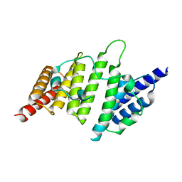
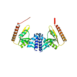
 | | Structure of RIP2 CARD domain | | 分子名称: | Receptor-interacting serine/threonine-protein kinase 2 | | 著者 | Wu, B, Gong, Q. | | 登録日 | 2017-11-09 | | 公開日 | 2018-11-14 | | 最終更新日 | 2024-03-27 | | 実験手法 | ELECTRON MICROSCOPY (4.1 Å) | | 主引用文献 | Structural basis of RIP2 activation and signaling.
Nat Commun, 9, 2018
|
|
5YZ0
 | | Cryo-EM Structure of human ATR-ATRIP complex | | 分子名称: | ATR-interacting protein, Serine/threonine-protein kinase ATR | | 著者 | Rao, Q, Liu, M, Tian, Y, Wu, Z, Wang, H, Wang, J, Xu, Y. | | 登録日 | 2017-12-11 | | 公開日 | 2018-01-31 | | 最終更新日 | 2019-11-06 | | 実験手法 | ELECTRON MICROSCOPY (4.7 Å) | | 主引用文献 | Cryo-EM structure of human ATR-ATRIP complex.
Cell Res., 28, 2018
|
|
5ENH
 | | Crystal structure of the second bromodomain of Pleckstrin homology domain interacting protein (PHIP) in complex with compound-12 N11528 (SGC - Diamond I04-1 fragment screening) | | 分子名称: | PH-interacting protein, ~{N}-[(2,6-dimethoxyphenyl)methyl]ethanamide | | 著者 | Krojer, T, Talon, R, Collins, P, Bradley, A, Cox, O, Szykowska, A, Burgess-Brown, N, Brennan, P, Bountra, C, Arrowsmith, C.H, Edwards, A, von Delft, F, Structural Genomics Consortium (SGC) | | 登録日 | 2015-11-09 | | 公開日 | 2016-04-27 | | 最終更新日 | 2024-01-10 | | 実験手法 | X-RAY DIFFRACTION (1.95 Å) | | 主引用文献 | A poised fragment library enables rapid synthetic expansion yielding the first reported inhibitors of PHIP(2), an atypical bromodomain.
Chem Sci, 7, 2016
|
|
5ENI
 | | Crystal structure of the second bromodomain of Pleckstrin homology domain interacting protein (PHIP) in complex with compound-13 N11537 (SGC - Diamond I04-1 fragment screening) | | 分子名称: | PH-interacting protein, ~{N}-[[2,6-bis(chloranyl)phenyl]methyl]-2-oxidanyl-ethanamide | | 著者 | Krojer, T, Talon, R, Collins, P, Bradley, A, Cox, O, Szykowska, A, Burgess-Brown, N, Brennan, P, Bountra, C, Arrowsmith, C.H, Edwards, A, von Delft, F, Structural Genomics Consortium (SGC) | | 登録日 | 2015-11-09 | | 公開日 | 2016-04-27 | | 最終更新日 | 2024-01-10 | | 実験手法 | X-RAY DIFFRACTION (1.69 Å) | | 主引用文献 | A poised fragment library enables rapid synthetic expansion yielding the first reported inhibitors of PHIP(2), an atypical bromodomain.
Chem Sci, 7, 2016
|
|
3M9V
 | |
4WDC
 | |
3VWI
 | | High resolution crystal structure of FraC in the monomeric form | | 分子名称: | AMMONIUM ION, CHLORIDE ION, Fragaceatoxin C, ... | | 著者 | Tanaka, K, Morante, K, Caaveiro, J.M.M, Gonzalez-Manas, J.M, Tsumoto, K. | | 登録日 | 2012-08-23 | | 公開日 | 2013-08-28 | | 最終更新日 | 2023-11-08 | | 実験手法 | X-RAY DIFFRACTION (1.7 Å) | | 主引用文献 | Structural basis for self-assembly of a cytolytic pore lined by protein and lipid
Nat Commun, 6, 2015
|
|
6LUF
 | |
4NQ0
 | | Structural insights into yeast histone chaperone Hif1: a scaffold protein recruiting protein complexes to core histones | | 分子名称: | HAT1-interacting factor 1 | | 著者 | Liu, H, Zhang, M, He, W, Zhu, Z, Teng, M, Gao, Y, Niu, L. | | 登録日 | 2013-11-23 | | 公開日 | 2014-07-16 | | 最終更新日 | 2024-03-20 | | 実験手法 | X-RAY DIFFRACTION (2.1 Å) | | 主引用文献 | Structural insights into yeast histone chaperone Hif1: a scaffold protein recruiting protein complexes to core histones
Biochem.J., 462, 2014
|
|
8JT3
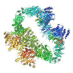
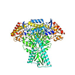 | | Crystal structure of aminotransferase CrmG from Actinoalloteichus sp. WH1-2216-6 in complex with amino donor L-Arg | | 分子名称: | (E)-N~2~-({3-hydroxy-2-methyl-5-[(phosphonooxy)methyl]pyridin-4-yl}methylidene)-L-arginine, ACETATE ION, CrmG, ... | | 著者 | Su, K, Zhang, Y, Xu, J, Liu, J. | | 登録日 | 2023-06-21 | | 公開日 | 2023-08-02 | | 最終更新日 | 2024-05-08 | | 実験手法 | X-RAY DIFFRACTION (2.35 Å) | | 主引用文献 | Co-crystal structure provides insights on transaminase CrmG recognition amino donor L-Arg.
Biochem.Biophys.Res.Commun., 675, 2023
|
|
1WS1
 | |
6OVM
 | |
5BT1
 | | histone chaperone Hif1 playing with histone H2A-H2B dimer | | 分子名称: | HAT1-interacting factor 1, Histone H2A.1, Histone H2B.1 | | 著者 | Liu, H, Zhang, M, Gao, Y, Teng, M, Niu, L. | | 登録日 | 2015-06-02 | | 公開日 | 2016-10-26 | | 最終更新日 | 2023-11-08 | | 実験手法 | X-RAY DIFFRACTION (2.62 Å) | | 主引用文献 | Structural Insights into the Association of Hif1 with Histones H2A-H2B Dimer and H3-H4 Tetramer
Structure, 24, 2016
|
|
2RT5
 | |
3A4S
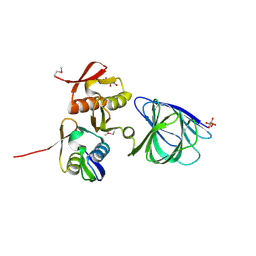
 | | The crystal structure of the SLD2:Ubc9 complex | | 分子名称: | NFATC2-interacting protein, SUMO-conjugating enzyme UBC9 | | 著者 | Sekiyama, N, Arita, K, Ikeda, Y, Ariyoshi, M, Tochio, H, Saitoh, H, Shirakawa, M. | | 登録日 | 2009-07-14 | | 公開日 | 2010-02-02 | | 最終更新日 | 2023-11-01 | | 実験手法 | X-RAY DIFFRACTION (2.7 Å) | | 主引用文献 | Structural basis for regulation of poly-SUMO chain by a SUMO-like domain of Nip45
Proteins, 78, 2009
|
|
5COS
 | |
1YSM
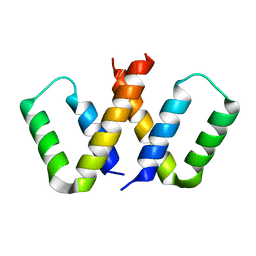
 | | NMR Structure of N-terminal domain (Residues 1-77) of Siah-Interacting Protein. | | 分子名称: | Calcyclin-binding protein | | 著者 | Bhattacharya, S, Lee, Y.T, Michowski, W, Jastrzebska, B, Filipek, A, Kuznicki, J, Chazin, W.J. | | 登録日 | 2005-02-08 | | 公開日 | 2005-07-26 | | 最終更新日 | 2024-05-22 | | 実験手法 | SOLUTION NMR | | 主引用文献 | The Modular Structure of SIP Facilitates Its Role in Stabilizing Multiprotein Assemblies.
Biochemistry, 44, 2005
|
|
4WHN
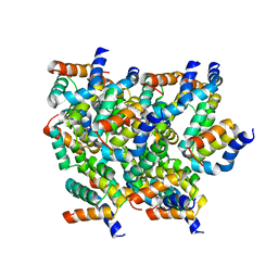
 | | Structure of toxin-activating acyltransferase (TAAT) | | 分子名称: | ApxC, CITRIC ACID | | 著者 | Crow, A, Greene, N.P, Hughes, C, Koronakis, V. | | 登録日 | 2014-09-23 | | 公開日 | 2015-06-03 | | 最終更新日 | 2024-05-08 | | 実験手法 | X-RAY DIFFRACTION (2.15 Å) | | 主引用文献 | Structure of a bacterial toxin-activating acyltransferase.
Proc.Natl.Acad.Sci.USA, 112, 2015
|
|
6OVK
 | |


