8HLN
 
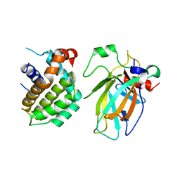 | | Crystal structure of p53/BCL2 fusion complex(complex3) | | 分子名称: | Apoptosis regulator Bcl-2, Cellular tumor antigen p53, ZINC ION | | 著者 | Guo, M, Wei, H, Wang, H, Chen, Y. | | 登録日 | 2022-11-30 | | 公開日 | 2023-07-26 | | 最終更新日 | 2024-05-29 | | 実験手法 | X-RAY DIFFRACTION (2.354 Å) | | 主引用文献 | Structures of p53/BCL-2 complex suggest a mechanism for p53 to antagonize BCL-2 activity.
Nat Commun, 14, 2023
|
|
8HLM
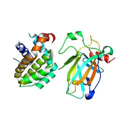 | | Crystal structure of p53/BCL2 fusion complex (complex 2) | | 分子名称: | Apoptosis regulator Bcl-2, Cellular tumor antigen p53, ZINC ION | | 著者 | Guo, M, Wang, H, Wei, H, Chen, Y. | | 登録日 | 2022-11-30 | | 公開日 | 2023-07-26 | | 最終更新日 | 2024-05-29 | | 実験手法 | X-RAY DIFFRACTION (2.522 Å) | | 主引用文献 | Structures of p53/BCL-2 complex suggest a mechanism for p53 to antagonize BCL-2 activity.
Nat Commun, 14, 2023
|
|
2VGC
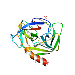 | | GAMMA-CHYMOTRYPSIN D-PARA-CHLORO-1-ACETAMIDO BORONIC ACID INHIBITOR COMPLEX | | 分子名称: | D-1-(4-CHLOROPHENYL)-2-(ACETAMIDO)ETHANE BORONIC ACID, GAMMA CHYMOTRYPSIN, SULFATE ION | | 著者 | Stoll, V.S, Eger, B.T, Hynes, R.C, Martichonok, V, Jones, J.B, Pai, E.F. | | 登録日 | 1997-05-01 | | 公開日 | 1997-11-12 | | 最終更新日 | 2023-08-09 | | 実験手法 | X-RAY DIFFRACTION (1.8 Å) | | 主引用文献 | Differences in binding modes of enantiomers of 1-acetamido boronic acid based protease inhibitors: crystal structures of gamma-chymotrypsin and subtilisin Carlsberg complexes.
Biochemistry, 37, 1998
|
|
2VYR
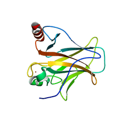
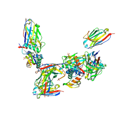 | | Structure of human MDM4 N-terminal domain bound to a single domain antibody | | 分子名称: | HUMAN SINGLE DOMAIN ANTIBODY, MDM4 PROTEIN, SULFATE ION | | 著者 | Yu, G.W, Vaysburd, M, Allen, M.D, Settanni, G, Fersht, A.R. | | 登録日 | 2008-07-28 | | 公開日 | 2008-11-25 | | 最終更新日 | 2013-03-06 | | 実験手法 | X-RAY DIFFRACTION (2 Å) | | 主引用文献 | Structure of Human Mdm4 N-Terminal Domain Bound to a Single-Domain Antibody.
J.Mol.Biol., 385, 2009
|
|
2XWR
 | |
8GCR
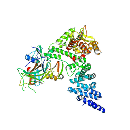 | | HPV16 E6-E6AP-p53 complex | | 分子名称: | Cellular tumor antigen p53, Maltose/maltodextrin-binding periplasmic protein,Protein E6, Ubiquitin-protein ligase E3A, ... | | 著者 | Bratkowski, M.A, Wang, J.C.K, Hao, Q, Nile, A.H. | | 登録日 | 2023-03-02 | | 公開日 | 2024-03-06 | | 実験手法 | ELECTRON MICROSCOPY (3.38 Å) | | 主引用文献 | Structure of the p53 degradation complex from HPV16.
Nat Commun, 15, 2024
|
|
6CHA
 | |
3BG4
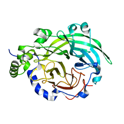
 | | The crystal structure of guamerin in complex with chymotrypsin and the development of an elastase-specific inhibitor | | 分子名称: | Chymotrypsin A chain A, Chymotrypsin A chain B, Chymotrypsin A chain C, ... | | 著者 | Kim, H, Chu, T.T.T, Kim, D.Y, Kim, D.R, Nguyen, C.M.T, Choi, J, Lee, J.R, Hahn, M.J, Kim, K.K. | | 登録日 | 2007-11-26 | | 公開日 | 2008-07-29 | | 最終更新日 | 2023-11-01 | | 実験手法 | X-RAY DIFFRACTION (2.5 Å) | | 主引用文献 | The crystal structure of guamerin in complex with chymotrypsin and the development of an elastase-specific inhibitor.
J.Mol.Biol., 376, 2008
|
|
3BYN
 | |
3BYK
 | |
6DI8
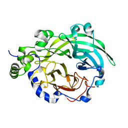
 | | Crystal structure of bovine alpha-chymotrypsin in space group P65 | | 分子名称: | Chymotrypsin A chain A, Chymotrypsin A chain B, Chymotrypsin A chain C, ... | | 著者 | Marshall, A.C, Keiller, B.G, Bruning, J.B. | | 登録日 | 2018-05-23 | | 公開日 | 2019-01-23 | | 最終更新日 | 2023-10-11 | | 実験手法 | X-RAY DIFFRACTION (1.859 Å) | | 主引用文献 | Crystal Structure of Bovine Alpha-Chymotrypsin in Space Group P65
Crystals, 8, 2018
|
|
3BYJ
 | |
3BYL
 | |
3TS8
 | |
1ACB
 | | CRYSTAL AND MOLECULAR STRUCTURE OF THE BOVINE ALPHA-CHYMOTRYPSIN-EGLIN C COMPLEX AT 2.0 ANGSTROMS RESOLUTION | | 分子名称: | ALPHA-CHYMOTRYPSIN, Eglin C | | 著者 | Bolognesi, M, Frigerio, F, Coda, A, Pugliese, L, Lionetti, C, Menegatti, E, Amiconi, G, Schnebli, H.P, Ascenzi, P. | | 登録日 | 1991-11-08 | | 公開日 | 1993-10-31 | | 最終更新日 | 2017-11-29 | | 実験手法 | X-RAY DIFFRACTION (2 Å) | | 主引用文献 | Crystal and molecular structure of the bovine alpha-chymotrypsin-eglin c complex at 2.0 A resolution.
J.Mol.Biol., 225, 1992
|
|
1CA0
 | | BOVINE CHYMOTRYPSIN COMPLEXED TO APPI | | 分子名称: | BOVINE CHYMOTRYPSIN, PROTEASE INHIBITOR DOMAIN OF ALZHEIMER'S AMYLOID BETA-PROTEIN PRECURSOR | | 著者 | Scheidig, A.J, Kossiakoff, A.A. | | 登録日 | 1997-01-23 | | 公開日 | 1997-07-23 | | 最終更新日 | 2011-07-13 | | 実験手法 | X-RAY DIFFRACTION (2.1 Å) | | 主引用文献 | Crystal structures of bovine chymotrypsin and trypsin complexed to the inhibitor domain of Alzheimer's amyloid beta-protein precursor (APPI) and basic pancreatic trypsin inhibitor (BPTI): engineering of inhibitors with altered specificities.
Protein Sci., 6, 1997
|
|
8E7B
 | | Crystal structure of the p53 (Y107H) core domain monoclinic P form | | 分子名称: | Cellular tumor antigen p53, ZINC ION | | 著者 | Lovell, S, Liu, L, Battaile, K.P, Miller, S, Karanicolas, J. | | 登録日 | 2022-08-23 | | 公開日 | 2023-05-17 | | 最終更新日 | 2023-10-25 | | 実験手法 | X-RAY DIFFRACTION (2.5 Å) | | 主引用文献 | An African-Specific Variant of TP53 Reveals PADI4 as a Regulator of p53-Mediated Tumor Suppression.
Cancer Discov, 13, 2023
|
|
8E7A
 | | Crystal structure of the p53 (Y107H) core domain orthorhombic P form | | 分子名称: | Cellular tumor antigen p53, ZINC ION | | 著者 | Lovell, S, Liu, L, Battaile, K.P, Miller, S, Karanicolas, J. | | 登録日 | 2022-08-23 | | 公開日 | 2023-05-17 | | 最終更新日 | 2023-10-25 | | 実験手法 | X-RAY DIFFRACTION (1.3 Å) | | 主引用文献 | An African-Specific Variant of TP53 Reveals PADI4 as a Regulator of p53-Mediated Tumor Suppression.
Cancer Discov, 13, 2023
|
|
1AB9
 | | CRYSTAL STRUCTURE OF BOVINE GAMMA-CHYMOTRYPSIN | | 分子名称: | GAMMA-CHYMOTRYPSIN, PENTAPEPTIDE (TPGVY), SULFATE ION | | 著者 | Sugio, S, Kashima, A, Inoue, Y, Maeda, I, Nose, T, Shimohigashi, Y. | | 登録日 | 1997-02-05 | | 公開日 | 1997-08-20 | | 最終更新日 | 2023-08-02 | | 実験手法 | X-RAY DIFFRACTION (1.6 Å) | | 主引用文献 | X-ray crystal structure of a dipeptide-chymotrypsin complex in an inhibitory interaction.
Eur.J.Biochem., 255, 1998
|
|
1AFQ
 | | CRYSTAL STRUCTURE OF BOVINE GAMMA-CHYMOTRYPSIN COMPLEXED WITH A SYNTHETIC INHIBITOR | | 分子名称: | BOVINE GAMMA-CHYMOTRYPSIN, D-leucyl-N-(4-fluorobenzyl)-L-phenylalaninamide, SULFATE ION | | 著者 | Sugio, S, Kashima, A, Inoue, Y, Maeda, I, Nose, T, Shimohigashi, Y. | | 登録日 | 1997-03-12 | | 公開日 | 1997-09-17 | | 最終更新日 | 2023-08-02 | | 実験手法 | X-RAY DIFFRACTION (1.8 Å) | | 主引用文献 | X-ray crystal structure of a dipeptide-chymotrypsin complex in an inhibitory interaction.
Eur.J.Biochem., 255, 1998
|
|
1CHO
 | | CRYSTAL AND MOLECULAR STRUCTURES OF THE COMPLEX OF ALPHA-*CHYMOTRYPSIN WITH ITS INHIBITOR TURKEY OVOMUCOID THIRD DOMAIN AT 1.8 ANGSTROMS RESOLUTION | | 分子名称: | ALPHA-CHYMOTRYPSIN A, TURKEY OVOMUCOID THIRD DOMAIN (OMTKY3) | | 著者 | Fujinaga, M, Sielecki, A.R, Read, R.J, Ardelt, W, Laskowskijunior, M, James, M.N.G. | | 登録日 | 1988-03-04 | | 公開日 | 1988-07-16 | | 最終更新日 | 2017-11-29 | | 実験手法 | X-RAY DIFFRACTION (1.8 Å) | | 主引用文献 | Crystal and molecular structures of the complex of alpha-chymotrypsin with its inhibitor turkey ovomucoid third domain at 1.8 A resolution.
J.Mol.Biol., 195, 1987
|
|
8F2I
 | | P53 monomer structure | | 分子名称: | Cellular tumor antigen p53 | | 著者 | Solares, M, Kelly, D.F. | | 登録日 | 2022-11-08 | | 公開日 | 2022-11-23 | | 最終更新日 | 2024-05-01 | | 実験手法 | ELECTRON MICROSCOPY (5 Å) | | 主引用文献 | High-Resolution Imaging of Human Cancer Proteins Using Microprocessor Materials.
Chembiochem, 23, 2022
|
|
8F2H
 | |
1CHG
 | | CHYMOTRYPSINOGEN,2.5 ANGSTROMS CRYSTAL STRUCTURE, COMPARISON WITH ALPHA-CHYMOTRYPSIN,AND IMPLICATIONS FOR ZYMOGEN ACTIVATION | | 分子名称: | CHYMOTRYPSINOGEN A | | 著者 | Freer, S.T, Kraut, J, Robertus, J.D, Wright, H.T, Xuong, N.H. | | 登録日 | 1975-03-01 | | 公開日 | 1976-11-22 | | 最終更新日 | 2023-09-27 | | 実験手法 | X-RAY DIFFRACTION (2.5 Å) | | 主引用文献 | Chymotrypsinogen: 2.5-angstrom crystal structure, comparison with alpha-chymotrypsin, and implications for zymogen activation.
Biochemistry, 9, 1970
|
|
1CBW
 | | BOVINE CHYMOTRYPSIN COMPLEXED TO BPTI | | 分子名称: | BOVINE CHYMOTRYPSIN, BPTI, SULFATE ION | | 著者 | Hynes, T.R, Scheidig, A.J, Kossiakoff, A.A. | | 登録日 | 1996-12-22 | | 公開日 | 1997-07-23 | | 最終更新日 | 2023-08-09 | | 実験手法 | X-RAY DIFFRACTION (2.6 Å) | | 主引用文献 | Crystal structures of bovine chymotrypsin and trypsin complexed to the inhibitor domain of Alzheimer's amyloid beta-protein precursor (APPI) and basic pancreatic trypsin inhibitor (BPTI): engineering of inhibitors with altered specificities.
Protein Sci., 6, 1997
|
|


