7QQM
 
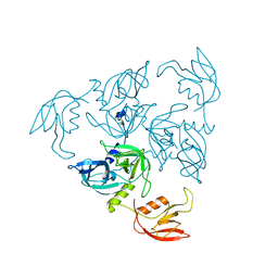
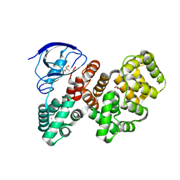 | | The PDZ domain of LRRC7 fused with ANXA2 | | 分子名称: | CALCIUM ION, GLYCEROL, Leucine-rich repeat-containing protein 7,Annexin A2 | | 著者 | Cousido-Siah, A, Trave, G, Gogl, G. | | 登録日 | 2022-01-10 | | 公開日 | 2022-04-20 | | 最終更新日 | 2024-01-31 | | 実験手法 | X-RAY DIFFRACTION (1.6 Å) | | 主引用文献 | A scalable strategy to solve structures of PDZ domains and their complexes.
Acta Crystallogr D Struct Biol, 78, 2022
|
|
5F3X
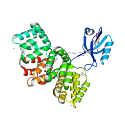
 | | Crystal structure of Harmonin NPDZ1 in complex with ANKS4B SAM-PBM | | 分子名称: | Ankyrin repeat and SAM domain-containing protein 4B, CHLORIDE ION, Harmonin | | 著者 | Li, J, He, Y, Lu, Q, Zhang, M. | | 登録日 | 2015-12-03 | | 公開日 | 2016-03-16 | | 最終更新日 | 2023-11-08 | | 実験手法 | X-RAY DIFFRACTION (2.649 Å) | | 主引用文献 | Mechanistic Basis of Organization of the Harmonin/USH1C-Mediated Brush Border Microvilli Tip-Link Complex
Dev.Cell, 36, 2016
|
|
8AEL
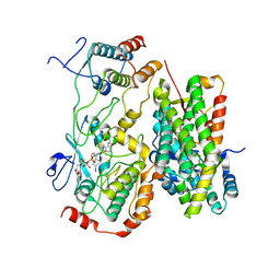
 | | SYNJ2BP complex with a synthetic Vangl2 peptide (3mer). | | 分子名称: | CALCIUM ION, GLY-GLY-GLY-THR-SER-VAL, GLYCEROL, ... | | 著者 | Carrasco, K, Cousido Siah, A, Gogl, G, Betzi, S, McEwen, A, Kostmann, C, Trave, G. | | 登録日 | 2022-07-13 | | 公開日 | 2023-08-16 | | 実験手法 | X-RAY DIFFRACTION (2.2 Å) | | 主引用文献 | SYNJ2BP PDZ domain in complex with a synthetic Vangl2 peptide.
To Be Published
|
|
8BW9
 | |
8BW8
 | |
2R3Y
 | |
1SOT
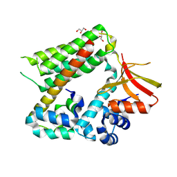
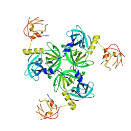
 | | Crystal Structure of the DegS stress sensor | | 分子名称: | Protease degS | | 著者 | Wilken, C, Kitzing, K, Kurzbauer, R, Ehrmann, M, Clausen, T. | | 登録日 | 2004-03-15 | | 公開日 | 2004-06-08 | | 最終更新日 | 2023-11-15 | | 実験手法 | X-RAY DIFFRACTION (2.3 Å) | | 主引用文献 | Crystal structure of the DegS stress sensor: How a PDZ domain recognizes misfolded protein and activates a protease
Cell(Cambridge,Mass.), 117, 2004
|
|
1TE0
 | |
1SOZ
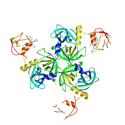 | | Crystal Structure of DegS protease in complex with an activating peptide | | 分子名称: | Protease degS, activating peptide | | 著者 | Wilken, C, Kitzing, K, Kurzbauer, R, Ehrmann, M, Clausen, T. | | 登録日 | 2004-03-16 | | 公開日 | 2004-06-08 | | 最終更新日 | 2024-04-03 | | 実験手法 | X-RAY DIFFRACTION (2.4 Å) | | 主引用文献 | Crystal structure of the DegS stress sensor: How a PDZ domain recognizes misfolded protein and activates a protease
Cell(Cambridge,Mass.), 117, 2004
|
|
1VCW
 | | Crystal structure of DegS after backsoaking the activating peptide | | 分子名称: | Protease degS | | 著者 | Wilken, C, Kitzing, K, Kurzbauer, R, Ehrmann, M, Clausen, T. | | 登録日 | 2004-03-16 | | 公開日 | 2004-06-08 | | 最終更新日 | 2023-12-27 | | 実験手法 | X-RAY DIFFRACTION (3.05 Å) | | 主引用文献 | Crystal structure of the DegS stress sensor: How a PDZ domain recognizes misfolded protein and activates a protease.
Cell(Cambridge,Mass.), 117, 2004
|
|


