1PEM
 
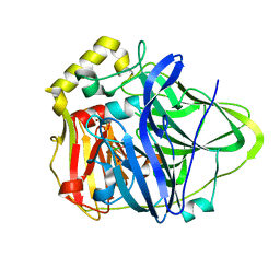
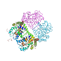
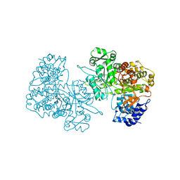 | | Ribonucleotide Reductase Protein R1E from Salmonella typhimurium | | 分子名称: | Ribonucleoside-diphosphate reductase 2 alpha chain | | 著者 | Uppsten, M, Farnegardh, M, Jordan, A, Eliasson, R, Eklund, H, Uhlin, U. | | 登録日 | 2003-05-22 | | 公開日 | 2004-05-25 | | 最終更新日 | 2024-04-03 | | 実験手法 | X-RAY DIFFRACTION (2.99 Å) | | 主引用文献 | Structure of the large subunit of class Ib ribonucleotide reductase from Salmonella typhimurium and its complexes with allosteric effectors.
J.Mol.Biol., 330, 2003
|
|
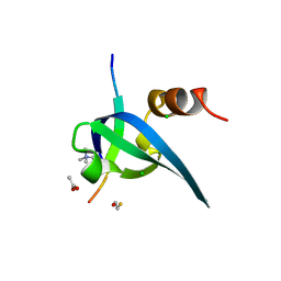
1PEN
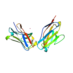
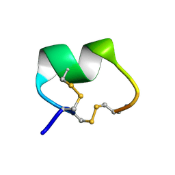 | | ALPHA-CONOTOXIN PNI1 | | 分子名称: | ALPHA-CONOTOXIN PNIA | | 著者 | Hu, S.-H, Gehrmann, J, Guddat, L.W, Alewood, P.F, Craik, D.J, Martin, J.L. | | 登録日 | 1996-01-29 | | 公開日 | 1997-04-21 | | 最終更新日 | 2011-07-13 | | 実験手法 | X-RAY DIFFRACTION (1.1 Å) | | 主引用文献 | The 1.1 A crystal structure of the neuronal acetylcholine receptor antagonist, alpha-conotoxin PnIA from Conus pennaceus.
Structure, 4, 1996
|
|
1PEO
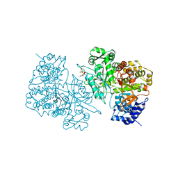 | | Ribonucleotide Reductase Protein R1E from Salmonella typhimurium | | 分子名称: | 2'-DEOXYCYTIDINE-5'-TRIPHOSPHATE, MAGNESIUM ION, Ribonucleoside-diphosphate reductase 2 alpha chain | | 著者 | Uppsten, M, Farnegardh, M, Jordan, A, Eliasson, R, Eklund, H, Uhlin, U. | | 登録日 | 2003-05-22 | | 公開日 | 2004-05-25 | | 最終更新日 | 2024-04-03 | | 実験手法 | X-RAY DIFFRACTION (3 Å) | | 主引用文献 | Structure of the large subunit of class Ib ribonucleotide reductase from Salmonella typhimurium and its complexes with allosteric effectors.
J.Mol.Biol., 330, 2003
|
|
1PEQ
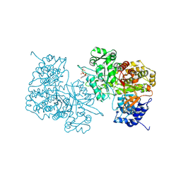 | | Ribonucleotide Reductase Protein R1E from Salmonella typhimurium | | 分子名称: | MAGNESIUM ION, Ribonucleoside-diphosphate reductase 2 alpha chain, THYMIDINE-5'-TRIPHOSPHATE | | 著者 | Uppsten, M, Farnegardh, M, Jordan, A, Eliasson, R, Eklund, H, Uhlin, U. | | 登録日 | 2003-05-22 | | 公開日 | 2004-05-25 | | 最終更新日 | 2024-04-03 | | 実験手法 | X-RAY DIFFRACTION (2.8 Å) | | 主引用文献 | Structure of the large subunit of class Ib ribonucleotide reductase from Salmonella typhimurium and its complexes with allosteric effectors.
J.Mol.Biol., 330, 2003
|
|
1PER
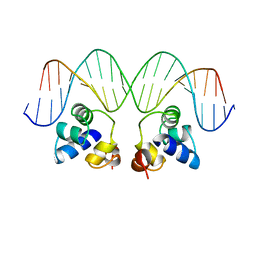 | |
1PES
 | | NMR SOLUTION STRUCTURE OF THE TETRAMERIC MINIMUM TRANSFORMING DOMAIN OF P53 | | 分子名称: | TUMOR SUPPRESSOR P53 | | 著者 | Lee, W, Harvey, T.S, Yin, Y, Yau, P, Litchfield, D, Arrowsmith, C.H. | | 登録日 | 1994-11-24 | | 公開日 | 1995-02-07 | | 最終更新日 | 2024-05-22 | | 実験手法 | SOLUTION NMR | | 主引用文献 | Solution structure of the tetrameric minimum transforming domain of p53.
Nat.Struct.Biol., 1, 1994
|
|
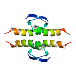
1PET
 | | NMR SOLUTION STRUCTURE OF THE TETRAMERIC MINIMUM TRANSFORMING DOMAIN OF P53 | | 分子名称: | TUMOR SUPPRESSOR P53 | | 著者 | Lee, W, Harvey, T.S, Yin, Y, Yau, P, Litchfield, D, Arrowsmith, C.H. | | 登録日 | 1994-11-24 | | 公開日 | 1995-02-07 | | 最終更新日 | 2024-05-22 | | 実験手法 | SOLUTION NMR | | 主引用文献 | Solution structure of the tetrameric minimum transforming domain of p53.
Nat.Struct.Biol., 1, 1994
|
|
1PEU
 | | Ribonucleotide Reductase Protein R1E from Salmonella typhimurium | | 分子名称: | 2'-DEOXYADENOSINE 5'-TRIPHOSPHATE, MAGNESIUM ION, Ribonucleoside-diphosphate reductase 2 alpha chain | | 著者 | Uppsten, M, Farnegardh, M, Jordan, A, Eliasson, R, Eklund, H, Uhlin, U. | | 登録日 | 2003-05-22 | | 公開日 | 2004-05-25 | | 最終更新日 | 2024-04-03 | | 実験手法 | X-RAY DIFFRACTION (3.2 Å) | | 主引用文献 | Structure of the large subunit of class Ib ribonucleotide reductase from Salmonella typhimurium and its complexes with allosteric effectors.
J.Mol.Biol., 330, 2003
|
|
1PEV
 | | Crystal Structure of the Actin Interacting Protein from Caenorhabditis Elegans | | 分子名称: | Actin interacting protein 1 | | 著者 | Vorobiev, S, Mohri, K, Fedorov, A.A, Ono, S, Almo, S.C, Burley, S.K, New York SGX Research Center for Structural Genomics (NYSGXRC) | | 登録日 | 2003-05-22 | | 公開日 | 2003-07-01 | | 最終更新日 | 2023-08-16 | | 実験手法 | X-RAY DIFFRACTION (2 Å) | | 主引用文献 | Identification of functional residues on Caenorhabditis elegans actin-interacting protein 1 (UNC-78) for disassembly of actin depolymerizing factor/cofilin-bound actin filaments.
J.Biol.Chem., 279, 2004
|
|
1PEW
 | | High Resolution Crystal Structure of Jto2, a mutant of the non-amyloidogenic Lamba6 Light Chain, Jto | | 分子名称: | CADMIUM ION, Jto2, a LAMBDA-6 TYPE IMMUNOGLOBULIN LIGHT CHAIN, ... | | 著者 | Dealwis, C, Gupta, V, Wilkerson, M. | | 登録日 | 2003-05-22 | | 公開日 | 2004-07-13 | | 最終更新日 | 2023-08-16 | | 実験手法 | X-RAY DIFFRACTION (1.6 Å) | | 主引用文献 | Structural basis of light chain amyloidogenicity: comparison of the thermodynamic properties, fibrillogenic potential and tertiary structural features of four V(lambda)6 proteins
J.Mol.Recog., 17, 2004
|
|
1PEX
 | | COLLAGENASE-3 (MMP-13) C-TERMINAL HEMOPEXIN-LIKE DOMAIN | | 分子名称: | CALCIUM ION, CHLORIDE ION, COLLAGENASE-3, ... | | 著者 | Gomis-Ruth, F.X, Gohlke, U, Betz, M, Knauper, V, Murphy, G, Lopez-Otin, C, Bode, W. | | 登録日 | 1996-05-24 | | 公開日 | 1996-12-23 | | 最終更新日 | 2024-06-05 | | 実験手法 | X-RAY DIFFRACTION (2.7 Å) | | 主引用文献 | The helping hand of collagenase-3 (MMP-13): 2.7 A crystal structure of its C-terminal haemopexin-like domain.
J.Mol.Biol., 264, 1996
|
|
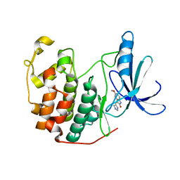
1PEY
 | | Crystal structure of the Response Regulator Spo0F complexed with Mn2+ | | 分子名称: | MANGANESE (II) ION, Sporulation initiation phosphotransferase F | | 著者 | Mukhopadhyay, D, Sen, U, Zapf, J, Varughese, K.I. | | 登録日 | 2003-05-22 | | 公開日 | 2004-05-18 | | 最終更新日 | 2024-02-14 | | 実験手法 | X-RAY DIFFRACTION (2.25 Å) | | 主引用文献 | Metals in the sporulation phosphorelay: manganese binding by the response regulator Spo0F.
Acta Crystallogr.,Sect.D, 60, 2004
|
|
1PEZ
 | | Bacillus circulans strain 251 mutant A230V | | 分子名称: | (4S)-2-METHYL-2,4-PENTANEDIOL, 4-(2-HYDROXYETHYL)-1-PIPERAZINE ETHANESULFONIC ACID, ACETIC ACID, ... | | 著者 | Rozeboom, H.J, Dijkstra, B.W. | | 登録日 | 2003-05-23 | | 公開日 | 2003-10-28 | | 最終更新日 | 2023-08-16 | | 実験手法 | X-RAY DIFFRACTION (2.32 Å) | | 主引用文献 | Conversion of Cyclodextrin Glycosyltransferase into a Starch Hydrolase by Directed Evolution: The Role of Alanine 230 in Acceptor Subsite +1
Biochemistry, 42, 2003
|
|
1PF3
 | | Crystal Structure of the M441L mutant of the multicopper oxidase CueO | | 分子名称: | Blue copper oxidase cueO, COPPER (II) ION, CU-CL-CU LINKAGE | | 著者 | Roberts, S.A, Wildner, G.F, Grass, G, Weichsel, A, Ambrus, A, Rensing, C, Montfort, W.R. | | 登録日 | 2003-05-23 | | 公開日 | 2003-06-24 | | 最終更新日 | 2023-08-16 | | 実験手法 | X-RAY DIFFRACTION (1.5 Å) | | 主引用文献 | A Labile Regulatory Copper Ion Lies Near the T1 Copper Site in the Multicopper Oxidase CueO.
J.Biol.Chem., 278, 2003
|
|
1PF5
 | | Structural Genomics, Protein YJGH | | 分子名称: | Hypothetical protein yjgH, MERCURY (II) ION | | 著者 | Zhang, R, Joachimiak, A, Edwards, A, Savchenko, A, Xu, L, Midwest Center for Structural Genomics (MCSG) | | 登録日 | 2003-05-23 | | 公開日 | 2003-12-09 | | 最終更新日 | 2024-02-14 | | 実験手法 | X-RAY DIFFRACTION (2.5 Å) | | 主引用文献 | The 2.5A crystal structure of protein YJGH from E. Coli
To be Published
|
|
1PF7
 | | CRYSTAL STRUCTURE OF HUMAN PNP COMPLEXED WITH IMMUCILLIN H | | 分子名称: | 1,4-DIDEOXY-4-AZA-1-(S)-(9-DEAZAHYPOXANTHIN-9-YL)-D-RIBITOL, PURINE NUCLEOSIDE PHOSPHORYLASE, SULFATE ION | | 著者 | De Azevedo Jr, W.F, Canduri, F, Dos Santos, D.M, Pereira, J.H, Dias, M.V.B, Silva, R.G, Mendes, M.A, Palma, M.S, Basso, L.A, Santos, D.S. | | 登録日 | 2003-05-24 | | 公開日 | 2004-06-01 | | 最終更新日 | 2023-09-20 | | 実験手法 | X-RAY DIFFRACTION (2.6 Å) | | 主引用文献 | Structural basis for inhibition of human PNP by immucillin-H
Biochem.Biophys.Res.Commun., 309, 2003
|
|
1PF8
 | | Crystal Structure of Human Cyclin-Dependent Kinase 2 Complexed with a Nucleoside Inhibitor | | 分子名称: | (3Z)-3-(1H-IMIDAZOL-5-YLMETHYLENE)-5-METHOXY-1H-INDOL-2(3H)-ONE, Cell division protein kinase 2 | | 著者 | Moshinsky, D.J, Bellamacina, R.C, Boisvert, D.C, Huang, P, Hui, T, Jancarik, J, Kim, S.H, Rice, A.G. | | 登録日 | 2003-05-24 | | 公開日 | 2003-12-23 | | 最終更新日 | 2023-08-16 | | 実験手法 | X-RAY DIFFRACTION (2.51 Å) | | 主引用文献 | SU9516: biochemical analysis of cdk inhibition and crystal structure in complex with cdk2.
Biochem.Biophys.Res.Commun., 310, 2003
|
|
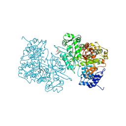
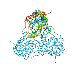
1PF9
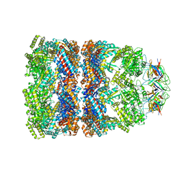 | | GroEL-GroES-ADP | | 分子名称: | ADENOSINE-5'-DIPHOSPHATE, MAGNESIUM ION, groEL protein, ... | | 著者 | Chaudhry, C, Farr, G.W, Todd, M.J, Rye, H.S, Brunger, A.T, Adams, P.D, Horwich, A.L, Sigler, P.B. | | 登録日 | 2003-05-24 | | 公開日 | 2003-11-04 | | 最終更新日 | 2024-02-14 | | 実験手法 | X-RAY DIFFRACTION (2.993 Å) | | 主引用文献 | Role of the gamma-phosphate of ATP in triggering protein folding by GroEL-GroES: function, structure and energetics.
Embo J., 22, 2003
|
|
1PFB
 | | Structural Basis for specific binding of polycomb chromodomain to histone H3 methylated at K27 | | 分子名称: | ACETIC ACID, BETA-MERCAPTOETHANOL, CHLORIDE ION, ... | | 著者 | Min, J.R, Zhang, Y, Xu, R.-M. | | 登録日 | 2003-05-24 | | 公開日 | 2003-10-07 | | 最終更新日 | 2017-10-11 | | 実験手法 | X-RAY DIFFRACTION (1.4 Å) | | 主引用文献 | Structural basis for specific binding of Polycomb chromodomain to histone H3 methylated at Lys 27.
Genes Dev., 17, 2003
|
|
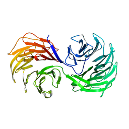
1PFC
 | | MOLECULAR-REPLACEMENT STRUCTURE OF GUINEA PIG IGG1 P*FC(PRIME) REFINED AT 3.1 ANGSTROMS RESOLUTION | | 分子名称: | IGG1 PFC' FC | | 著者 | Bryant, S.H, Amzel, L.M, Poljak, R.J, Phizackerley, R.P. | | 登録日 | 1981-10-28 | | 公開日 | 1982-02-03 | | 最終更新日 | 2024-06-05 | | 実験手法 | X-RAY DIFFRACTION (3.125 Å) | | 主引用文献 | Molecular-Replacement Structure of Guinea Pig Igg1 Pfc(Prime) Refined at 3.1 Angstroms Resolution
Acta Crystallogr.,Sect.B, 41, 1985
|
|
1PFD
 | | THE SOLUTION STRUCTURE OF HIGH PLANT PARSLEY [2FE-2S] FERREDOXIN, NMR, 18 STRUCTURES | | 分子名称: | FE2/S2 (INORGANIC) CLUSTER, FERREDOXIN | | 著者 | Im, S.-C, Liu, G, Luchinat, C, Sykes, A.G, Bertini, I. | | 登録日 | 1998-05-05 | | 公開日 | 1999-05-11 | | 最終更新日 | 2024-05-22 | | 実験手法 | SOLUTION NMR | | 主引用文献 | The solution structure of parsley [2Fe-2S]ferredoxin.
Eur.J.Biochem., 258, 1998
|
|
1PFE
 | | Echinomycin-(gcgtacgc)2 complex | | 分子名称: | 2-CARBOXYQUINOXALINE, 5'-D(*GP*CP*GP*TP*AP*CP*GP*C)-3', CHLORIDE ION, ... | | 著者 | Cuesta-Seijo, J.A. | | 登録日 | 2003-05-26 | | 公開日 | 2004-06-08 | | 最終更新日 | 2024-04-03 | | 実験手法 | X-RAY DIFFRACTION (1.1 Å) | | 主引用文献 | Structures of Complexes between Echinomycin and Duplex DNA.
Acta Crystallogr.,Sect.D, 61, 2005
|
|
1PFF
 | | Crystal Structure of Homocysteine alpha-, gamma-lyase at 1.8 Angstroms | | 分子名称: | 1,2-ETHANEDIOL, DI(HYDROXYETHYL)ETHER, methionine gamma-lyase | | 著者 | Allen, T.W, Sridhar, V, Prasad, S.G, Han, Q, Xu, M, Tan, Y, Hoffman, R.M, Ramaswamy, S. | | 登録日 | 2003-05-26 | | 公開日 | 2004-08-10 | | 最終更新日 | 2023-08-16 | | 実験手法 | X-RAY DIFFRACTION (2.5 Å) | | 主引用文献 |
|
|
1PFG
 | | Strategy to design inhibitors: Structure of a complex of Proteinase K with a designed octapeptide inhibitor N-Ac-Pro-Ala-Pro-Phe-DAla-Ala-Ala-Ala-NH2 at 2.5A resolution | | 分子名称: | N-Ac-PAPFAAAA-NH2, Proteinase K | | 著者 | Saxena, A.K, Singh, T.P, Peters, K, Fittkau, S, Betzel, C. | | 登録日 | 2003-05-27 | | 公開日 | 2003-06-10 | | 最終更新日 | 2023-10-25 | | 実験手法 | X-RAY DIFFRACTION (2.5 Å) | | 主引用文献 | Strategy to design peptide inhibitors: structure of a complex of proteinase K with a designed octapeptide inhibitor N-Ac-Pro-Ala-Pro-Phe-DAla-Ala-Ala-Ala-NH2 at 2.5 A resolution.
Protein Sci., 5, 1996
|
|
1PFH
 | |


