Loading
PDBj
MenuPDBj@FacebookPDBj@TwitterPDBj@YouTubewwPDB FoundationwwPDB
RCSB PDBPDBeBMRBAdv. SearchSearch help

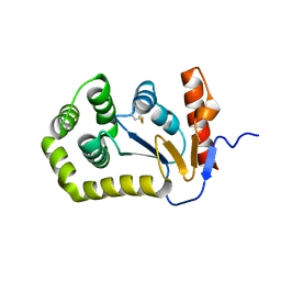
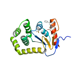
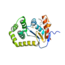
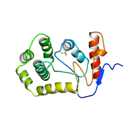
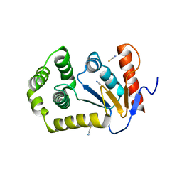
5QMN
DownloadVisualize
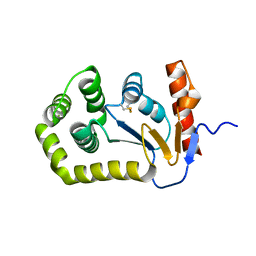
BU of 5qmn by Molmil
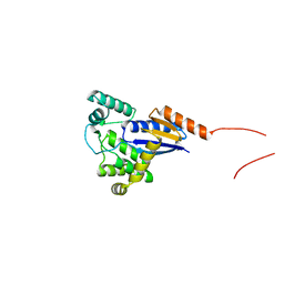
Group deposition of library data - Crystal Structure of EcDsbA after initial refinement with no ligand modelled (structure F5_1)
Descriptor: Thiol:disulfide interchange protein
Authors:Ilyichova, O.V, Bentley, M.R, Doak, B.C, Scanlon, M.J.
Deposit date:2019-01-27
Release date:2020-02-05
Last modified:2020-07-22
Method:X-RAY DIFFRACTION (1.976 Å)
Cite:Rapid Elaboration of Fragments into Leads by X-ray Crystallographic Screening of Parallel Chemical Libraries (REFiLX).
J.Med.Chem., 63, 2020
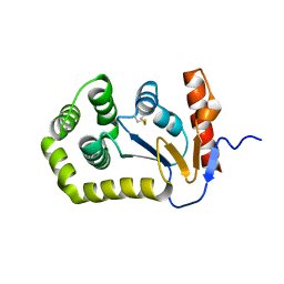
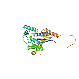
4OCF
DownloadVisualize
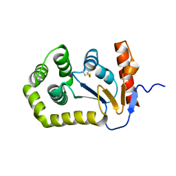
BU of 4ocf by Molmil
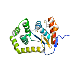
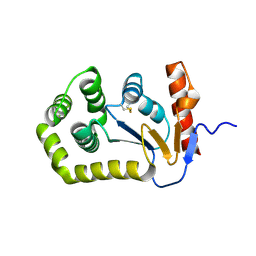
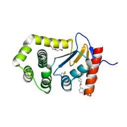
Crystal structure of the disulfide oxidoreductase DsbA (S30XXC33) active site mutant from Proteus mirabilis
Descriptor: THIOCYANATE ION, Thiol:disulfide interchange protein
Authors:Kurth, F, Martin, J.L.
Deposit date:2014-01-09
Release date:2014-05-28
Last modified:2023-09-20
Method:X-RAY DIFFRACTION (1.979 Å)
Cite:Crystal Structure of the Dithiol Oxidase DsbA Enzyme from Proteus Mirabilis Bound Non-covalently to an Active Site Peptide Ligand.
J.Biol.Chem., 289, 2014
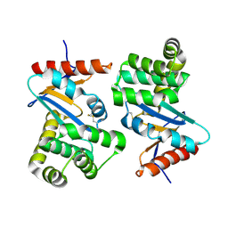
5QOA
DownloadVisualize
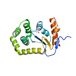
BU of 5qoa by Molmil
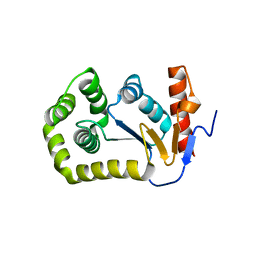
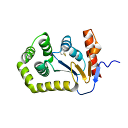
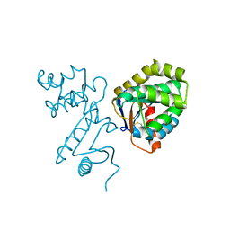
Group deposition of apo datasets for PANDDA analysis - Crystal Structure of apo EcDsbA after initial refinement (apo_dataset_40)
Descriptor: Thiol:disulfide interchange protein
Authors:Ilyichova, O.V, Bentley, M.R, Doak, B.C, Scanlon, M.J.
Deposit date:2019-01-27
Release date:2020-02-05
Last modified:2020-07-22
Method:X-RAY DIFFRACTION (1.979 Å)
Cite:Rapid Elaboration of Fragments into Leads by X-ray Crystallographic Screening of Parallel Chemical Libraries (REFiLX).
J.Med.Chem., 63, 2020
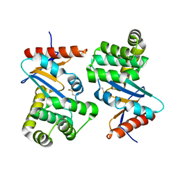
5QM3
DownloadVisualize
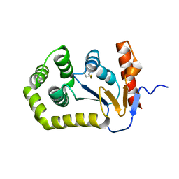
BU of 5qm3 by Molmil
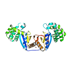
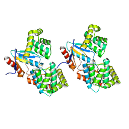
Group deposition of library data - Crystal Structure of EcDsbA after initial refinement with no ligand modelled (structure E1_1)
Descriptor: Thiol:disulfide interchange protein
Authors:Ilyichova, O.V, Bentley, M.R, Doak, B.C, Scanlon, M.J.
Deposit date:2019-01-27
Release date:2020-02-05
Last modified:2020-07-22
Method:X-RAY DIFFRACTION (1.982 Å)
Cite:Rapid Elaboration of Fragments into Leads by X-ray Crystallographic Screening of Parallel Chemical Libraries (REFiLX).
J.Med.Chem., 63, 2020
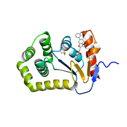
6XT3
DownloadVisualize
BU of 6xt3 by Molmil
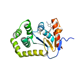
Crystal structure of E.coli DsbA in complex with 3-(3-(carboxymethyl)-6-(3-methoxyphenyl)benzofuran-2-yl)benzoic acid
Descriptor: 3-[3-(carboxymethyl)-6-(3-methoxyphenyl)-1-benzofuran-2-yl]benzoic acid, COPPER (II) ION, Thiol:disulfide interchange protein DsbA
Authors:Wang, G, Heras, B.
Deposit date:2020-07-17
Release date:2021-08-11
Last modified:2023-10-18
Method:X-RAY DIFFRACTION (1.99 Å)
Cite:Elaboration of a benzofuran scaffold and evaluation of binding affinity and inhibition of Escherichia coli DsbA: A fragment-based drug design approach to novel antivirulence compounds.
Bioorg.Med.Chem., 45, 2021
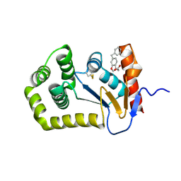
6PVZ
DownloadVisualize
BU of 6pvz by Molmil
E.coli DsbA in complex with benzofuran compound 28 ((6-benzyl-1-benzofuran-3-yl)acetic acid)
Descriptor: (6-benzyl-1-benzofuran-3-yl)acetic acid, COPPER (II) ION, Thiol:disulfide interchange protein DsbA
Authors:Wang, G, Heras, B.
Deposit date:2019-07-21
Release date:2019-12-25
Last modified:2020-10-07
Method:X-RAY DIFFRACTION (1.99 Å)
Cite:The Fragment-Based Development of a Benzofuran Hit as a New Class of Escherichia coli DsbA Inhibitors.
Molecules, 24, 2019
7TTV
DownloadVisualize
BU of 7ttv by Molmil
E.coli DsbA in complex with 4-phenyl-2-(3-phenylpropyl)thiazole-5-carboxylic acid
Descriptor: 4-phenyl-2-(3-phenylpropyl)-1,3-thiazole-5-carboxylic acid, COPPER (II) ION, Thiol:disulfide interchange protein DsbA
Authors:Wang, G, Heras, B.
Deposit date:2022-02-02
Release date:2023-04-12
Last modified:2023-10-25
Method:X-RAY DIFFRACTION (1.99 Å)
Cite:Methyl probes in proteins for determining ligand binding mode in weak protein-ligand complexes.
Sci Rep, 12, 2022
6BR4
DownloadVisualize
BU of 6br4 by Molmil
Crystal structure of Escherichia coli DsbA in complex with {N}-methyl-1-(3-thiophen-2-ylphenyl)methanamine
Descriptor: COPPER (II) ION, Thiol:disulfide interchange protein DsbA, ~{N}-methyl-1-(3-thiophen-2-ylphenyl)methanamine
Authors:Heras, B, Totsika, M, Paxman, J.J, Wang, G, Scanlon, M.J, Martin, J.L.
Deposit date:2017-11-29
Release date:2017-12-27
Last modified:2020-01-01
Method:X-RAY DIFFRACTION (1.99 Å)
Cite:Inhibition of Diverse DsbA Enzymes in Multi-DsbA Encoding Pathogens.
Antioxid. Redox Signal., 29, 2018
6WHD
DownloadVisualize
BU of 6whd by Molmil
Crystal structure of E.coli DsbA in complex with diaryl ether analogue 2
Descriptor: COPPER (II) ION, Thiol:disulfide interchange protein DsbA, [4-(4-cyano-3-methylphenoxy)phenyl]acetic acid
Authors:Wang, G, Heras, B.
Deposit date:2020-04-08
Release date:2020-06-24
Last modified:2023-10-18
Method:X-RAY DIFFRACTION (1.99 Å)
Cite:Rapid Elaboration of Fragments into Leads by X-ray Crystallographic Screening of Parallel Chemical Libraries (REFiLX).
J.Med.Chem., 63, 2020
4MCU
DownloadVisualize
BU of 4mcu by Molmil
Crystal structure of disulfide oxidoreductase from Klebsiella pneumoniae in reduced state
Descriptor: Thiol:disulfide interchange protein
Authors:Kurth, F, Premkumar, L, Martin, J.L.
Deposit date:2013-08-21
Release date:2013-11-27
Last modified:2023-09-20
Method:X-RAY DIFFRACTION (1.99 Å)
Cite:Comparative Sequence, Structure and Redox Analyses of Klebsiella pneumoniae DsbA Show That Anti-Virulence Target DsbA Enzymes Fall into Distinct Classes.
Plos One, 8, 2013
6BQX
DownloadVisualize
BU of 6bqx by Molmil
Crystal structure of Escherichia coli DsbA in complex with N-methyl-1-(4-phenoxyphenyl)methanamine
Descriptor: N-methyl-1-(4-phenoxyphenyl)methanamine, Thiol:disulfide interchange protein DsbA
Authors:Heras, B, Totsika, M, Paxman, J.J, Wang, G, Scanlon, M.J.
Deposit date:2017-11-29
Release date:2017-12-27
Last modified:2020-01-01
Method:X-RAY DIFFRACTION (1.992 Å)
Cite:Inhibition of Diverse DsbA Enzymes in Multi-DsbA Encoding Pathogens.
Antioxid. Redox Signal., 29, 2018
5QLK
DownloadVisualize
BU of 5qlk by Molmil
Group deposition of library data - Crystal Structure of EcDsbA after initial refinement with no ligand modelled (structure C7_1)
Descriptor: Thiol:disulfide interchange protein
Authors:Ilyichova, O.V, Bentley, M.R, Doak, B.C, Scanlon, M.J.
Deposit date:2019-01-27
Release date:2020-02-05
Last modified:2020-07-22
Method:X-RAY DIFFRACTION (1.993 Å)
Cite:Rapid Elaboration of Fragments into Leads by X-ray Crystallographic Screening of Parallel Chemical Libraries (REFiLX).
J.Med.Chem., 63, 2020
5QLZ
DownloadVisualize
BU of 5qlz by Molmil
Group deposition of library data - Crystal Structure of EcDsbA after initial refinement with no ligand modelled (structure D6_1)
Descriptor: Thiol:disulfide interchange protein
Authors:Ilyichova, O.V, Bentley, M.R, Doak, B.C, Scanlon, M.J.
Deposit date:2019-01-27
Release date:2020-02-05
Last modified:2020-07-22
Method:X-RAY DIFFRACTION (1.996 Å)
Cite:Rapid Elaboration of Fragments into Leads by X-ray Crystallographic Screening of Parallel Chemical Libraries (REFiLX).
J.Med.Chem., 63, 2020
5XUR
DownloadVisualize
BU of 5xur by Molmil
Crystal Structure of Rv2466c C22S Mutant
Descriptor: 1,2-ETHANEDIOL, CHLORIDE ION, Thioredoxin-like reductase Rv2466c
Authors:Zhang, X, Li, H.
Deposit date:2017-06-25
Release date:2018-03-14
Last modified:2023-11-22
Method:X-RAY DIFFRACTION (1.996 Å)
Cite:Identification of a Mycothiol-Dependent Nitroreductase from Mycobacterium tuberculosis.
ACS Infect Dis, 4, 2018
5XWH
DownloadVisualize
BU of 5xwh by Molmil
Structure of FrnE, a novel disulfide oxidoreductase from Deinococcus radiodurans crystallized in the presence of GSH
Descriptor: FrnE protein
Authors:Bihani, S.C, Panicker, L, Kumar, V.
Deposit date:2017-06-29
Release date:2018-07-04
Last modified:2023-11-22
Method:X-RAY DIFFRACTION (2 Å)
Cite:Structure of FrnE, a novel disulfide oxidoreductase from Deinococcus radiodurans crystallized in the presence of GSH
To Be Published
6PML
DownloadVisualize
BU of 6pml by Molmil
Crystal Structure of EcDsbA in complex benzyl ether 23
Descriptor: COPPER (II) ION, DI(HYDROXYETHYL)ETHER, Thiol:disulfide interchange protein DsbA, ...
Authors:Ilyichova, O.V, Scanlon, M.J.
Deposit date:2019-07-02
Release date:2019-11-06
Last modified:2023-10-11
Method:X-RAY DIFFRACTION (2 Å)
Cite:The Fragment-Based Development of a Benzofuran Hit as a New Class of Escherichia coli DsbA Inhibitors.
Molecules, 24, 2019
1BED
DownloadVisualize
BU of 1bed by Molmil
STRUCTURE OF DISULFIDE OXIDOREDUCTASE
Descriptor: DSBA OXIDOREDUCTASE
Authors:Hu, S.-H, Martin, J.L.
Deposit date:1996-09-16
Release date:1997-10-08
Last modified:2023-08-02
Method:X-RAY DIFFRACTION (2 Å)
Cite:Structure of TcpG, the DsbA protein folding catalyst from Vibrio cholerae.
J.Mol.Biol., 268, 1997
1U3A
DownloadVisualize
BU of 1u3a by Molmil
mutant DsbA
Descriptor: 3,6,9,12,15,18,21,24-OCTAOXAHEXACOSAN-1-OL, Thiol: disulfide interchange protein dsbA
Authors:Serre, L.
Deposit date:2004-07-21
Release date:2005-05-03
Last modified:2023-08-23
Method:X-RAY DIFFRACTION (2 Å)
Cite:Intriguing conformation changes associated with the trans/cis isomerization of a prolyl residue in the active site of the DsbA C33A mutant
J.Mol.Biol., 347, 2005
1A2J
DownloadVisualize
BU of 1a2j by Molmil
OXIDIZED DSBA CRYSTAL FORM II
Descriptor: DISULFIDE BOND FORMATION PROTEIN
Authors:Martin, J.L, Guddat, L.W.
Deposit date:1998-01-06
Release date:1998-09-16
Last modified:2023-08-02
Method:X-RAY DIFFRACTION (2 Å)
Cite:Crystal structures of reduced and oxidized DsbA: investigation of domain motion and thiolate stabilization.
Structure, 6, 1998
5E59
DownloadVisualize
BU of 5e59 by Molmil
Crystal structure of reduced state of a novel disulfide oxidoreductase from Deinococcus radiodurans
Descriptor: FrnE protein, GLYCEROL
Authors:Bihani, S.C, Panicker, L, Kumar, V.
Deposit date:2015-10-08
Release date:2016-10-12
Last modified:2023-11-08
Method:X-RAY DIFFRACTION (2 Å)
Cite:drFrnE Represents a Hitherto Unknown Class of Eubacterial Cytoplasmic Disulfide Oxido-Reductases.
Antioxid. Redox Signal., 28, 2018
1AC1
DownloadVisualize
BU of 1ac1 by Molmil
DSBA MUTANT H32L
Descriptor: DSBA
Authors:Guddat, L.W, Martin, J.L.
Deposit date:1997-02-10
Release date:1997-10-15
Last modified:2023-08-02
Method:X-RAY DIFFRACTION (2 Å)
Cite:Structural analysis of three His32 mutants of DsbA: support for an electrostatic role of His32 in DsbA stability.
Protein Sci., 6, 1997
1DSB
DownloadVisualize
BU of 1dsb by Molmil
CRYSTAL STRUCTURE OF THE DSBA PROTEIN REQUIRED FOR DISULPHIDE BOND FORMATION IN VIVO
Descriptor: DSBA
Authors:Martin, J.L, Bardwell, J.C.A, Kuriyan, J.
Deposit date:1993-05-24
Release date:1994-01-31
Last modified:2024-06-05
Method:X-RAY DIFFRACTION (2 Å)
Cite:Crystal structure of the DsbA protein required for disulphide bond formation in vivo.
Nature, 365, 1993
5QLF
DownloadVisualize
BU of 5qlf by Molmil
Group deposition of library data - Crystal Structure of EcDsbA after initial refinement with no ligand modelled (structure C3_2)
Descriptor: Thiol:disulfide interchange protein
Authors:Ilyichova, O.V, Bentley, M.R, Doak, B.C, Scanlon, M.J.
Deposit date:2019-01-27
Release date:2020-02-05
Last modified:2020-07-22
Method:X-RAY DIFFRACTION (2.003 Å)
Cite:Rapid Elaboration of Fragments into Leads by X-ray Crystallographic Screening of Parallel Chemical Libraries (REFiLX).
J.Med.Chem., 63, 2020
5QKO
DownloadVisualize
BU of 5qko by Molmil
Group deposition of library data - Crystal Structure of EcDsbA after initial refinement with no ligand modelled (structure A6_1)
Descriptor: Thiol:disulfide interchange protein
Authors:Ilyichova, O.V, Bentley, M.R, Doak, B.C, Scanlon, M.J.
Deposit date:2019-01-27
Release date:2020-02-05
Last modified:2020-07-22
Method:X-RAY DIFFRACTION (2.004 Å)
Cite:Rapid Elaboration of Fragments into Leads by X-ray Crystallographic Screening of Parallel Chemical Libraries (REFiLX).
J.Med.Chem., 63, 2020
5QMR
DownloadVisualize
BU of 5qmr by Molmil
Group deposition of library data - Crystal Structure of EcDsbA after initial refinement with no ligand modelled (structure F9_1)
Descriptor: Thiol:disulfide interchange protein
Authors:Ilyichova, O.V, Bentley, M.R, Doak, B.C, Scanlon, M.J.
Deposit date:2019-01-27
Release date:2020-02-05
Last modified:2020-07-22
Method:X-RAY DIFFRACTION (2.004 Å)
Cite:Rapid Elaboration of Fragments into Leads by X-ray Crystallographic Screening of Parallel Chemical Libraries (REFiLX).
J.Med.Chem., 63, 2020

221051

PDB entries from 2024-06-12

PDB statisticsPDBj update infoContact PDBjnumon