Loading
PDBj
MenuPDBj@FacebookPDBj@X(formerly Twitter)PDBj@BlueSkyPDBj@YouTubewwPDB FoundationwwPDB
RCSB PDBPDBeBMRBAdv. SearchSearch help

7DW8
DownloadVisualize
BU of 7dw8 by Molmil
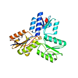
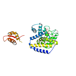
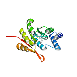
Structure of a novel beta-mannanase BaMan113A with mannobiose, N236Y mutation.
Descriptor: Endo-beta-1,4-mannanase, beta-D-mannopyranose-(1-4)-beta-D-mannopyranose
Authors:Liu, W.T, Liu, W.D, Zheng, Y.Y.
Deposit date:2021-01-15
Release date:2021-06-02
Last modified:2023-11-29
Method:X-RAY DIFFRACTION (1.9 Å)
Cite:Functional and structural investigation of a novel beta-mannanase BaMan113A from Bacillus sp. N16-5.
Int.J.Biol.Macromol., 182, 2021
3HRG
DownloadVisualize
BU of 3hrg by Molmil
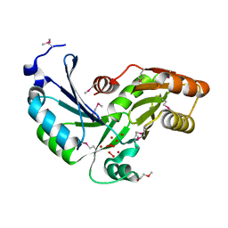
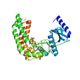
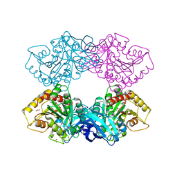
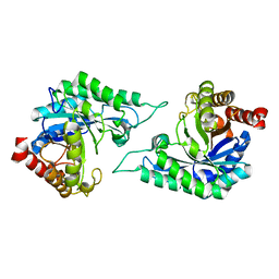
Crystal structure of Bacteroides thetaiotaomicron BT_3980, protein with actin-like ATPase fold and unknown function (NP_812891.1) from BACTEROIDES THETAIOTAOMICRON VPI-5482 at 1.85 A resolution
Descriptor: 1,2-ETHANEDIOL, DI(HYDROXYETHYL)ETHER, UNKNOWN LIGAND, ...
Authors:Joint Center for Structural Genomics (JCSG)
Deposit date:2009-06-09
Release date:2009-06-23
Last modified:2024-10-30
Method:X-RAY DIFFRACTION (1.85 Å)
Cite:Crystal structure of Bacteroides thetaiotaomicron BT_3980, protein with actin-like ATPase fold and unknown function (NP_812891.1) from BACTEROIDES THETAIOTAOMICRON VPI-5482 at 1.85 A resolution
To be Published
3HM4
DownloadVisualize
BU of 3hm4 by Molmil
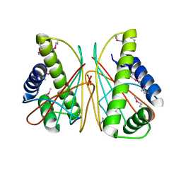
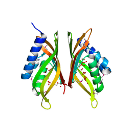
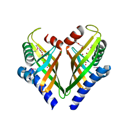
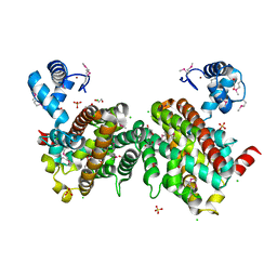
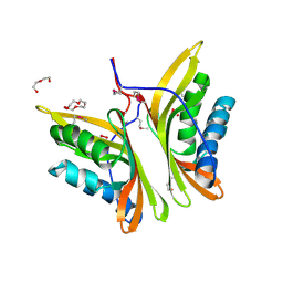
CRYSTAL STRUCTURE OF A CHEMOTAXIS PROTEIN CHEX (DDE_0281) FROM DESULFOVIBRIO DESULFURICANS SUBSP. AT 1.30 A RESOLUTION
Descriptor: Chemotaxis protein CheX, GLYCEROL, UNKNOWN LIGAND
Authors:Joint Center for Structural Genomics (JCSG)
Deposit date:2009-05-28
Release date:2009-06-23
Last modified:2023-02-01
Method:X-RAY DIFFRACTION (1.3 Å)
Cite:Crystal structure of Chemotaxis protein CheX (YP_386777.1) from DESULFOVIBRIO DESULFURICANS G20 at 1.30 A resolution
To be published
7LD3
DownloadVisualize
BU of 7ld3 by Molmil
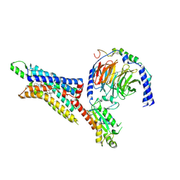
Cryo-EM structure of the human adenosine A1 receptor-Gi2-protein complex bound to its endogenous agonist and an allosteric ligand
Descriptor: ADENOSINE, Chimera protein of Muscarinic acetylcholine receptor M4 and Adenosine receptor A1, Guanine nucleotide-binding protein G(I)/G(S)/G(O) subunit gamma-2, ...
Authors:Draper-Joyce, C.J, Danev, R, Thal, D.M, Christopoulos, A, Glukhova, A.
Deposit date:2021-01-12
Release date:2021-09-08
Last modified:2024-11-13
Method:ELECTRON MICROSCOPY (3.2 Å)
Cite:Positive allosteric mechanisms of adenosine A 1 receptor-mediated analgesia.
Nature, 597, 2021
2OEJ
DownloadVisualize
BU of 2oej by Molmil
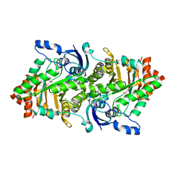
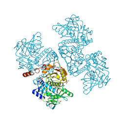
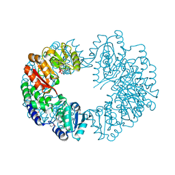
Crystal structure of a rubisco-like protein from Geobacillus kaustophilus (tetramutant form), liganded with phosphate ions
Descriptor: 2,3-diketo-5-methylthiopentyl-1-phosphate enolase, PHOSPHATE ION
Authors:Fedorov, A.A, Imker, H.J, Fedorov, E.V, Almo, S.C, Gerlt, J.A.
Deposit date:2006-12-30
Release date:2007-03-20
Last modified:2023-12-27
Method:X-RAY DIFFRACTION (2.55 Å)
Cite:Mechanistic Diversity in the RuBisCO Superfamily: The "Enolase" in the Methionine Salvage Pathway in Geobacillus kaustophilus.
Biochemistry, 46, 2007
4H57
DownloadVisualize
BU of 4h57 by Molmil
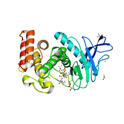
Thermolysin inhibition
Descriptor: CALCIUM ION, DIMETHYL SULFOXIDE, GLYCEROL, ...
Authors:Englert, L, Biela, A, Heine, A, Klebe, G.
Deposit date:2012-09-18
Release date:2012-10-03
Last modified:2023-09-20
Method:X-RAY DIFFRACTION (1.56 Å)
Cite:Dissecting the hydrophobic effect on the molecular level: the role of water, enthalpy, and entropy in ligand binding to thermolysin.
Angew.Chem.Int.Ed.Engl., 52, 2013
3HX8
DownloadVisualize
BU of 3hx8 by Molmil
Crystal structure of putative ketosteroid isomerase (NP_103587.1) from Mesorhizobium loti at 1.45 A resolution
Descriptor: IMIDAZOLE, TETRAETHYLENE GLYCOL, UNKNOWN LIGAND, ...
Authors:Joint Center for Structural Genomics (JCSG)
Deposit date:2009-06-19
Release date:2009-07-07
Last modified:2024-10-16
Method:X-RAY DIFFRACTION (1.45 Å)
Cite:Crystal structure of putative ketosteroid isomerase (NP_103587.1) from MESORHIZOBIUM LOTI at 1.45 A resolution
To be published
4D5G
DownloadVisualize
BU of 4d5g by Molmil
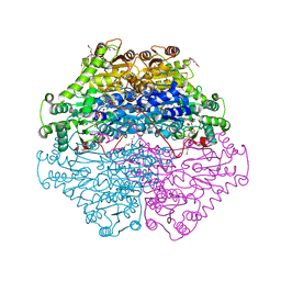
Structure of recombinant CDH-H28AN484A
Descriptor: 2-(2-{2-[2-(2-METHOXY-ETHOXY)-ETHOXY]-ETHOXY}-ETHOXY)-ETHANOL, CYCLOHEXANE-1,2-DIONE HYDROLASE, DI(HYDROXYETHYL)ETHER, ...
Authors:Loschonsky, S, Wacker, T, Waltzer, S, Giovannini, P.P, McLeish, M.J, Andrade, S.L.A, Mueller, M.
Deposit date:2014-11-04
Release date:2015-01-21
Last modified:2024-05-08
Method:X-RAY DIFFRACTION (2 Å)
Cite:Extended Reaction Scope of Thiamine Diphosphate Dependent Cyclohexane-1,2-Dione Hydrolase: From C-C Bond Cleavage to C-C Bond Ligation.
Angew.Chem.Int.Ed.Engl., 53, 2014
4CVD
DownloadVisualize
BU of 4cvd by Molmil
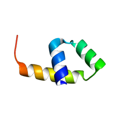
Crystal structure of the central repeat of cell wall binding module of Cpl7
Descriptor: LYSOZYME
Authors:Silva-Martin, N, Uson, I, Rodriguez, D.D, Hermoso, J.A.
Deposit date:2014-03-25
Release date:2015-04-08
Last modified:2024-05-08
Method:X-RAY DIFFRACTION (1.666 Å)
Cite:Deciphering how Cpl-7 cell wall-binding repeats recognize the bacterial peptidoglycan.
Sci Rep, 7, 2017
4AR7
DownloadVisualize
BU of 4ar7 by Molmil
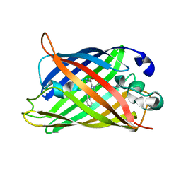
X-ray structure of the cyan fluorescent protein mTurquoise
Descriptor: GREEN FLUORESCENT PROTEIN
Authors:von Stetten, D, Noirclerc-Savoye, M, Goedhart, J, Gadella, T.W.J, Royant, A.
Deposit date:2012-04-21
Release date:2012-08-08
Last modified:2024-10-23
Method:X-RAY DIFFRACTION (1.23 Å)
Cite:Structure of a Fluorescent Protein from Aequorea Victoria Bearing the Obligate-Monomer Mutation A206K.
Acta Crystallogr.,Sect.F, 68, 2012
3KL7
DownloadVisualize
BU of 3kl7 by Molmil
Crystal structure of Putative metal-dependent hydrolase (YP_001302908.1) from Parabacteroides distasonis ATCC 8503 at 2.30 A resolution
Descriptor: ACETATE ION, GLYCEROL, Putative metal-dependent hydrolase, ...
Authors:Joint Center for Structural Genomics (JCSG)
Deposit date:2009-11-06
Release date:2010-02-02
Last modified:2019-07-17
Method:X-RAY DIFFRACTION (2.3 Å)
Cite:Crystal structure of Putative metal-dependent hydrolase (YP_001302908.1) from Parabacteroides distasonis ATCC 8503 at 2.30 A resolution
To be published
4NW4
DownloadVisualize
BU of 4nw4 by Molmil
Crystal structure of a DUF4822 family protein (EF0375) from Enterococcus faecalis V583 at 1.85 A resolution
Descriptor: CHLORIDE ION, GLYCEROL, Lipoprotein S-layer protein, ...
Authors:Joint Center for Structural Genomics (JCSG)
Deposit date:2013-12-05
Release date:2014-02-19
Last modified:2024-11-06
Method:X-RAY DIFFRACTION (1.85 Å)
Cite:Crystal structure of a hypothetical protein (EF0375) from Enterococcus faecalis V583 at 1.85 A resolution
To be published
1HCZ
DownloadVisualize
BU of 1hcz by Molmil
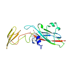
LUMEN-SIDE DOMAIN OF REDUCED CYTOCHROME F AT-35 DEGREES CELSIUS
Descriptor: CYTOCHROME F, PROTOPORPHYRIN IX CONTAINING FE
Authors:Martinez, S.E, Huang, D, Szczepaniak, A, Cramer, W.A, Smith, J.L.
Deposit date:1996-09-18
Release date:1997-03-12
Last modified:2024-10-30
Method:X-RAY DIFFRACTION (1.96 Å)
Cite:The heme redox center of chloroplast cytochrome f is linked to a buried five-water chain.
Protein Sci., 5, 1996
3HOI
DownloadVisualize
BU of 3hoi by Molmil
Crystal structure of FMN-dependent nitroreductase BF3017 from Bacteroides fragilis NCTC 9343 (YP_212631.1) from Bacteroides fragilis NCTC 9343 at 1.55 A resolution
Descriptor: CITRATE ANION, DI(HYDROXYETHYL)ETHER, FLAVIN MONONUCLEOTIDE, ...
Authors:Joint Center for Structural Genomics (JCSG)
Deposit date:2009-06-02
Release date:2009-06-23
Last modified:2024-10-16
Method:X-RAY DIFFRACTION (1.55 Å)
Cite:Crystal structure of FMN-dependent nitroreductase BF3017 from Bacteroides fragilis NCTC 9343 (YP_212631.1) from Bacteroides fragilis NCTC 9343 at 1.55 A resolution
To be published
4EB3
DownloadVisualize
BU of 4eb3 by Molmil
Crystal structure of IspH in complex with iso-HMBPP
Descriptor: 3-(hydroxymethyl)but-3-en-1-yl trihydrogen diphosphate, 4-hydroxy-3-methylbut-2-enyl diphosphate reductase, IRON/SULFUR CLUSTER
Authors:Wang, W, Wang, K, Span, I, Bacher, A, Groll, M, Oldfield, E.
Deposit date:2012-03-23
Release date:2013-02-06
Last modified:2023-09-13
Method:X-RAY DIFFRACTION (1.9 Å)
Cite:Are free radicals involved in IspH catalysis? An EPR and crystallographic investigation.
J.Am.Chem.Soc., 134, 2012
3I0Y
DownloadVisualize
BU of 3i0y by Molmil
Crystal structure of a putative polyketide cyclase (xcc0381) from xanthomonas campestris pv. campestris at 1.50 A resolution
Descriptor: CHLORIDE ION, DI(HYDROXYETHYL)ETHER, Putative polyketide cyclase, ...
Authors:Joint Center for Structural Genomics (JCSG)
Deposit date:2009-06-25
Release date:2009-07-21
Last modified:2024-11-06
Method:X-RAY DIFFRACTION (1.5 Å)
Cite:Crystal structure of putative polyketide cyclase (NP_635776.1) from XANTHOMONAS CAMPESTRIS at 1.50 A resolution
To be published
6DA9
DownloadVisualize
BU of 6da9 by Molmil
Crystal structure of the TtnD decarboxylase from the tautomycetin biosynthesis pathway of Streptomyces griseochromogenes with FMN bound at 2.05 A resolution
Descriptor: FLAVIN MONONUCLEOTIDE, GLYCEROL, MANGANESE (II) ION, ...
Authors:Han, L, Rudolf, J.D, Chang, C.-Y, Miller, M.D, Soman, J, Xu, W, Phillips Jr, G.N, Shen, B, Enzyme Discovery for Natural Product Biosynthesis (NatPro)
Deposit date:2018-05-01
Release date:2018-10-03
Last modified:2023-10-04
Method:X-RAY DIFFRACTION (2.05 Å)
Cite:Biochemical and Structural Characterization of TtnD, a Prenylated FMN-Dependent Decarboxylase from the Tautomycetin Biosynthetic Pathway.
ACS Chem. Biol., 13, 2018
2Q83
DownloadVisualize
BU of 2q83 by Molmil
Crystal structure of ytaA (2635576) from Bacillus subtilis at 2.50 A resolution
Descriptor: 1,2-ETHANEDIOL, ADENOSINE, CITRIC ACID, ...
Authors:Joint Center for Structural Genomics (JCSG)
Deposit date:2007-06-08
Release date:2007-06-26
Last modified:2024-10-16
Method:X-RAY DIFFRACTION (2.5 Å)
Cite:Genomics, evolution, and crystal structure of a new family of bacterial spore kinases.
Proteins, 78, 2010
4RGL
DownloadVisualize
BU of 4rgl by Molmil
Crystal structure of a Fic family protein (Dde_2494) from Desulfovibrio desulfuricans G20 at 2.70 A resolution
Descriptor: Filamentation induced by cAMP protein Fic, UNKNOWN LIGAND
Authors:Joint Center for Structural Genomics (JCSG)
Deposit date:2014-09-30
Release date:2014-10-29
Last modified:2024-10-30
Method:X-RAY DIFFRACTION (2.7 Å)
Cite:Crystal structure of a Fic family protein (Dde_2494) from Desulfovibrio desulfuricans G20 at 2.70 A resolution
To be published
3L2N
DownloadVisualize
BU of 3l2n by Molmil
Crystal structure of Putative carboxypeptidase A (YP_562911.1) from SHEWANELLA DENITRIFICANS OS-217 at 2.39 A resolution
Descriptor: ACETATE ION, CALCIUM ION, GLYCEROL, ...
Authors:Joint Center for Structural Genomics (JCSG)
Deposit date:2009-12-15
Release date:2009-12-29
Last modified:2023-02-01
Method:X-RAY DIFFRACTION (2.39 Å)
Cite:Crystal structure of Putative carboxypeptidase A (YP_562911.1) from SHEWANELLA DENITRIFICANS OS-217 at 2.39 A resolution
To be published
4LQX
DownloadVisualize
BU of 4lqx by Molmil
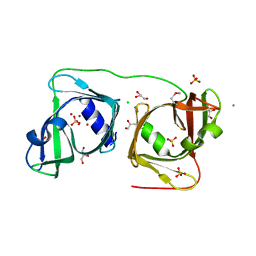
Crystal structure of a TENA/THI-4 domain-containing protein (SSO2700) from Sulfolobus solfataricus P2 at 2.34 A resolution
Descriptor: 1,2-ETHANEDIOL, CHLORIDE ION, SULFATE ION, ...
Authors:Joint Center for Structural Genomics (JCSG)
Deposit date:2013-07-19
Release date:2013-09-04
Last modified:2024-10-16
Method:X-RAY DIFFRACTION (2.34 Å)
Cite:Crystal structure of a TENA/THI-4 domain-containing protein (SSO2700) from Sulfolobus solfataricus P2 at 2.34 A resolution
To be published
2PY6
DownloadVisualize
BU of 2py6 by Molmil
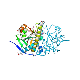
Crystal structure of Methyltransferase FkbM (YP_546752.1) from Methylobacillus flagellatus KT at 2.20 A resolution
Descriptor: 1,2-ETHANEDIOL, CHLORIDE ION, DI(HYDROXYETHYL)ETHER, ...
Authors:Joint Center for Structural Genomics (JCSG)
Deposit date:2007-05-15
Release date:2007-05-29
Last modified:2024-10-30
Method:X-RAY DIFFRACTION (2.15 Å)
Cite:Crystal structure of Methyltransferase FkbM (YP_546752.1) from Methylobacillus flagellatus KT at 2.20 A resolution
To be published
5K09
DownloadVisualize
BU of 5k09 by Molmil
Crystal Structure of COMT in complex with a thiazole ligand
Descriptor: 5-{3-[(4-methoxyphenyl)methyl]-1H-pyrazol-5-yl}-2,4-dimethyl-1,3-thiazole, Catechol O-methyltransferase, PHOSPHATE ION, ...
Authors:Ehler, A, Rodriguez-Sarmiento, R.M, Rudolph, M.G.
Deposit date:2016-05-17
Release date:2016-09-07
Last modified:2024-05-08
Method:X-RAY DIFFRACTION (2.7 Å)
Cite:Design of Potent and Druglike Nonphenolic Inhibitors for Catechol O-Methyltransferase Derived from a Fragment Screening Approach Targeting the S-Adenosyl-l-methionine Pocket.
J. Med. Chem., 59, 2016
3H51
DownloadVisualize
BU of 3h51 by Molmil
Crystal structure of Putative calcium/calmodulin dependent protein kinase II association domain (NP_636218.1) from XANTHOMONAS CAMPESTRIS at 1.70 A resolution
Descriptor: DI(HYDROXYETHYL)ETHER, Putative calcium/calmodulin dependent protein kinase II association domain, TETRAETHYLENE GLYCOL, ...
Authors:Joint Center for Structural Genomics (JCSG)
Deposit date:2009-04-21
Release date:2009-05-05
Last modified:2023-02-01
Method:X-RAY DIFFRACTION (1.7 Å)
Cite:Crystal structure of Putative calcium/calmodulin dependent protein kinase II association domain (NP_636218.1) from XANTHOMONAS CAMPESTRIS at 1.70 A resolution
To be published
3GPT
DownloadVisualize
BU of 3gpt by Molmil
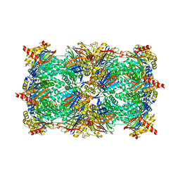
Crystal structure of the yeast 20S proteasome in complex with Salinosporamide derivatives: slow substrate ligand
Descriptor: (2R,3S,4R)-2-[(S)-(1S)-cyclohex-2-en-1-yl(hydroxy)methyl]-4-(2-fluoroethyl)-3-hydroxy-3-methyl-5-oxopyrrolidine-2-carbaldehyde, Proteasome component C1, Proteasome component C11, ...
Authors:Groll, M, Macherla, V.R, Manam, R.R, Arthur, K.A.M, Potts, C.B.
Deposit date:2009-03-23
Release date:2009-09-15
Last modified:2024-11-06
Method:X-RAY DIFFRACTION (2.41 Å)
Cite:Snapshots of the fluorosalinosporamide/20S complex offer mechanistic insights for fine tuning proteasome inhibition
J.Med.Chem., 52, 2009

227344

PDB entries from 2024-11-13

PDB statisticsPDBj update infoContact PDBjnumon