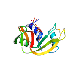
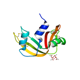
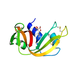
Loading
PDBj
MenuPDBj@FacebookPDBj@TwitterPDBj@YouTubewwPDB FoundationwwPDB
RCSB PDBPDBeBMRBAdv. SearchSearch help

2P45
DownloadVisualize
BU of 2p45 by Molmil
Complex of a camelid single-domain vhh antibody fragment with RNASE A at 1.1A resolution: SE5B-ORTHO-1 crystal form with five se-met sites (L4M, M34, M51, F68M, M83) in vhh scaffold.
Descriptor: ANTIBODY CAB-RN05, Ribonuclease pancreatic, SULFATE ION
Authors:Tereshko, V, Uysal, S, Koide, A, Margalef, K, Koide, S, Kossiakoff, A.A.
Deposit date:2007-03-11
Release date:2008-03-11
Last modified:2011-07-13
Method:X-RAY DIFFRACTION (1.1 Å)
Cite:Toward chaperone-assisted crystallography: protein engineering enhancement of crystal packing and X-ray phasing capabilities of a camelid single-domain antibody (VHH) scaffold
Protein Sci., 17, 2008
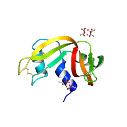
1U1B
DownloadVisualize
BU of 1u1b by Molmil
Structure of bovine pancreatic Ribonuclease A in complex with 3'-phosphothymidine (3'-5')-pyrophosphate adenosine 3'-phosphate
Descriptor: 5'-PHOSPHOTHYMIDINE (3'-5')-PYROPHOSPHATE ADENOSINE 3'-PHOSPHATE, Ribonuclease, pancreatic
Authors:Beach, H, Cole, R, Gill, M.L, Loria, J.P.
Deposit date:2004-07-15
Release date:2005-06-28
Last modified:2023-08-23
Method:X-RAY DIFFRACTION (2 Å)
Cite:Conservation of mus-ms enzyme motions in the apo- and substrate-mimicked state.
J.Am.Chem.Soc., 127, 2005
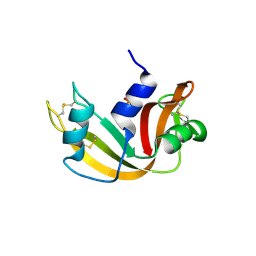
1SSC
DownloadVisualize
BU of 1ssc by Molmil
THE 1.6 ANGSTROMS STRUCTURE OF A SEMISYNTHETIC RIBONUCLEASE CRYSTALLIZED FROM AQUEOUS ETHANOL. COMPARISON WITH CRYSTALS FROM SALT SOLUTIONS AND WITH RNASE A FROM AQUEOUS ALCOHOL SOLUTIONS
Descriptor: PHOSPHATE ION, RIBONUCLEASE A
Authors:De Mel, V.S.J, Doscher, M.S, Martin, P.D, Rodier, F, Edwards, B.F.P.
Deposit date:1994-10-05
Release date:1995-01-26
Last modified:2011-07-13
Method:X-RAY DIFFRACTION (2 Å)
Cite:1.6 A structure of semisynthetic ribonuclease crystallized from aqueous ethanol. Comparison with crystals from salt solutions and with ribonuclease A from aqueous alcohol solutions.
Acta Crystallogr.,Sect.D, 51, 1995
7QPZ
DownloadVisualize
BU of 7qpz by Molmil
X-ray structure of the adduct obtained upon reaction of [cis-Rh2(OCOCH3)2(OCOCF3)2] with RNase A (4)
Descriptor: Ribonuclease pancreatic, cis-bis(mi2-acetato-O, O')-(mi2-trifluoroacetato-O, ...
Authors:Loreto, D, Merlino, A.
Deposit date:2022-01-05
Release date:2022-03-02
Last modified:2024-01-31
Method:X-RAY DIFFRACTION (1.45 Å)
Cite:Reactivity of a fluorine-containing dirhodium tetracarboxylate compound with proteins.
Dalton Trans, 51, 2022
7QPW
DownloadVisualize
BU of 7qpw by Molmil
X-ray structure of the adduct obtained upon reaction of [cis-Rh2(OCOCH3)2(OCOCF3)2] with RNase A (1)
Descriptor: (mi2-acetato-O, O')-hexaaquo-dirhodium (II), Ribonuclease pancreatic, ...
Authors:Loreto, D, Merlino, A.
Deposit date:2022-01-05
Release date:2022-03-16
Last modified:2024-01-31
Method:X-RAY DIFFRACTION (1.15 Å)
Cite:Reactivity of a fluorine-containing dirhodium tetracarboxylate compound with proteins.
Dalton Trans, 51, 2022
7QPY
DownloadVisualize
BU of 7qpy by Molmil
X-ray structure of the adduct obtained upon reaction of [cis-Rh2(OCOCH3)2(OCOCF3)2] with RNase A (3)
Descriptor: (mi2-acetato-O, O')-hexaaquo-dirhodium (II), Ribonuclease pancreatic, ...
Authors:Loreto, D, Merlino, A.
Deposit date:2022-01-05
Release date:2022-03-16
Last modified:2024-05-01
Method:X-RAY DIFFRACTION (1.42 Å)
Cite:Reactivity of a fluorine-containing dirhodium tetracarboxylate compound with proteins.
Dalton Trans, 51, 2022
7QQ0
DownloadVisualize
BU of 7qq0 by Molmil
X-ray structure of the adduct obtained upon reaction of [cis-Rh2(OCOCH3)2(OCOCF3)2] with RNase A (2)
Descriptor: (mi2-acetato-O, O')-hexaaquo-dirhodium (II), RHODIUM(II) ION, ...
Authors:Loreto, D, Merlino, A.
Deposit date:2022-01-05
Release date:2022-12-14
Last modified:2024-01-31
Method:X-RAY DIFFRACTION (1.32 Å)
Cite:Reactivity of a fluorine-containing dirhodium tetracarboxylate compound with proteins.
Dalton Trans, 51, 2022
4WYZ
DownloadVisualize
BU of 4wyz by Molmil
The crystal structure of the A109G mutant of RNase A in complex with 3'UMP
Descriptor: 3'-URIDINEMONOPHOSPHATE, Ribonuclease pancreatic
Authors:French, R.L, Gagne, D, Doucet, N, Simonovic, M.
Deposit date:2014-11-18
Release date:2015-11-18
Last modified:2019-12-25
Method:X-RAY DIFFRACTION (1.449 Å)
Cite:Perturbation of the Conformational Dynamics of an Active-Site Loop Alters Enzyme Activity.
Structure, 23, 2015
6XVX
DownloadVisualize
BU of 6xvx by Molmil
X-ray structure obtained upon reaction of dirhodium tetraacetate with RNase A (high resolution)
Descriptor: ACETATE ION, RHODIUM(III) ION, Ribonuclease pancreatic
Authors:Merlino, A, Ferraro, G.
Deposit date:2020-01-22
Release date:2020-02-19
Last modified:2024-01-24
Method:X-RAY DIFFRACTION (1.4 Å)
Cite:Protein interactions of dirhodium tetraacetate: a structural study.
Dalton Trans, 49, 2020
6XW0
DownloadVisualize
BU of 6xw0 by Molmil
X-ray structure obtained upon reaction of dirhodium tetraacetate with RNase A (low resolution)
Descriptor: ACETATE ION, RHODIUM(III) ION, Ribonuclease pancreatic
Authors:Merlino, A, Ferraro, G.
Deposit date:2020-01-22
Release date:2020-02-19
Last modified:2024-01-24
Method:X-RAY DIFFRACTION (1.8 Å)
Cite:Protein interactions of dirhodium tetraacetate: a structural study.
Dalton Trans, 49, 2020
1O0M
DownloadVisualize
BU of 1o0m by Molmil
Ribonuclease A in complex with uridine-2'-phosphate
Descriptor: PHOSPHORIC ACID MONO-[2-(2,4-DIOXO-3,4-DIHYDRO-2H-PYRIMIDIN-1-YL)-4-HYDROXY-5-HYDROXYMETHYL-TETRAHYDRO-FURAN-3-YL] ESTER, Ribonuclease pancreatic
Authors:Leonidas, D.D, Oikonomakos, N.G, Chrysina, E.D, Kosmopoulou, M.N, Vlassi, M.
Deposit date:2003-02-24
Release date:2003-12-09
Last modified:2023-10-25
Method:X-RAY DIFFRACTION (1.5 Å)
Cite:High-resolution crystal structures of ribonuclease A complexed with adenylic and uridylic nucleotide inhibitors. Implications for structure-based design of ribonucleolytic inhibitors
PROTEIN SCI., 12, 2003
1O0N
DownloadVisualize
BU of 1o0n by Molmil
Ribonuclease A in complex with uridine-3'-phosphate
Descriptor: 3'-URIDINEMONOPHOSPHATE, Ribonuclease pancreatic
Authors:Leonidas, D.D, Oikonomakos, N.G, Chrysina, E.D, Kosmopoulou, M.N, Vlassi, M.
Deposit date:2003-02-24
Release date:2003-12-09
Last modified:2023-10-25
Method:X-RAY DIFFRACTION (1.5 Å)
Cite:High-resolution crystal structures of ribonuclease A complexed with adenylic and uridylic nucleotide inhibitors. Implications for structure-based design of ribonucleolytic inhibitors
PROTEIN SCI., 12, 2003
4WYN
DownloadVisualize
BU of 4wyn by Molmil
The crystal structure of the A109G mutant of RNase A
Descriptor: Ribonuclease pancreatic
Authors:French, R.L, Gagne, D, Doucet, N, Simonovic, M.
Deposit date:2014-11-17
Release date:2015-11-18
Last modified:2019-12-25
Method:X-RAY DIFFRACTION (1.805 Å)
Cite:Perturbation of the Conformational Dynamics of an Active-Site Loop Alters Enzyme Activity.
Structure, 23, 2015
1ROB
DownloadVisualize
BU of 1rob by Molmil
STRUCTURE OF THE CRYSTALLINE COMPLEX OF CYTIDYLIC ACID (2'-CMP) WITH RIBONUCLEASE AT 1.6 ANGSTROMS RESOLUTION
Descriptor: CYTIDINE-2'-MONOPHOSPHATE, RIBONUCLEASE A
Authors:Lisgarten, J.N, Palmer, R.A.
Deposit date:1993-08-23
Release date:1994-01-31
Last modified:2024-06-05
Method:X-RAY DIFFRACTION (1.6 Å)
Cite:Structure of the crystalline complex of cytidylic acid (2'-CMP) with ribonuclease at 1.6 A resolution. Conservation of solvent sites in RNase-A high-resolution structures.
Acta Crystallogr.,Sect.D, 49, 1993
4WYP
DownloadVisualize
BU of 4wyp by Molmil
The crystal structure of the A109G mutant of RNase A in complex with 5'AMP
Descriptor: ADENOSINE MONOPHOSPHATE, Ribonuclease pancreatic
Authors:French, R.L, Gagne, D, Doucet, N, Simonovic, M.
Deposit date:2014-11-17
Release date:2015-11-18
Last modified:2023-09-27
Method:X-RAY DIFFRACTION (1.502 Å)
Cite:Perturbation of the Conformational Dynamics of an Active-Site Loop Alters Enzyme Activity.
Structure, 23, 2015
1RCA
DownloadVisualize
BU of 1rca by Molmil
STRUCTURE OF THE CRYSTALLINE COMPLEX OF DEOXYCYTIDYLYL-3',5'-GUANOSINE (3',5'-DCPDG) CO-CRYSTALISED WITH RIBONUCLEASE AT 1.9 ANGSTROMS RESOLUTION. RETROBINDING IN PANCREATIC RNASEA IS INDEPENDENT OF MODE OF INHIBITOR INTROMISSION
Descriptor: 2'-DEOXYCYTIDINE-2'-DEOXYGUANOSINE-3',5'-MONOPHOSPHATE, PHOSPHATE ION, RIBONUCLEASE A
Authors:Lisgarten, J.N, Palmer, R.A.
Deposit date:1994-07-18
Release date:1995-09-15
Last modified:2024-06-05
Method:X-RAY DIFFRACTION (1.9 Å)
Cite:Structure of the crystalline complex of deoxycytidylyl-3',5'-guanosine (3',5'-dCpdG) cocrystallized with ribonuclease at 1.9 A resolution.
Acta Crystallogr.,Sect.D, 51, 1995
3D6O
DownloadVisualize
BU of 3d6o by Molmil
The RNase A- 5'-Deoxy-5'-N-(ethyl isonipecotatyl)uridine complex
Descriptor: 1-{5-deoxy-5-[4-(ethoxycarbonyl)piperidin-1-yl]-alpha-L-arabinofuranosyl}pyrimidine-2,4(1H,3H)-dione, Ribonuclease pancreatic
Authors:Leonidas, D.D, Zographos, S.E, Oikonomakos, N.G.
Deposit date:2008-05-20
Release date:2009-02-10
Last modified:2023-08-30
Method:X-RAY DIFFRACTION (1.58 Å)
Cite:Morpholino, piperidino, and pyrrolidino derivatives of pyrimidine nucleosides as inhibitors of ribonuclease A: synthesis, biochemical, and crystallographic evaluation.
J.Med.Chem., 52, 2009
3D6Q
DownloadVisualize
BU of 3d6q by Molmil
The RNase A- 5'-Deoxy-5'-N-piperidinouridine complex
Descriptor: 1-(5-deoxy-5-piperidin-1-yl-alpha-L-arabinofuranosyl)pyrimidine-2,4(1H,3H)-dione, CITRATE ANION, Ribonuclease pancreatic
Authors:Leonidas, D.D, Zographos, S.E, Oikonomakos, N.G.
Deposit date:2008-05-20
Release date:2009-02-10
Last modified:2023-08-30
Method:X-RAY DIFFRACTION (1.6 Å)
Cite:Morpholino, piperidino, and pyrrolidino derivatives of pyrimidine nucleosides as inhibitors of ribonuclease A: synthesis, biochemical, and crystallographic evaluation.
J.Med.Chem., 52, 2009
1FEV
DownloadVisualize
BU of 1fev by Molmil
CRYSTAL STRUCTURE OF THE ALA4AIB MUTATION IN RNASE S
Descriptor: S PEPTIDE, S PROTEIN, SULFATE ION
Authors:Ratnaparkhi, G.S, Varadarajan, R.
Deposit date:2000-07-23
Release date:2000-08-09
Last modified:2021-11-03
Method:X-RAY DIFFRACTION (2.25 Å)
Cite:Structural and thermodynamic consequences of introducing alpha-aminoisobutyric acid in the S peptide of ribonuclease S.
Protein Eng., 13, 2000
6ETL
DownloadVisualize
BU of 6etl by Molmil
Atomic resolution structure of RNase A (data collection 2)
Descriptor: ISOPROPYL ALCOHOL, Ribonuclease pancreatic
Authors:Caterino, M, Vergara, A, Merlino, A.
Deposit date:2017-10-27
Release date:2018-02-21
Method:X-RAY DIFFRACTION (0.85 Å)
Cite:Raman-markers of X-ray radiation damage of proteins.
Int. J. Biol. Macromol., 111, 2018
6ETM
DownloadVisualize
BU of 6etm by Molmil
Atomic resolution structure of RNase A (data collection 3)
Descriptor: ISOPROPYL ALCOHOL, Ribonuclease pancreatic
Authors:Caterino, M, Vergara, A, Merlino, A.
Deposit date:2017-10-27
Release date:2018-02-21
Method:X-RAY DIFFRACTION (0.92 Å)
Cite:Raman-markers of X-ray radiation damage of proteins.
Int. J. Biol. Macromol., 111, 2018
6ETR
DownloadVisualize
BU of 6etr by Molmil
Atomic resolution structure of RNase A (data collection 8)
Descriptor: ISOPROPYL ALCOHOL, Ribonuclease pancreatic
Authors:Caterino, M, Vergara, A, Merlino, A.
Deposit date:2017-10-27
Release date:2018-02-21
Last modified:2024-01-17
Method:X-RAY DIFFRACTION (1.17 Å)
Cite:Raman-markers of X-ray radiation damage of proteins.
Int. J. Biol. Macromol., 111, 2018
6ETP
DownloadVisualize
BU of 6etp by Molmil
Atomic resolution structure of RNase A (data collection 6)
Descriptor: ISOPROPYL ALCOHOL, Ribonuclease pancreatic
Authors:Caterino, M, Vergara, A, Merlino, A.
Deposit date:2017-10-27
Release date:2018-02-21
Last modified:2024-01-17
Method:X-RAY DIFFRACTION (1.02 Å)
Cite:Raman-markers of X-ray radiation damage of proteins.
Int. J. Biol. Macromol., 111, 2018
6ETK
DownloadVisualize
BU of 6etk by Molmil
Atomic resolution structure of RNase A (data collection 1)
Descriptor: ISOPROPYL ALCOHOL, Ribonuclease pancreatic
Authors:Caterino, M, Vergara, A, Merlino, A.
Deposit date:2017-10-27
Release date:2018-02-21
Last modified:2024-01-17
Method:X-RAY DIFFRACTION (0.85 Å)
Cite:Raman-markers of X-ray radiation damage of proteins.
Int. J. Biol. Macromol., 111, 2018
6ETQ
DownloadVisualize
BU of 6etq by Molmil
Atomic resolution structure of RNase A (data collection 7)
Descriptor: ISOPROPYL ALCOHOL, Ribonuclease pancreatic
Authors:Caterino, M, Vergara, A, Merlino, A.
Deposit date:2017-10-27
Release date:2018-02-21
Last modified:2024-01-17
Method:X-RAY DIFFRACTION (1.08 Å)
Cite:Raman-markers of X-ray radiation damage of proteins.
Int. J. Biol. Macromol., 111, 2018

223790

PDB entries from 2024-08-14

PDB statisticsPDBj update infoContact PDBjnumon