Loading
PDBj
MenuPDBj@FacebookPDBj@TwitterPDBj@YouTubewwPDB FoundationwwPDB
RCSB PDBPDBeBMRBAdv. SearchSearch help

7JT9
DownloadVisualize
BU of 7jt9 by Molmil
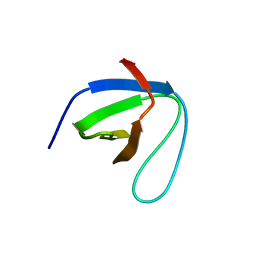
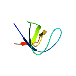
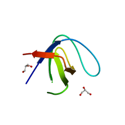
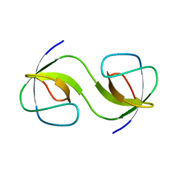
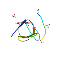
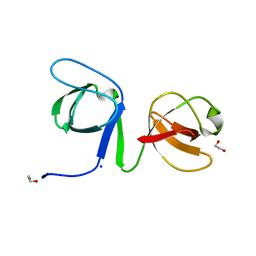
Fgr SH3 domain crystal structure
Descriptor: Tyrosine-protein kinase Fgr
Authors:Perez, I, Berndt, S, Gurevich, V.V, Iverson, T.M.
Deposit date:2020-08-17
Release date:2022-01-26
Last modified:2023-10-18
Method:X-RAY DIFFRACTION (1.93 Å)
Cite:A Model for the Signal Initiation Complex Between Arrestin-3 and the Src Family Kinase Fgr.
J.Mol.Biol., 434, 2022
7CSR
DownloadVisualize
BU of 7csr by Molmil
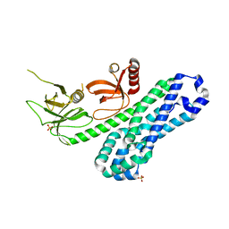
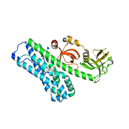
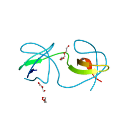
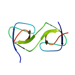
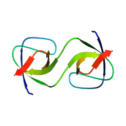
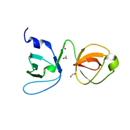
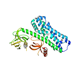
Structure of Ephexin4 R676L
Descriptor: Rho guanine nucleotide exchange factor 16
Authors:Zhang, M, Lin, L, Wang, C, Zhu, J.
Deposit date:2020-08-17
Release date:2021-02-24
Last modified:2023-11-29
Method:X-RAY DIFFRACTION (3 Å)
Cite:Double inhibition and activation mechanisms of Ephexin family RhoGEFs.
Proc.Natl.Acad.Sci.USA, 118, 2021
7CSO
DownloadVisualize
BU of 7cso by Molmil
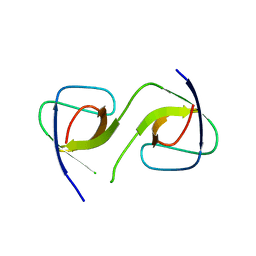
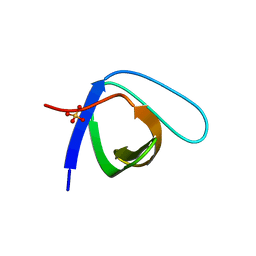
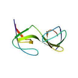
Structure of Ephexin4 DH-PH-SH3
Descriptor: Rho guanine nucleotide exchange factor 16, SULFATE ION
Authors:Zhang, M, Lin, L, Wang, C, Zhu, J.
Deposit date:2020-08-15
Release date:2021-02-24
Last modified:2024-03-27
Method:X-RAY DIFFRACTION (2.39 Å)
Cite:Double inhibition and activation mechanisms of Ephexin family RhoGEFs.
Proc.Natl.Acad.Sci.USA, 118, 2021
7CSP
DownloadVisualize
BU of 7csp by Molmil
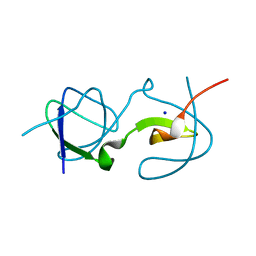
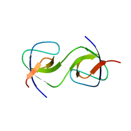
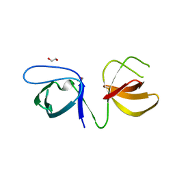
Structure of Ephexin4 IDPSH
Descriptor: Rho guanine nucleotide exchange factor 16
Authors:Zhang, M, Lin, L, Wang, C, Zhu, J.
Deposit date:2020-08-15
Release date:2021-02-24
Last modified:2023-11-29
Method:X-RAY DIFFRACTION (3 Å)
Cite:Double inhibition and activation mechanisms of Ephexin family RhoGEFs.
Proc.Natl.Acad.Sci.USA, 118, 2021
7CFZ
DownloadVisualize
BU of 7cfz by Molmil
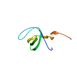
SH3 domain of NADPH oxidase activator 1
Descriptor: NADPH oxidase activator 1
Authors:Kim, M, Park, J.H, Attri, P, Lee, W.
Deposit date:2020-06-29
Release date:2021-07-07
Last modified:2023-11-29
Method:X-RAY DIFFRACTION (1.89 Å)
Cite:Structural modification of NADPH oxidase activator (Noxa 1) by oxidative stress: An experimental and computational study.
Int.J.Biol.Macromol., 163, 2020
6XX4
DownloadVisualize
BU of 6xx4 by Molmil
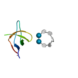
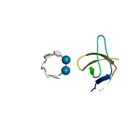
Crystal structure of the c-Src SH3 domain H122R-Q128E mutant in complex with Ni(II) at pH 7.5 co-crystallized with methyl beta-cyclodextrin
Descriptor: Cyclic 2,3-di-O-methyl-alpha-D-glucopyranose-(1-4)-2-O-methyl-alpha-D-glucopyranose-(1-4)-2,6-di-O-methyl-alpha-D-glucopyranose-(1-4)-2-O-methyl-alpha-D-glucopyranose-(1-4)-alpha-D-glucopyranose-(1-4)-alpha-D-glucopyranose-(1-4)-3-O-methyl-alpha-D-glucopyranose, NICKEL (II) ION, Proto-oncogene tyrosine-protein kinase Src
Authors:Camara-Artigas, A.
Deposit date:2020-01-26
Release date:2020-04-22
Last modified:2024-01-24
Method:X-RAY DIFFRACTION (1.05 Å)
Cite:The effect of an engineered ATCUN motif on the structure and biophysical properties of the SH3 domain of c-Src tyrosine kinase.
J.Biol.Inorg.Chem., 25, 2020
6XX3
DownloadVisualize
BU of 6xx3 by Molmil
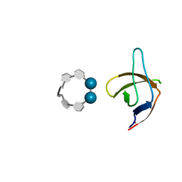
Crystal structure of the c-Src SH3 domain H122R-Q128E mutant in complex with Cu(II) at pH 6.5 co-crystallized with methyl beta-cyclodextrin
Descriptor: COPPER (II) ION, Cyclic 2,3-di-O-methyl-alpha-D-glucopyranose-(1-4)-2-O-methyl-alpha-D-glucopyranose-(1-4)-2,6-di-O-methyl-alpha-D-glucopyranose-(1-4)-2-O-methyl-alpha-D-glucopyranose-(1-4)-alpha-D-glucopyranose-(1-4)-alpha-D-glucopyranose-(1-4)-3-O-methyl-alpha-D-glucopyranose, Proto-oncogene tyrosine-protein kinase Src
Authors:Camara-Artigas, A.
Deposit date:2020-01-26
Release date:2020-04-22
Last modified:2024-01-24
Method:X-RAY DIFFRACTION (1.36 Å)
Cite:The effect of an engineered ATCUN motif on the structure and biophysical properties of the SH3 domain of c-Src tyrosine kinase.
J.Biol.Inorg.Chem., 25, 2020
6XX5
DownloadVisualize
BU of 6xx5 by Molmil
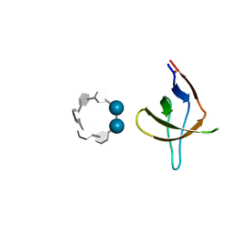
Crystal structure of the c-Src SH3 domain H122R-Q128K mutant in complex with Ni(II) at pH 7.5 co-crystallized with methyl beta-cyclodextrin
Descriptor: Cyclic 2,3-di-O-methyl-alpha-D-glucopyranose-(1-4)-2-O-methyl-alpha-D-glucopyranose-(1-4)-2,6-di-O-methyl-alpha-D-glucopyranose-(1-4)-2-O-methyl-alpha-D-glucopyranose-(1-4)-alpha-D-glucopyranose-(1-4)-alpha-D-glucopyranose-(1-4)-3-O-methyl-alpha-D-glucopyranose, NICKEL (II) ION, Proto-oncogene tyrosine-protein kinase Src
Authors:Camara-Artigas, A.
Deposit date:2020-01-26
Release date:2020-05-27
Last modified:2024-01-24
Method:X-RAY DIFFRACTION (1.3 Å)
Cite:The effect of an engineered ATCUN motif on the structure and biophysical properties of the SH3 domain of c-Src tyrosine kinase.
J.Biol.Inorg.Chem., 25, 2020
6XX2
DownloadVisualize
BU of 6xx2 by Molmil
Crystal structure of the c-Src SH3 domain H122R-Q128K mutant in complex with Cu(II) at pH 7.5 co-crystallized with methyl beta-cyclodextrin
Descriptor: COPPER (II) ION, Cyclic 2,3-di-O-methyl-alpha-D-glucopyranose-(1-4)-2-O-methyl-alpha-D-glucopyranose-(1-4)-2,6-di-O-methyl-alpha-D-glucopyranose-(1-4)-2-O-methyl-alpha-D-glucopyranose-(1-4)-alpha-D-glucopyranose-(1-4)-alpha-D-glucopyranose-(1-4)-3-O-methyl-alpha-D-glucopyranose, Proto-oncogene tyrosine-protein kinase Src
Authors:Camara-Artigas, A.
Deposit date:2020-01-26
Release date:2020-04-22
Last modified:2024-01-24
Method:X-RAY DIFFRACTION (1.25 Å)
Cite:The effect of an engineered ATCUN motif on the structure and biophysical properties of the SH3 domain of c-Src tyrosine kinase.
J.Biol.Inorg.Chem., 25, 2020
6XVN
DownloadVisualize
BU of 6xvn by Molmil
Crystal structure of c-Src SH3 domain without ATCUN motif: monomer 1
Descriptor: Proto-oncogene tyrosine-protein kinase Src
Authors:Camara-Artigas, A, Plaza-Garrido, M.
Deposit date:2020-01-22
Release date:2020-04-22
Last modified:2024-01-24
Method:X-RAY DIFFRACTION (1.7 Å)
Cite:The effect of an engineered ATCUN motif on the structure and biophysical properties of the SH3 domain of c-Src tyrosine kinase.
J.Biol.Inorg.Chem., 25, 2020
6XVM
DownloadVisualize
BU of 6xvm by Molmil
Crystal structure of c-Src SH3 domain without ATCUN motif: monomer 2
Descriptor: GLYCEROL, Proto-oncogene tyrosine-protein kinase Src
Authors:Camara-Artigas, A, Plaza-Garrido, M.
Deposit date:2020-01-22
Release date:2020-04-22
Last modified:2024-01-24
Method:X-RAY DIFFRACTION (0.9 Å)
Cite:The effect of an engineered ATCUN motif on the structure and biophysical properties of the SH3 domain of c-Src tyrosine kinase.
J.Biol.Inorg.Chem., 25, 2020
6XVO
DownloadVisualize
BU of 6xvo by Molmil
Crystal structure of the intertwined dimer of the c-Src SH3 domain without ATCUN motif
Descriptor: DI(HYDROXYETHYL)ETHER, Proto-oncogene tyrosine-protein kinase Src, TRIETHYLENE GLYCOL
Authors:Camara-Artigas, A, Plaza-Garrido, M.
Deposit date:2020-01-22
Release date:2020-04-22
Last modified:2024-01-24
Method:X-RAY DIFFRACTION (1.7 Å)
Cite:The effect of an engineered ATCUN motif on the structure and biophysical properties of the SH3 domain of c-Src tyrosine kinase.
J.Biol.Inorg.Chem., 25, 2020
5QU6
DownloadVisualize
BU of 5qu6 by Molmil
Crystal Structure of swapped human Nck SH3.1 domain, 1.8A, triclinic
Descriptor: Cytoplasmic protein NCK1
Authors:Rudolph, M.G.
Deposit date:2019-12-13
Release date:2020-02-12
Last modified:2024-04-03
Method:X-RAY DIFFRACTION (1.816 Å)
Cite:Small molecule AX-024 reduces T cell proliferation independently of CD3ε/Nck1 interaction, which is governed by a domain swap in the Nck1-SH3.1 domain.
J.Biol.Chem., 295, 2020
5QU8
DownloadVisualize
BU of 5qu8 by Molmil
Crystal Structure of symmetric swapped human Nck SH3.1 domain, 0.93A, orthorhombic form IV
Descriptor: Cytoplasmic protein NCK1, SODIUM ION
Authors:Rudolph, M.G.
Deposit date:2019-12-13
Release date:2020-02-12
Last modified:2024-04-03
Method:X-RAY DIFFRACTION (0.93 Å)
Cite:Small molecule AX-024 reduces T cell proliferation independently of CD3ε/Nck1 interaction, which is governed by a domain swap in the Nck1-SH3.1 domain.
J.Biol.Chem., 295, 2020
5QU3
DownloadVisualize
BU of 5qu3 by Molmil
Crystal Structure of domain swapped human Nck SH3.1, 1.01A, monoclinic
Descriptor: Cytoplasmic protein NCK1
Authors:Rudolph, M.G.
Deposit date:2019-12-13
Release date:2020-02-12
Last modified:2024-04-03
Method:X-RAY DIFFRACTION (1.02 Å)
Cite:Small molecule AX-024 reduces T cell proliferation independently of CD3ε/Nck1 interaction, which is governed by a domain swap in the Nck1-SH3.1 domain.
J.Biol.Chem., 295, 2020
5QUA
DownloadVisualize
BU of 5qua by Molmil
Crystal Structure of swapped human Nck SH3.1 domain, 1.5A, C2221
Descriptor: Cytoplasmic protein NCK1
Authors:Rudolph, M.G.
Deposit date:2019-12-13
Release date:2020-02-12
Last modified:2024-04-03
Method:X-RAY DIFFRACTION (1.51 Å)
Cite:Small molecule AX-024 reduces T cell proliferation independently of CD3ε/Nck1 interaction, which is governed by a domain swap in the Nck1-SH3.1 domain.
J.Biol.Chem., 295, 2020
5QU1
DownloadVisualize
BU of 5qu1 by Molmil
Crystal Structure of the monomeric human Nck SH3.1 domain, triclinic, 1.08A
Descriptor: Cytoplasmic protein NCK1, SULFATE ION
Authors:Burger, D, Ruf, A, Benz, J, Schlatter, D, Rudolph, M.G.
Deposit date:2019-12-13
Release date:2020-02-12
Last modified:2024-04-03
Method:X-RAY DIFFRACTION (1.08 Å)
Cite:Small molecule AX-024 reduces T cell proliferation independently of CD3ε/Nck1 interaction, which is governed by a domain swap in the Nck1-SH3.1 domain.
J.Biol.Chem., 295, 2020
5QU5
DownloadVisualize
BU of 5qu5 by Molmil
Domain Swap in the first SH3 domain of human Nck1
Descriptor: Cytoplasmic protein NCK1
Authors:Burger, D, Ruf, A, Benz, J, Schlatter, D, Rudolph, M.G.
Deposit date:2019-12-13
Release date:2020-02-12
Last modified:2024-04-03
Method:X-RAY DIFFRACTION (1.11 Å)
Cite:Small molecule AX-024 reduces T cell proliferation independently of CD3ε/Nck1 interaction, which is governed by a domain swap in the Nck1-SH3.1 domain.
J.Biol.Chem., 295, 2020
5QU2
DownloadVisualize
BU of 5qu2 by Molmil
Crystal Structure of human Nck SH3.1 in complex with peptide PPPVPNPDY
Descriptor: ACE-PRO-PRO-PRO-VAL-PRO-ASN-PRO-ASP-TYR-NH2, Cytoplasmic protein NCK1, SULFATE ION
Authors:Rudolph, M.G.
Deposit date:2019-12-13
Release date:2020-02-12
Last modified:2024-04-03
Method:X-RAY DIFFRACTION (1.04 Å)
Cite:Small molecule AX-024 reduces T cell proliferation independently of CD3ε/Nck1 interaction, which is governed by a domain swap in the Nck1-SH3.1 domain.
J.Biol.Chem., 295, 2020
5QU7
DownloadVisualize
BU of 5qu7 by Molmil
Crystal Structure of swapped human Nck SH3.1 domain, 1.3A, orthorhombic form III
Descriptor: Cytoplasmic protein NCK1
Authors:Rudolph, M.G.
Deposit date:2019-12-13
Release date:2020-02-12
Last modified:2024-04-03
Method:X-RAY DIFFRACTION (1.27 Å)
Cite:Small molecule AX-024 reduces T cell proliferation independently of CD3ε/Nck1 interaction, which is governed by a domain swap in the Nck1-SH3.1 domain.
J.Biol.Chem., 295, 2020
5QU4
DownloadVisualize
BU of 5qu4 by Molmil
Crystal Structure of swapped human Nck SH3.1 domain, 1.05A, orthorhombic form I
Descriptor: Cytoplasmic protein NCK1
Authors:Rudolph, M.G.
Deposit date:2019-12-13
Release date:2020-02-12
Last modified:2024-04-03
Method:X-RAY DIFFRACTION (1.05 Å)
Cite:Small molecule AX-024 reduces T cell proliferation independently of CD3ε/Nck1 interaction, which is governed by a domain swap in the Nck1-SH3.1 domain.
J.Biol.Chem., 295, 2020
6UY7
DownloadVisualize
BU of 6uy7 by Molmil
Crystal structure of the STAC3 tandem SH3 domains - P269R
Descriptor: 1,2-ETHANEDIOL, SH3 and cysteine-rich domain-containing protein 3
Authors:Rufenach, B, Van Petegem, F.
Deposit date:2019-11-12
Release date:2020-06-17
Last modified:2023-10-11
Method:X-RAY DIFFRACTION (2.105 Å)
Cite:Multiple Sequence Variants in STAC3 Affect Interactions with CaV1.1 and Excitation-Contraction Coupling.
Structure, 28, 2020
6UY9
DownloadVisualize
BU of 6uy9 by Molmil
Crystal structure of the STAC3 tandem SH3 domains - P269R, W284S
Descriptor: 1,2-ETHANEDIOL, SH3 and cysteine-rich domain-containing protein 3, SODIUM ION
Authors:Rufenach, B, Van Petegem, F.
Deposit date:2019-11-12
Release date:2020-06-17
Last modified:2023-10-11
Method:X-RAY DIFFRACTION (1.6 Å)
Cite:Multiple Sequence Variants in STAC3 Affect Interactions with CaV1.1 and Excitation-Contraction Coupling.
Structure, 28, 2020
6UY8
DownloadVisualize
BU of 6uy8 by Molmil
Crystal structure of the STAC3 tandem SH3 domains - P269R, K329N
Descriptor: 1,2-ETHANEDIOL, CHLORIDE ION, SH3 and cysteine-rich domain-containing protein 3
Authors:Rufenach, B, Van Petegem, F.
Deposit date:2019-11-12
Release date:2020-06-17
Last modified:2023-10-11
Method:X-RAY DIFFRACTION (1.649 Å)
Cite:Multiple Sequence Variants in STAC3 Affect Interactions with CaV1.1 and Excitation-Contraction Coupling.
Structure, 28, 2020
6SDF
DownloadVisualize
BU of 6sdf by Molmil
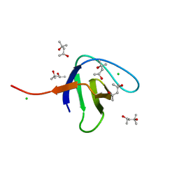
N-terminal SH3 domain of Grb2 protein
Descriptor: (4S)-2-METHYL-2,4-PENTANEDIOL, CHLORIDE ION, Growth factor receptor-bound protein 2
Authors:Bolgov, A.A, Korban, S.A, Luzik, D.A, Rogacheva, O.N, Zhemkov, V.A, Kim, M, Skrynnikov, N.R, Bezprozvanny, I.B.
Deposit date:2019-07-26
Release date:2020-01-29
Last modified:2024-01-24
Method:X-RAY DIFFRACTION (2.5 Å)
Cite:Crystal structure of the SH3 domain of growth factor receptor-bound protein 2.
Acta Crystallogr.,Sect.F, 76, 2020

221051

PDB entries from 2024-06-12

PDB statisticsPDBj update infoContact PDBjnumon