Loading
PDBj
MenuPDBj@FacebookPDBj@X(formerly Twitter)PDBj@BlueSkyPDBj@YouTubewwPDB FoundationwwPDB
RCSB PDBPDBeBMRBAdv. SearchSearch help

7NES
DownloadVisualize
BU of 7nes by Molmil
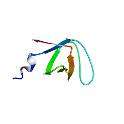
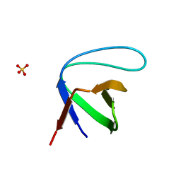
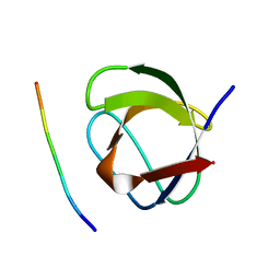
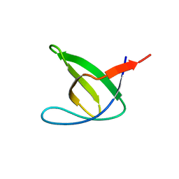
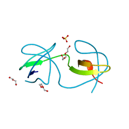
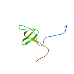
Crystal structure of the v-Src SH3 domain N117D-V124L mutant
Descriptor: GLYCINE, v-Src SH3 domain
Authors:Camara-Artigas, A, Salinas-Garcia, M.C.
Deposit date:2021-02-04
Release date:2021-06-02
Last modified:2024-05-15
Method:X-RAY DIFFRACTION (1.35 Å)
Cite:The impact of oncogenic mutations of the viral Src kinase on the structure and stability of the SH3 domain.
Acta Crystallogr D Struct Biol, 77, 2021
6IPZ
DownloadVisualize
BU of 6ipz by Molmil
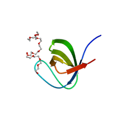
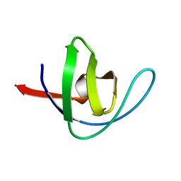
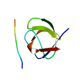
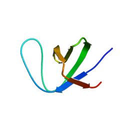
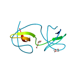
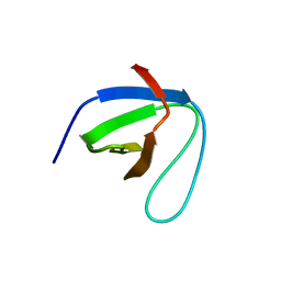
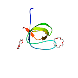
Fyn SH3 domain R96W mutant, crystallized with 18-crown-6
Descriptor: 1,4,7,10,13,16-HEXAOXACYCLOOCTADECANE, Tyrosine-protein kinase Fyn
Authors:Arold, S.T, Aljedani, S.S, Shahul Hameed, U.F.
Deposit date:2018-11-05
Release date:2018-11-28
Last modified:2023-11-22
Method:X-RAY DIFFRACTION (1.576 Å)
Cite:Synergy and allostery in ligand binding by HIV-1 Nef.
Biochem.J., 478, 2021
5XG9
DownloadVisualize
BU of 5xg9 by Molmil
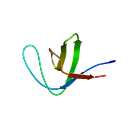
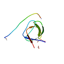
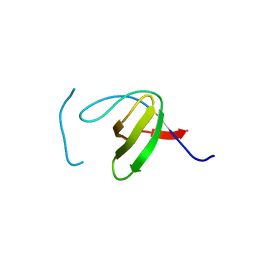
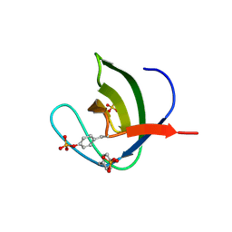
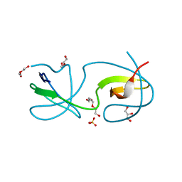
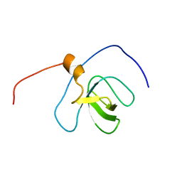
Crystal Structure of PEG-bound SH3 domain of Myosin IB from Entamoeba histolytica
Descriptor: 1-(2-METHOXY-ETHOXY)-2-{2-[2-(2-METHOXY-ETHOXY]-ETHOXY}-ETHANE, 2,5,8,11,14,17,20,23,26,29,32,35,38,41,44,47,50,53,56,59,62,65,68,71,74,77,80-HEPTACOSAOXADOOCTACONTAN-82-OL, PENTAETHYLENE GLYCOL, ...
Authors:Gautam, G, Gourinath, S.
Deposit date:2017-04-13
Release date:2017-08-16
Last modified:2023-11-22
Method:X-RAY DIFFRACTION (1.78 Å)
Cite:Crystal structure of the PEG-bound SH3 domain of myosin IB from Entamoeba histolytica reveals its mode of ligand recognition
Acta Crystallogr D Struct Biol, 73, 2017
8CHH
DownloadVisualize
BU of 8chh by Molmil
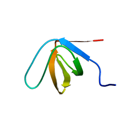
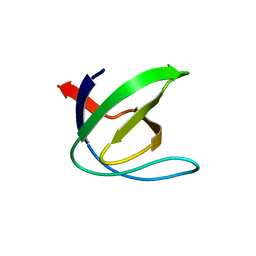
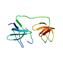
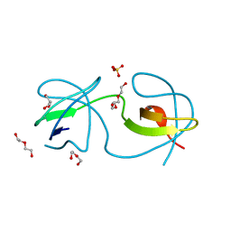
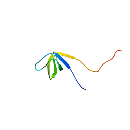
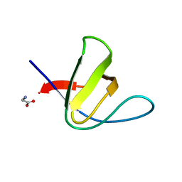
SH3 domain solved by the exact solid-state method from the Bruker Dynamics Center using the correction method for spin-diffusion with PDB 2NUZ as reference
Descriptor: Spectrin alpha chain, non-erythrocytic 1
Authors:Soeldner, B.
Deposit date:2023-02-08
Release date:2023-03-08
Last modified:2024-06-19
Method:SOLID-STATE NMR
Cite:Integrated Assessment of the Structure and Dynamics of Solid Proteins.
J Phys Chem Lett, 14, 2023
8CF4
DownloadVisualize
BU of 8cf4 by Molmil
SH3 domain solved by the exact solid-state method from the Bruker Dynamics Center using the combined correction method with PDB 2NUZ
Descriptor: Spectrin alpha chain, non-erythrocytic 1
Authors:Soeldner, B.
Deposit date:2023-02-02
Release date:2023-03-08
Last modified:2024-06-19
Method:SOLID-STATE NMR
Cite:Integrated Assessment of the Structure and Dynamics of Solid Proteins.
J Phys Chem Lett, 14, 2023
8CHG
DownloadVisualize
BU of 8chg by Molmil
SH3 domain solved by the exact solid-state method from the Bruker Dynamics Center using the correction for dipolar truncation with PDB 2NUZ
Descriptor: Spectrin alpha chain, non-erythrocytic 1
Authors:Soeldner, B.
Deposit date:2023-02-08
Release date:2023-03-08
Last modified:2024-06-19
Method:SOLID-STATE NMR
Cite:Integrated Assessment of the Structure and Dynamics of Solid Proteins.
J Phys Chem Lett, 14, 2023
3I9Q
DownloadVisualize
BU of 3i9q by Molmil
Crystal Structure of the triple mutant S19G-P20D-R21S of alpha spectrin SH3 domain
Descriptor: SULFATE ION, Spectrin alpha chain
Authors:Camara-Artigas, A, Gavira, J.A.
Deposit date:2009-07-12
Release date:2009-12-15
Last modified:2023-09-06
Method:X-RAY DIFFRACTION (1.45 Å)
Cite:The effect of a proline residue on the rate of growth and the space group of alpha-spectrin SH3-domain crystals.
Acta Crystallogr.,Sect.D, 65, 2009
1BK2
DownloadVisualize
BU of 1bk2 by Molmil
A-SPECTRIN SH3 DOMAIN D48G MUTANT
Descriptor: A-SPECTRIN
Authors:Martinez, J.C, Pisabarro, M.T, Serrano, L.
Deposit date:1998-07-14
Release date:1999-02-16
Last modified:2024-05-22
Method:X-RAY DIFFRACTION (2.01 Å)
Cite:Obligatory steps in protein folding and the conformational diversity of the transition state.
Nat.Struct.Biol., 5, 1998
5L23
DownloadVisualize
BU of 5l23 by Molmil
Crystal structure of the complex between the N-terminal SH3 domain of CrkII and a proline-rich ligand
Descriptor: Adapter molecule crk, C3G derived peptide, DI(HYDROXYETHYL)ETHER
Authors:Bhatt, V.S, Krieger, I, Sacchettini, J, Cho, J.-H.
Deposit date:2016-07-30
Release date:2016-09-07
Last modified:2024-10-23
Method:X-RAY DIFFRACTION (1.77 Å)
Cite:Crystal structure of the complex between the N-terminal SH3 domain of CrkII and a proline-rich ligand
To Be Published
1CSK
DownloadVisualize
BU of 1csk by Molmil
THE CRYSTAL STRUCTURE OF HUMAN CSKSH3: STRUCTURAL DIVERSITY NEAR THE RT-SRC AND N-SRC LOOP
Descriptor: C-SRC SH3 DOMAIN
Authors:Mathieu, M, Wierenga, R.K.
Deposit date:1994-03-22
Release date:1994-07-31
Last modified:2024-02-07
Method:X-RAY DIFFRACTION (2.5 Å)
Cite:The crystal structure of human CskSH3: structural diversity near the RT-Src and n-Src loop.
FEBS Lett., 341, 1994
1CKB
DownloadVisualize
BU of 1ckb by Molmil
STRUCTURAL BASIS FOR THE SPECIFIC INTERACTION OF LYSINE-CONTAINING PROLINE-RICH PEPTIDES WITH THE N-TERMINAL SH3 DOMAIN OF C-CRK
Descriptor: C-CRK N-TERMINAL SH3 DOMAIN, SOS PEPTIDE (PRO-PRO-PRO-VAL-PRO-PRO-ARG-ARG-ARG-ARG)
Authors:Wu, X, Kuriyan, J.
Deposit date:1995-01-24
Release date:1995-05-08
Last modified:2024-02-07
Method:X-RAY DIFFRACTION (1.9 Å)
Cite:Structural basis for the specific interaction of lysine-containing proline-rich peptides with the N-terminal SH3 domain of c-Crk.
Structure, 3, 1995
1CKA
DownloadVisualize
BU of 1cka by Molmil
STRUCTURAL BASIS FOR THE SPECIFIC INTERACTION OF LYSINE-CONTAINING PROLINE-RICH PEPTIDES WITH THE N-TERMINAL SH3 DOMAIN OF C-CRK
Descriptor: C-CRK N-TERMINAL SH3 DOMAIN, C3G PEPTIDE (PRO-PRO-PRO-ALA-LEU-PRO-PRO-LYS-LYS-ARG)
Authors:Wu, X, Kuriyan, J.
Deposit date:1995-01-24
Release date:1995-05-08
Last modified:2024-02-07
Method:X-RAY DIFFRACTION (1.5 Å)
Cite:Structural basis for the specific interaction of lysine-containing proline-rich peptides with the N-terminal SH3 domain of c-Crk.
Structure, 3, 1995
8KDX
DownloadVisualize
BU of 8kdx by Molmil
Tau-S214 Phosphorylation Inhibits Fyn Kinase Interaction and Increases the Decay Time of NMDAR-mediated Current
Descriptor: Microtubule-associated protein tau, Tyrosine-protein kinase Fyn
Authors:Padavattan, S, Jos, S.
Deposit date:2023-08-10
Release date:2024-02-28
Method:X-RAY DIFFRACTION (1.01 Å)
Cite:Tau-S214 Phosphorylation Inhibits Fyn Kinase Interaction and Increases the Decay Time of NMDAR-mediated Current.
J.Mol.Biol., 436, 2024
7YXW
DownloadVisualize
BU of 7yxw by Molmil
Structure of the p22phox A200G mutant in complex with p47phox peptide
Descriptor: Cytochrome b-245 light chain, Neutrophil cytosol factor 1
Authors:Cukier, C.D, Vuillard, L.M, Komjati, B, Szlavik, Z.
Deposit date:2022-02-16
Release date:2022-03-30
Last modified:2024-01-31
Method:X-RAY DIFFRACTION (2.5 Å)
Cite:Targeting NOX2 via p47/phox-p22/phox Inhibition with Novel Triproline Mimetics
Acs Med.Chem.Lett., 13, 2022
5NP3
DownloadVisualize
BU of 5np3 by Molmil
Abl2 SH3
Descriptor: Abelson tyrosine-protein kinase 2
Authors:Mero, B, Radnai, L, Gogl, G, Leveles, I, Buday, L.
Deposit date:2017-04-13
Release date:2018-05-16
Last modified:2024-01-17
Method:X-RAY DIFFRACTION (2 Å)
Cite:Structural insights into the tyrosine phosphorylation-mediated inhibition of SH3 domain-ligand interactions.
J.Biol.Chem., 294, 2019
7Y4N
DownloadVisualize
BU of 7y4n by Molmil
Insight into the C-terminal SH3 domain mediated binding of Drosophila Drk to Sos and Dos
Descriptor: Growth factor receptor-bound protein 2
Authors:Pooppadi, M.S, Ikeya, T, Sugasawa, H, Watanabe, R, Mishima, M, Inomata, K, Ito, Y.
Deposit date:2022-06-15
Release date:2022-08-31
Last modified:2024-05-15
Method:SOLUTION NMR
Cite:Insight into the C-terminal SH3 domain mediated binding of Drosophila Drk to Sos and Dos.
Biochem.Biophys.Res.Commun., 625, 2022
5NP5
DownloadVisualize
BU of 5np5 by Molmil
Abl2 SH3 pTyr116/161
Descriptor: Abelson tyrosine-protein kinase 2, SULFATE ION
Authors:Mero, B, Radnai, L, Gogl, G, Leveles, I, Buday, L.
Deposit date:2017-04-13
Release date:2018-05-16
Last modified:2024-10-23
Method:X-RAY DIFFRACTION (1.4 Å)
Cite:Structural insights into the tyrosine phosphorylation-mediated inhibition of SH3 domain-ligand interactions.
J.Biol.Chem., 294, 2019
4OMN
DownloadVisualize
BU of 4omn by Molmil
Crystal structure of the intertwined dimer of the c-Src tyrosine kinase SH3 domain mutant Q128E
Descriptor: DI(HYDROXYETHYL)ETHER, GLYCEROL, Proto-oncogene tyrosine-protein kinase Src, ...
Authors:Camara-Artigas, A, Bacarizo, J.
Deposit date:2014-01-27
Release date:2014-12-10
Last modified:2023-09-20
Method:X-RAY DIFFRACTION (1.5 Å)
Cite:Electrostatic Effects in the Folding of the SH3 Domain of the c-Src Tyrosine Kinase: pH-Dependence in 3D-Domain Swapping and Amyloid Formation.
Plos One, 9, 2014
4OMQ
DownloadVisualize
BU of 4omq by Molmil
Crystal structure of the intertwined dimer of the c-Src tyrosine kinase SH3 domain mutant S94A
Descriptor: DI(HYDROXYETHYL)ETHER, Proto-oncogene tyrosine-protein kinase Src, SULFATE ION, ...
Authors:Camara-Artigas, A, Bacarizo, J.
Deposit date:2014-01-27
Release date:2015-01-28
Last modified:2023-09-20
Method:X-RAY DIFFRACTION (2 Å)
Cite:The role of Gutamine 128 of the SH3 domain of the c-Src tyrosine kinase in 3D domain swapping and amyloid formation
To be Published
4OMP
DownloadVisualize
BU of 4omp by Molmil
Crystal structure of the intertwined dimer of the c-Src tyrosine kinase SH3 domain mutant Q128K
Descriptor: DI(HYDROXYETHYL)ETHER, Proto-oncogene tyrosine-protein kinase Src, TRIETHYLENE GLYCOL
Authors:Camara-Artigas, A, Bacarizo, J.
Deposit date:2014-01-27
Release date:2014-12-10
Last modified:2023-09-20
Method:X-RAY DIFFRACTION (2 Å)
Cite:Electrostatic Effects in the Folding of the SH3 Domain of the c-Src Tyrosine Kinase: pH-Dependence in 3D-Domain Swapping and Amyloid Formation.
Plos One, 9, 2014
4OMM
DownloadVisualize
BU of 4omm by Molmil
Crystal structure of the intertwined dimer of the c-Src tyrosine kinase SH3 domain mutant N113S
Descriptor: DI(HYDROXYETHYL)ETHER, GLYCEROL, Proto-oncogene tyrosine-protein kinase Src, ...
Authors:Camara-Artigas, A, Bacarizo, J.
Deposit date:2014-01-27
Release date:2015-01-28
Last modified:2023-09-20
Method:X-RAY DIFFRACTION (1.9 Å)
Cite:The role of Gutamine 128 of the SH3 domain of the c-Src tyrosine kinase in 3D domain swapping and amyloid formation
To be Published
2YUO
DownloadVisualize
BU of 2yuo by Molmil
Solution structure of the SH3 domain of mouse RUN and TBC1 domain containing 3
Descriptor: RUN and TBC1 domain containing 3
Authors:Abe, H, Tochio, N, Miyamoto, K, Saito, K, Sasagawa, A, Koshiba, S, Inoue, M, Kigawa, T, Yokoyama, S, RIKEN Structural Genomics/Proteomics Initiative (RSGI)
Deposit date:2007-04-06
Release date:2007-10-09
Last modified:2024-05-29
Method:SOLUTION NMR
Cite:Solution structure of the SH3 domain of mouse RUN and TBC1 domain containing 3
To be Published
2YUQ
DownloadVisualize
BU of 2yuq by Molmil
Solution structure of the SH3 domain of human Tyrosine-protein kinase ITK/TSK
Descriptor: Tyrosine-protein kinase ITK/TSK
Authors:Abe, H, Miyamoto, K, Tochio, N, Sato, M, Koshiba, S, Harada, T, Watanabe, S, Kigawa, T, Yokoyama, S, RIKEN Structural Genomics/Proteomics Initiative (RSGI)
Deposit date:2007-04-06
Release date:2007-10-09
Last modified:2024-05-29
Method:SOLUTION NMR
Cite:Solution structure of the SH3 domain of human Tyrosine-protein kinase ITK/TSK
To be Published
7CFZ
DownloadVisualize
BU of 7cfz by Molmil
SH3 domain of NADPH oxidase activator 1
Descriptor: NADPH oxidase activator 1
Authors:Kim, M, Park, J.H, Attri, P, Lee, W.
Deposit date:2020-06-29
Release date:2021-07-07
Last modified:2023-11-29
Method:X-RAY DIFFRACTION (1.89 Å)
Cite:Structural modification of NADPH oxidase activator (Noxa 1) by oxidative stress: An experimental and computational study.
Int.J.Biol.Macromol., 163, 2020
2YT6
DownloadVisualize
BU of 2yt6 by Molmil
Solution structure of the SH3_1 domain of Yamaguchi sarcoma viral (v-yes) oncogene homolog 1
Descriptor: Adult male urinary bladder cDNA, RIKEN full-length enriched library, clone:9530076O17 product:Yamaguchi sarcoma viral (v-yes) oncogene homolog
Authors:Dang, W, Muto, Y, Inoue, M, Kigawa, T, Shirouzu, M, Tarada, T, Yokoyama, S, RIKEN Structural Genomics/Proteomics Initiative (RSGI)
Deposit date:2007-04-05
Release date:2008-04-08
Last modified:2024-05-29
Method:SOLUTION NMR
Cite:Solution structure of the SH3_1 domain of Yamaguchi sarcoma viral (v-yes) oncogene homolog 1
To be Published

227344

PDB entries from 2024-11-13

PDB statisticsPDBj update infoContact PDBjnumon