Loading
PDBj
MenuPDBj@FacebookPDBj@TwitterPDBj@YouTubewwPDB FoundationwwPDB
RCSB PDBPDBeBMRBAdv. SearchSearch help

1RVC
DownloadVisualize
BU of 1rvc by Molmil
MG2+ BINDING TO THE ACTIVE SITE OF ECO RV ENDONUCLEASE: A CRYSTALLOGRAPHIC STUDY OF COMPLEXES WITH SUBSTRATE AND PRODUCT DNA AT 2 ANGSTROMS RESOLUTION
Descriptor: DNA (5'-D(*AP*AP*AP*GP*AP*T)-3'), DNA (5'-D(*AP*TP*CP*TP*T)-3'), MAGNESIUM ION, ...
Authors:Kostrewa, D, Winkler, F.K.
Deposit date:1994-10-21
Release date:1995-01-26
Last modified:2024-02-14
Method:X-RAY DIFFRACTION (2.1 Å)
Cite:Mg2+ binding to the active site of EcoRV endonuclease: a crystallographic study of complexes with substrate and product DNA at 2 A resolution.
Biochemistry, 34, 1995
1HTR
DownloadVisualize
BU of 1htr by Molmil
CRYSTAL AND MOLECULAR STRUCTURES OF HUMAN PROGASTRICSIN AT 1.62 ANGSTROMS RESOLUTION
Descriptor: GASTRICSIN, PROGASTRICSIN (PRO SEGMENT)
Authors:Moore, S.A, Sielecki, A.R, James, M.N.G.
Deposit date:1994-10-21
Release date:1995-01-26
Last modified:2024-06-05
Method:X-RAY DIFFRACTION (1.62 Å)
Cite:Crystal and molecular structures of human progastricsin at 1.62 A resolution.
J.Mol.Biol., 247, 1995
1CYE
DownloadVisualize
BU of 1cye by Molmil
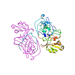
THREE DIMENSIONAL STRUCTURE OF CHEMOTACTIC CHE Y PROTEIN IN AQUEOUS SOLUTION BY NUCLEAR MAGNETIC RESONANCE METHODS
Descriptor: CHEY
Authors:Santoro, J, Bruix, M, Pascual, J, Lopez, E, Serrano, L, Rico, M.
Deposit date:1994-10-21
Release date:1995-02-07
Last modified:2024-04-10
Method:SOLUTION NMR
Cite:Three-dimensional structure of chemotactic Che Y protein in aqueous solution by nuclear magnetic resonance methods.
J.Mol.Biol., 247, 1995
1HDC
DownloadVisualize
BU of 1hdc by Molmil
MECHANISM OF INHIBITION OF 3ALPHA,20BETA-HYDROXYSTEROID DEHYDROGENASE BY A LICORICE-DERIVED STEROIDAL INHIBITOR
Descriptor: 3-ALPHA, 20 BETA-HYDROXYSTEROID DEHYDROGENASE, CARBENOXOLONE
Authors:Ghosh, D.
Deposit date:1994-10-21
Release date:1995-02-07
Last modified:2024-02-07
Method:X-RAY DIFFRACTION (2.2 Å)
Cite:Mechanism of inhibition of 3 alpha, 20 beta-hydroxysteroid dehydrogenase by a licorice-derived steroidal inhibitor.
Structure, 2, 1994
1ELD
DownloadVisualize
BU of 1eld by Molmil
Structural analysis of the active site of porcine pancreatic elastase based on the x-ray crystal structures of complexes with trifluoroacetyl-dipeptide-anilide inhibitors
Descriptor: ACETIC ACID, CALCIUM ION, ELASTASE, ...
Authors:Mattos, C, Petsko, G.A, Ringe, D.
Deposit date:1994-10-24
Release date:1995-02-14
Last modified:2024-06-05
Method:X-RAY DIFFRACTION (2 Å)
Cite:Structural analysis of the active site of porcine pancreatic elastase based on the X-ray crystal structures of complexes with trifluoroacetyl-dipeptide-anilide inhibitors.
Biochemistry, 34, 1995
1ELE
DownloadVisualize
BU of 1ele by Molmil
STRUCTURAL ANALYSIS OF THE ACTIVE SITE OF PORCINE PANCREATIC ELASTASE BASED ON THE X-RAY CRYSTAL STRUCTURES OF COMPLEXES WITH TRIFLUOROACETYL-DIPEPTIDE-ANILIDE INHIBITORS
Descriptor: CALCIUM ION, ELASTASE, N-(trifluoroacetyl)-L-valyl-N-[4-(trifluoromethyl)phenyl]-L-alaninamide, ...
Authors:Mattos, C, Petsko, G.A, Ringe, D.
Deposit date:1994-10-24
Release date:1995-02-14
Last modified:2024-06-05
Method:X-RAY DIFFRACTION (2 Å)
Cite:Structural analysis of the active site of porcine pancreatic elastase based on the X-ray crystal structures of complexes with trifluoroacetyl-dipeptide-anilide inhibitors.
Biochemistry, 34, 1995
1RPL
DownloadVisualize
BU of 1rpl by Molmil
2.3 ANGSTROMS CRYSTAL STRUCTURE OF THE CATALYTIC DOMAIN OF DNA POLYMERASE BETA
Descriptor: DNA POLYMERASE BETA
Authors:Davies II, J.F, Almassy, R.J.
Deposit date:1994-10-25
Release date:1995-01-26
Last modified:2024-02-14
Method:X-RAY DIFFRACTION (2.3 Å)
Cite:2.3 A crystal structure of the catalytic domain of DNA polymerase beta.
Cell(Cambridge,Mass.), 76, 1994
1LMP
DownloadVisualize
BU of 1lmp by Molmil
THE CRYSTAL STRUCTURES OF THREE COMPLEXES BETWEEN CHITOOLIGOSACCHARIDES AND LYSOZYME FROM THE RAINBOW TROUT
Descriptor: 2-acetamido-2-deoxy-beta-D-glucopyranose-(1-4)-2-acetamido-2-deoxy-beta-D-glucopyranose-(1-4)-2-acetamido-2-deoxy-alpha-D-glucopyranose, LYSOZYME
Authors:Karlsen, S, Hough, E.
Deposit date:1994-10-25
Release date:1996-01-01
Last modified:2023-07-26
Method:X-RAY DIFFRACTION (2 Å)
Cite:Crystal structures of three complexes between chito-oligosaccharides and lysozyme from the rainbow trout. How distorted is the NAG sugar in site D?
Acta Crystallogr.,Sect.D, 51, 1995
1LMQ
DownloadVisualize
BU of 1lmq by Molmil
THE CRYSTAL STRUCTURES OF THREE COMPLEXES BETWEEN CHITOOLIGOSACCHARIDES AND LYSOZYME FROM THE RAINBOW TROUT
Descriptor: 2-acetamido-2-deoxy-beta-D-glucopyranose-(1-4)-2-acetamido-2-deoxy-beta-D-glucopyranose-(1-4)-2-acetamido-2-deoxy-beta-D-glucopyranose-(1-4)-2-acetamido-2-deoxy-alpha-D-glucopyranose, LYSOZYME
Authors:Karlsen, S, Hough, E.
Deposit date:1994-10-25
Release date:1996-01-01
Last modified:2023-07-26
Method:X-RAY DIFFRACTION (1.6 Å)
Cite:Crystal structures of three complexes between chito-oligosaccharides and lysozyme from the rainbow trout. How distorted is the NAG sugar in site D?
Acta Crystallogr.,Sect.D, 51, 1995
1IBG
DownloadVisualize
BU of 1ibg by Molmil
STRUCTURE AND SPECIFICITY OF THE ANTI-DIGOXIN ANTIBODY 40-50
Descriptor: COPPER (II) ION, IGG2B-KAPPA 40-50 FAB (HEAVY CHAIN), IGG2B-KAPPA 40-50 FAB (LIGHT CHAIN), ...
Authors:Jeffrey, P.D, Sheriff, S.
Deposit date:1994-10-25
Release date:1995-05-08
Last modified:2024-06-05
Method:X-RAY DIFFRACTION (2.7 Å)
Cite:Structure and Specificity of the Anti-Digoxin Antibody 40-50
J.Mol.Biol., 248, 1995
1LSG
DownloadVisualize
BU of 1lsg by Molmil
THREE-DIMENSIONAL STRUCTURE OF THE PLATELET INTEGRIN RECOGNITION SEGMENT OF THE FIBRINOGEN GAMMA CHAIN OBTAINED BY CARRIER PROTEIN-DRIVEN CRYSTALLIZATION
Descriptor: HEN EGG WHITE LYSOZYME
Authors:Patel, H, Anderson, W.F.
Deposit date:1994-10-25
Release date:1995-09-15
Last modified:2024-06-05
Method:X-RAY DIFFRACTION (2.4 Å)
Cite:Three-dimensional structure of the platelet integrin recognition segment of the fibrinogen gamma chain obtained by carrier protein-driven crystallization.
Proc.Natl.Acad.Sci.USA, 91, 1994
1LMO
DownloadVisualize
BU of 1lmo by Molmil
THE CRYSTAL STRUCTURES OF THREE COMPLEXES BETWEEN CHITOOLIGOSACCHARIDES AND LYSOZYME FROM THE RAINBOW TROUT
Descriptor: 2-acetamido-2-deoxy-beta-D-glucopyranose-(1-4)-2-acetamido-2-deoxy-beta-D-glucopyranose, LYSOZYME
Authors:Karlsen, S, Hough, E.
Deposit date:1994-10-25
Release date:1996-01-01
Last modified:2023-07-26
Method:X-RAY DIFFRACTION (1.8 Å)
Cite:Crystal structures of three complexes between chito-oligosaccharides and lysozyme from the rainbow trout. How distorted is the NAG sugar in site D?
Acta Crystallogr.,Sect.D, 51, 1995
1DYJ
DownloadVisualize
BU of 1dyj by Molmil
ISOMORPHOUS CRYSTAL STRUCTURES OF ESCHERICHIA COLI DIHYDROFOLATE REDUCTASE COMPLEXED WITH FOLATE, 5-DEAZAFOLATE AND 5,10-DIDEAZATETRAHYDROFOLATE: MECHANISTIC IMPLICATIONS
Descriptor: 5,10-DIDEAZATETRAHYDROFOLIC ACID, CALCIUM ION, CHLORIDE ION, ...
Authors:Reyes, V.M, Kraut, J.
Deposit date:1994-10-26
Release date:1995-06-03
Last modified:2024-02-07
Method:X-RAY DIFFRACTION (1.85 Å)
Cite:Isomorphous crystal structures of Escherichia coli dihydrofolate reductase complexed with folate, 5-deazafolate, and 5,10-dideazatetrahydrofolate: mechanistic implications.
Biochemistry, 34, 1995
1DYH
DownloadVisualize
BU of 1dyh by Molmil
ISOMORPHOUS CRYSTAL STRUCTURES OF ESCHERICHIA COLI DIHYDROFOLATE REDUCTASE COMPLEXED WITH FOLATE, 5-DEAZAFOLATE AND 5,10-DIDEAZATETRAHYDROFOLATE: MECHANISTIC IMPLICATIONS
Descriptor: 5-DEAZAFOLIC ACID, CALCIUM ION, CHLORIDE ION, ...
Authors:Reyes, V.M, Kraut, J.
Deposit date:1994-10-26
Release date:1995-06-03
Last modified:2024-02-07
Method:X-RAY DIFFRACTION (1.9 Å)
Cite:Isomorphous crystal structures of Escherichia coli dihydrofolate reductase complexed with folate, 5-deazafolate, and 5,10-dideazatetrahydrofolate: mechanistic implications.
Biochemistry, 34, 1995
1DYI
DownloadVisualize
BU of 1dyi by Molmil
ISOMORPHOUS CRYSTAL STRUCTURES OF ESCHERICHIA COLI DIHYDROFOLATE REDUCTASE COMPLEXED WITH FOLATE, 5-DEAZAFOLATE AND 5,10-DIDEAZATETRAHYDROFOLATE: MECHANISTIC IMPLICATIONS
Descriptor: CALCIUM ION, CHLORIDE ION, DIHYDROFOLATE REDUCTASE, ...
Authors:Reyes, V.M, Kraut, J.
Deposit date:1994-10-26
Release date:1995-06-03
Last modified:2024-02-07
Method:X-RAY DIFFRACTION (1.9 Å)
Cite:Isomorphous crystal structures of Escherichia coli dihydrofolate reductase complexed with folate, 5-deazafolate, and 5,10-dideazatetrahydrofolate: mechanistic implications.
Biochemistry, 34, 1995
1STG
DownloadVisualize
BU of 1stg by Molmil
TWO DISTINCTLY DIFFERENT METAL BINDING MODES ARE SEEN IN X-RAY CRYSTAL STRUCTURES OF STAPHYLOCOCCAL NUCLEASE-COBALT(II)-NUCLEOTIDE COMPLEXES
Descriptor: COBALT (II) ION, STAPHYLOCOCCAL NUCLEASE, THYMIDINE-3',5'-DIPHOSPHATE
Authors:Loll, P.J, Quirk, S, Lattman, E.E.
Deposit date:1994-10-27
Release date:1995-01-26
Last modified:2024-02-14
Method:X-RAY DIFFRACTION (1.7 Å)
Cite:X-ray crystal structures of staphylococcal nuclease complexed with the competitive inhibitor cobalt(II) and nucleotide.
Biochemistry, 34, 1995
1IVA
DownloadVisualize
BU of 1iva by Molmil
STRUCTURE-ACTIVITY RELATIONSHIPS FOR P-TYPE CALCIUM CHANNEL SELECTIVE OMEGA-AGATOXINS
Descriptor: OMEGA-AGATOXIN-IVA
Authors:Reily, M.D, Holub, K.E.
Deposit date:1994-10-27
Release date:1995-02-07
Last modified:2022-02-23
Method:SOLUTION NMR
Cite:Structure-activity relationships for P-type calcium channel-selective omega-agatoxins.
Nat.Struct.Biol., 1, 1994
1STH
DownloadVisualize
BU of 1sth by Molmil
TWO DISTINCTLY DIFFERENT METAL BINDING MODES ARE SEEN IN X-RAY CRYSTAL STRUCTURES OF STAPHYLOCOCCAL NUCLEASE-COBALT(II)-NUCLEOTIDE COMPLEXES
Descriptor: COBALT (II) ION, STAPHYLOCOCCAL NUCLEASE, THYMIDINE-3',5'-DIPHOSPHATE
Authors:Loll, P.J, Quirk, S, Lattman, E.E.
Deposit date:1994-10-27
Release date:1995-02-27
Last modified:2024-02-14
Method:X-RAY DIFFRACTION (1.85 Å)
Cite:X-ray crystal structures of staphylococcal nuclease complexed with the competitive inhibitor cobalt(II) and nucleotide.
Biochemistry, 34, 1995
1AHL
DownloadVisualize
BU of 1ahl by Molmil
ANTHOPLEURIN-A,NMR, 20 STRUCTURES
Descriptor: ANTHOPLEURIN-A
Authors:Pallaghy, P.K, Scanlon, M.J, Monks, S.A, Norton, R.S.
Deposit date:1994-10-28
Release date:1995-11-14
Last modified:2017-11-29
Method:SOLUTION NMR
Cite:Three-dimensional structure in solution of the polypeptide cardiac stimulant anthopleurin-A.
Biochemistry, 34, 1995
1HAR
DownloadVisualize
BU of 1har by Molmil
2.2 ANGSTROMS RESOLUTION STRUCTURE OF THE AMINO-TERMINAL HALF OF HIV-1 REVERSE TRANSCRIPTASE (FINGERS AND PALM SUBDOMAINS)
Descriptor: HIV-1 REVERSE TRANSCRIPTASE (FINGERS AND PALM SUBDOMAINS)
Authors:Unge, T, Knight, S, Strandberg, B.
Deposit date:1994-10-28
Release date:1995-04-20
Last modified:2024-02-07
Method:X-RAY DIFFRACTION (2.2 Å)
Cite:2.2 A resolution structure of the amino-terminal half of HIV-1 reverse transcriptase (fingers and palm subdomains).
Structure, 2, 1994
1TIN
DownloadVisualize
BU of 1tin by Molmil
THREE-DIMENSIONAL STRUCTURE IN SOLUTION OF CUCURBITA MAXIMA TRYPSIN INHIBITOR-V DETERMINED BY NMR SPECTROSCOPY
Descriptor: TRYPSIN INHIBITOR V
Authors:Cai, M, Gong, Y, Kao, J, Krishnamoorthi, R.
Deposit date:1994-10-28
Release date:1995-01-26
Last modified:2017-11-29
Method:SOLUTION NMR
Cite:Three-dimensional solution structure of Cucurbita maxima trypsin inhibitor-V determined by NMR spectroscopy.
Biochemistry, 34, 1995
1TCP
DownloadVisualize
BU of 1tcp by Molmil
NMR STRUCTURE DETERMINATION OF TICK ANTICOAGULANT PEPTIDE (TAP)
Descriptor: TICK ANTICOAGULANT PEPTIDE
Authors:Brunck, T.K, Lim-Wilby, M.S.L.
Deposit date:1994-10-31
Release date:1995-10-31
Last modified:2017-11-29
Method:SOLUTION NMR
Cite:NMR structure determination of tick anticoagulant peptide (TAP).
Protein Sci., 4, 1995
1NDH
DownloadVisualize
BU of 1ndh by Molmil
CRYSTAL STRUCTURE OF NADH-CYTOCHROME B5 REDUCTASE FROM PIG LIVER AT 2.4 ANGSTROMS RESOLUTION
Descriptor: CYTOCHROME B5 REDUCTASE, FLAVIN-ADENINE DINUCLEOTIDE
Authors:Nishida, H, Miki, K.
Deposit date:1994-10-31
Release date:1995-02-14
Last modified:2024-02-14
Method:X-RAY DIFFRACTION (2.1 Å)
Cite:Crystal structure of NADH-cytochrome b5 reductase from pig liver at 2.4 A resolution.
Biochemistry, 34, 1995
1RCF
DownloadVisualize
BU of 1rcf by Molmil
STRUCTURE OF THE TRIGONAL FORM OF RECOMBINANT OXIDIZED FLAVODOXIN FROM ANABAENA 7120 AT 1.40 ANGSTROMS RESOLUTION
Descriptor: FLAVIN MONONUCLEOTIDE, FLAVODOXIN, SULFATE ION
Authors:Burkhart, B, Ramakrishnan, B, Yan, H, Reedstrom, R, Markley, J, Straus, N, Sundaralingam, M.
Deposit date:1994-10-31
Release date:1995-01-26
Last modified:2024-02-14
Method:X-RAY DIFFRACTION (1.4 Å)
Cite:Structure of the trigonal form of recombinant oxidized flavodoxin from Anabaena 7120 at 1.40 A resolution.
Acta Crystallogr.,Sect.D, 51, 1995
1EBH
DownloadVisualize
BU of 1ebh by Molmil
OCTAHEDRAL COORDINATION AT THE HIGH AFFINITY METAL SITE IN ENOLASE; CRYSTALLOGRAPHIC ANALYSIS OF THE MG++-ENZYME FROM YEAST AT 1.9 ANGSTROMS RESOLUTION
Descriptor: CHLORIDE ION, ENOLASE, MAGNESIUM ION
Authors:Wedekind, J.E, Reed, G.H, Rayment, I.
Deposit date:1994-11-01
Release date:1995-04-27
Last modified:2024-02-07
Method:X-RAY DIFFRACTION (1.9 Å)
Cite:Octahedral coordination at the high-affinity metal site in enolase: crystallographic analysis of the MgII--enzyme complex from yeast at 1.9 A resolution.
Biochemistry, 34, 1995

222415

PDB entries from 2024-07-10

PDB statisticsPDBj update infoContact PDBjnumon