Loading
PDBj
MenuPDBj@FacebookPDBj@TwitterPDBj@YouTubewwPDB FoundationwwPDB
RCSB PDBPDBeBMRBAdv. SearchSearch help
PDB: 143 results

6UY7
DownloadVisualize
BU of 6uy7 by Molmil
Crystal structure of the STAC3 tandem SH3 domains - P269R
Descriptor: 1,2-ETHANEDIOL, SH3 and cysteine-rich domain-containing protein 3
Authors:Rufenach, B, Van Petegem, F.
Deposit date:2019-11-12
Release date:2020-06-17
Last modified:2023-10-11
Method:X-RAY DIFFRACTION (2.105 Å)
Cite:Multiple Sequence Variants in STAC3 Affect Interactions with CaV1.1 and Excitation-Contraction Coupling.
Structure, 28, 2020
6VHO
DownloadVisualize
BU of 6vho by Molmil
Glycoside hydrolase family 16 endo-glucanase from Bacteroides ovatus in complex with G4G4G3G-NHCOCH2Br
Descriptor: Glycosyl hydrolase family 16, beta-D-glucopyranose-(1-4)-beta-D-glucopyranose-(1-3)-N-acetyl-beta-D-glucopyranosylamine
Authors:Tamura, K, Brumer, H, van Petegem, F.
Deposit date:2020-01-10
Release date:2021-01-13
Last modified:2023-10-11
Method:X-RAY DIFFRACTION (2.148 Å)
Cite:Orthogonal Active-Site Labels for Mixed-Linkage endo-beta-Glucanases.
Acs Chem.Biol., 16, 2021
6E60
DownloadVisualize
BU of 6e60 by Molmil
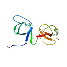
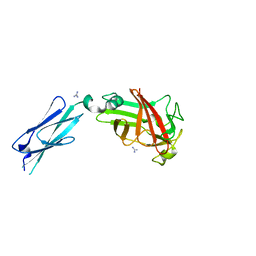
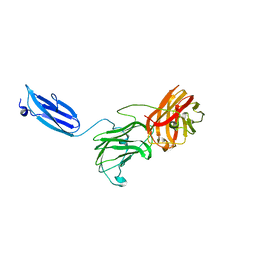
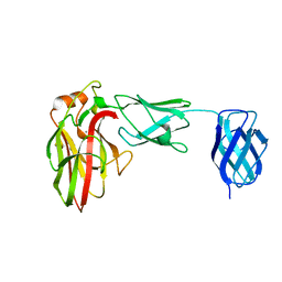
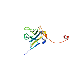
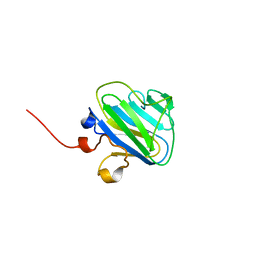
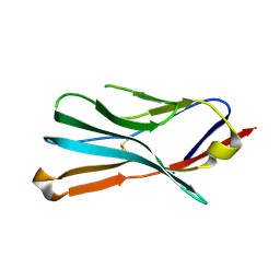
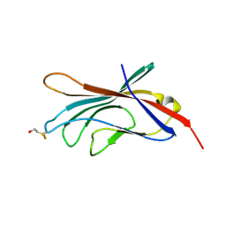
Bacteroides ovatus mixed-linkage glucan utilization locus (MLGUL) SGBP-A
Descriptor: 1,2-ETHANEDIOL, MAGNESIUM ION, mixed-linkage glucan utilization locus (MLGUL) SGBP-B
Authors:Tamura, K, Gardill, B.R, Brumer, H, Van Petegem, F.
Deposit date:2018-07-23
Release date:2019-05-15
Last modified:2024-04-03
Method:X-RAY DIFFRACTION (1.5 Å)
Cite:Surface glycan-binding proteins are essential for cereal beta-glucan utilization by the human gut symbiont Bacteroides ovatus.
Cell.Mol.Life Sci., 76, 2019
6DAF
DownloadVisualize
BU of 6daf by Molmil
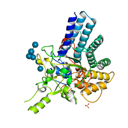
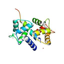
2.4 Angstrom crystal structure of the F141L Ca/CaM:CaV1.2 IQ domain complex
Descriptor: CALCIUM ION, Calmodulin-1, Voltage-dependent L-type calcium channel subunit alpha-1C
Authors:Wang, K, Van Petegem, F.
Deposit date:2018-05-01
Release date:2018-10-17
Last modified:2023-10-04
Method:X-RAY DIFFRACTION (2.4 Å)
Cite:Arrhythmia mutations in calmodulin cause conformational changes that affect interactions with the cardiac voltage-gated calcium channel.
Proc. Natl. Acad. Sci. U.S.A., 115, 2018
6UY9
DownloadVisualize
BU of 6uy9 by Molmil
Crystal structure of the STAC3 tandem SH3 domains - P269R, W284S
Descriptor: 1,2-ETHANEDIOL, SH3 and cysteine-rich domain-containing protein 3, SODIUM ION
Authors:Rufenach, B, Van Petegem, F.
Deposit date:2019-11-12
Release date:2020-06-17
Last modified:2023-10-11
Method:X-RAY DIFFRACTION (1.6 Å)
Cite:Multiple Sequence Variants in STAC3 Affect Interactions with CaV1.1 and Excitation-Contraction Coupling.
Structure, 28, 2020
7KV6
DownloadVisualize
BU of 7kv6 by Molmil
Surface glycan-binding protein B from Bacteroides fluxus in complex with mixed-linkage glucotriose
Descriptor: GUANIDINE, PKD domain protein, beta-D-glucopyranose-(1-4)-beta-D-glucopyranose-(1-3)-beta-D-glucopyranose
Authors:Tamura, K, Brumer, H, Van Petegem, F.
Deposit date:2020-11-26
Release date:2021-02-17
Last modified:2024-03-06
Method:X-RAY DIFFRACTION (1.6 Å)
Cite:Distinct protein architectures mediate species-specific beta-glucan binding and metabolism in the human gut microbiota.
J.Biol.Chem., 296, 2021
7KWB
DownloadVisualize
BU of 7kwb by Molmil
Surface glycan-binding protein B from Bacteroides thetaiotaomicron
Descriptor: BtSGBP-B
Authors:Tamura, K, Brumer, H, Van Petegem, F.
Deposit date:2020-11-30
Release date:2021-02-17
Last modified:2024-03-06
Method:X-RAY DIFFRACTION (2.6 Å)
Cite:Distinct protein architectures mediate species-specific beta-glucan binding and metabolism in the human gut microbiota.
J.Biol.Chem., 296, 2021
7KV3
DownloadVisualize
BU of 7kv3 by Molmil
Surface glycan-binding protein A from Bacteroides thetaiotaomicron in complex with laminarihexaose
Descriptor: RagB/SusD family nutrient uptake outer membrane protein, SULFATE ION, beta-D-glucopyranose-(1-3)-beta-D-glucopyranose-(1-3)-beta-D-glucopyranose-(1-3)-beta-D-glucopyranose-(1-3)-beta-D-glucopyranose-(1-3)-beta-D-glucopyranose
Authors:Tamura, K, Brumer, H, Van Petegem, F.
Deposit date:2020-11-26
Release date:2021-02-17
Last modified:2024-03-06
Method:X-RAY DIFFRACTION (2.05 Å)
Cite:Distinct protein architectures mediate species-specific beta-glucan binding and metabolism in the human gut microbiota.
J.Biol.Chem., 296, 2021
7KV4
DownloadVisualize
BU of 7kv4 by Molmil
Surface glycan-binding protein A from Bacteroides fluxus
Descriptor: SusD family protein
Authors:Tamura, K, Brumer, H, Van Petegem, F.
Deposit date:2020-11-26
Release date:2021-02-17
Last modified:2024-03-06
Method:X-RAY DIFFRACTION (1.84 Å)
Cite:Distinct protein architectures mediate species-specific beta-glucan binding and metabolism in the human gut microbiota.
J.Biol.Chem., 296, 2021
7KWC
DownloadVisualize
BU of 7kwc by Molmil
Surface glycan-binding protein B (truncated) from Bacteroides thetaiotaomicron
Descriptor: BtSGBP-B_31-285
Authors:Tamura, K, Brumer, H, Van Petegem, F.
Deposit date:2020-11-30
Release date:2021-02-17
Last modified:2024-03-06
Method:X-RAY DIFFRACTION (2.611 Å)
Cite:Distinct protein architectures mediate species-specific beta-glucan binding and metabolism in the human gut microbiota.
J.Biol.Chem., 296, 2021
3OXQ
DownloadVisualize
BU of 3oxq by Molmil
Crystal Structure of Ca2+/CaM-CaV1.2 pre-IQ/IQ domain complex
Descriptor: CALCIUM ION, Calmodulin, Voltage-dependent L-type calcium channel subunit alpha-1C
Authors:Kim, E.Y, Rumpf, C.H, Van Petegem, F, Arant, R, Findeisen, F, Cooley, E.S, Isacoff, E.Y, Minor, D.L.
Deposit date:2010-09-21
Release date:2010-11-03
Last modified:2023-09-06
Method:X-RAY DIFFRACTION (2.55 Å)
Cite:Multiple C-terminal tail Ca(2+)/CaMs regulate Ca(V)1.2 function but do not mediate channel dimerization.
Embo J., 29, 2010
3DVJ
DownloadVisualize
BU of 3dvj by Molmil
Crystal Structure of Ca2+/CaM-CaV2.2 IQ domain (without cloning artifact, HM to TV) complex
Descriptor: CALCIUM ION, Calmodulin, Voltage-dependent N-type calcium channel subunit alpha-1B
Authors:Kim, E.Y, Rumpf, C.H, Fujiwara, Y, Cooley, E.S, Van Petegem, F, Minor, D.L.
Deposit date:2008-07-18
Release date:2008-11-04
Last modified:2024-02-21
Method:X-RAY DIFFRACTION (2.8 Å)
Cite:Structures of Ca(V)2 Ca(2+)/CaM-IQ Domain Complexes Reveal Binding Modes that Underlie Calcium-Dependent Inactivation and Facilitation.
Structure, 16, 2008
3DVE
DownloadVisualize
BU of 3dve by Molmil
Crystal Structure of Ca2+/CaM-CaV2.2 IQ domain complex
Descriptor: CALCIUM ION, Calmodulin, NICKEL (II) ION, ...
Authors:Kim, E.Y, Rumpf, C.H, Fujiwara, Y, Cooley, E.S, Van Petegem, F, Minor, D.L.
Deposit date:2008-07-18
Release date:2008-11-04
Last modified:2024-02-21
Method:X-RAY DIFFRACTION (2.35 Å)
Cite:Structures of Ca(V)2 Ca(2+)/CaM-IQ Domain Complexes Reveal Binding Modes that Underlie Calcium-Dependent Inactivation and Facilitation.
Structure, 16, 2008
3DVM
DownloadVisualize
BU of 3dvm by Molmil
Crystal Structure of Ca2+/CaM-CaV2.1 IQ domain complex
Descriptor: CALCIUM ION, Calmodulin, Voltage-dependent P/Q-type calcium channel subunit alpha-1A
Authors:Kim, E.Y, Rumpf, C.H, Fujiwara, Y, Cooley, E.S, Van Petegem, F, Minor, D.L.
Deposit date:2008-07-18
Release date:2008-11-04
Last modified:2024-02-21
Method:X-RAY DIFFRACTION (2.6 Å)
Cite:Structures of Ca(V)2 Ca(2+)/CaM-IQ Domain Complexes Reveal Binding Modes that Underlie Calcium-Dependent Inactivation and Facilitation.
Structure, 16, 2008
3DVK
DownloadVisualize
BU of 3dvk by Molmil
Crystal Structure of Ca2+/CaM-CaV2.3 IQ domain complex
Descriptor: CALCIUM ION, Calmodulin, Voltage-dependent R-type calcium channel subunit alpha-1E
Authors:Kim, E.Y, Rumpf, C.H, Fujiwara, Y, Cooley, E.S, Van Petegem, F, Minor, D.L.
Deposit date:2008-07-18
Release date:2008-11-04
Last modified:2024-02-21
Method:X-RAY DIFFRACTION (2.3 Å)
Cite:Structures of Ca(V)2 Ca(2+)/CaM-IQ Domain Complexes Reveal Binding Modes that Underlie Calcium-Dependent Inactivation and Facilitation.
Structure, 16, 2008
7K44
DownloadVisualize
BU of 7k44 by Molmil
SGBP-B from a complex xyloglucan utilization locus in Bacteroides uniformis
Descriptor: CALCIUM ION, SGBP-B
Authors:Brumer, H, Van Petegem, F, Grondin, J.M.
Deposit date:2020-09-14
Release date:2021-09-29
Last modified:2023-10-18
Method:X-RAY DIFFRACTION (1.45 Å)
Cite:Cell Surface Xyloglucan Recognition and Hydrolysis by the Human Gut Commensal Bacteroides uniformis.
Appl.Environ.Microbiol., 88, 2022
3QR5
DownloadVisualize
BU of 3qr5 by Molmil
Structure of the first domain of a cardiac Ryanodine Receptor mutant with exon 3 deleted
Descriptor: Cardiac Ca2+ release channel
Authors:Lobo, P.A, Van Petegem, F.
Deposit date:2011-02-17
Release date:2011-06-08
Last modified:2023-07-26
Method:X-RAY DIFFRACTION (2.3 Å)
Cite:The deletion of exon 3 in the cardiac ryanodine receptor is rescued by beta strand switching.
Structure, 19, 2011
4P9I
DownloadVisualize
BU of 4p9i by Molmil
Crystal Structure of mouse Ryanodine Receptor 2 SPRY2 Domain (1080-1253)
Descriptor: Ryanodine receptor 2
Authors:Lau, K, Van Petegem, F.
Deposit date:2014-04-04
Release date:2014-11-05
Last modified:2023-12-27
Method:X-RAY DIFFRACTION (1.3401 Å)
Cite:Crystal structures of wild type and disease mutant forms of the ryanodine receptor SPRY2 domain.
Nat Commun, 5, 2014
4P9J
DownloadVisualize
BU of 4p9j by Molmil
Crystal Structure of rabbit Ryanodine Receptor 1 SPRY2 Domain (1070-1246)
Descriptor: Ryanodine receptor 1
Authors:Lau, K, Van Petegem, F.
Deposit date:2014-04-04
Release date:2014-11-05
Last modified:2023-12-27
Method:X-RAY DIFFRACTION (1.84 Å)
Cite:Crystal structures of wild type and disease mutant forms of the ryanodine receptor SPRY2 domain.
Nat Commun, 5, 2014
4P9L
DownloadVisualize
BU of 4p9l by Molmil
Crystal Structure of mouse Ryanodine Receptor 2 SPRY2 Domain (1080-1253) disease mutant A1107M
Descriptor: Ryanodine receptor 2
Authors:Lau, K, Van Petegem, F.
Deposit date:2014-04-04
Release date:2014-11-05
Last modified:2023-12-27
Method:X-RAY DIFFRACTION (1.4361 Å)
Cite:Crystal structures of wild type and disease mutant forms of the ryanodine receptor SPRY2 domain.
Nat Commun, 5, 2014
4MZ2
DownloadVisualize
BU of 4mz2 by Molmil
Crystal structure of the voltage-gated sodium channel beta 4 subunit extracellular domain
Descriptor: Sodium channel subunit beta-4
Authors:Das, S, Van Petegem, F.
Deposit date:2013-09-28
Release date:2013-12-04
Last modified:2023-09-20
Method:X-RAY DIFFRACTION (1.722 Å)
Cite:Crystallographic insights into sodium-channel modulation by the beta 4 subunit.
Proc.Natl.Acad.Sci.USA, 110, 2013
4MZ3
DownloadVisualize
BU of 4mz3 by Molmil
Crystal structure of the voltage-gated sodium channel beta 4 subunit extracellular domain, C131W mutant
Descriptor: Sodium channel subunit beta-4
Authors:Das, S, Van Petegem, F.
Deposit date:2013-09-28
Release date:2013-12-04
Last modified:2023-09-20
Method:X-RAY DIFFRACTION (1.741 Å)
Cite:Crystallographic insights into sodium-channel modulation by the beta 4 subunit.
Proc.Natl.Acad.Sci.USA, 110, 2013
3ILA
DownloadVisualize
BU of 3ila by Molmil
Crystal structure of rabbit ryanodine receptor 1 N-terminal domain (9-205)
Descriptor: Ryanodine receptor 1
Authors:Lobo, P.A, Van Petegem, F.
Deposit date:2009-08-06
Release date:2009-11-17
Last modified:2024-04-03
Method:X-RAY DIFFRACTION (2.9 Å)
Cite:Crystal structures of the N-terminal domains of cardiac and skeletal muscle ryanodine receptors: insights into disease mutations.
Structure, 17, 2009
4KEI
DownloadVisualize
BU of 4kei by Molmil
Crystal structure of mouse Ryanodine Receptor 2 (1-217) disease mutant P164S
Descriptor: Ryanodine receptor 2
Authors:Kimlicka, L, Van Petegem, F.
Deposit date:2013-04-25
Release date:2013-09-04
Last modified:2023-09-20
Method:X-RAY DIFFRACTION (2.406 Å)
Cite:Type 2 Ryanodine Receptor Domain A Contains a Unique and Dynamic alpha-Helix That Transitions to a beta-Strand in a Mutant Linked with a Heritable Cardiomyopathy.
J.Mol.Biol., 425, 2013
4KEK
DownloadVisualize
BU of 4kek by Molmil
Crystal structure of mouse Ryanodine Receptor 2 (1-217) disease mutant R176Q
Descriptor: Ryanodine receptor 2, SULFATE ION
Authors:Kimlicka, L, Van Petegem, F.
Deposit date:2013-04-25
Release date:2013-09-04
Last modified:2023-09-20
Method:X-RAY DIFFRACTION (2.146 Å)
Cite:Type 2 Ryanodine Receptor Domain A Contains a Unique and Dynamic alpha-Helix That Transitions to a beta-Strand in a Mutant Linked with a Heritable Cardiomyopathy.
J.Mol.Biol., 425, 2013

223532

PDB entries from 2024-08-07

PDB statisticsPDBj update infoContact PDBjnumon