7V8E
 
 | |
7V8F
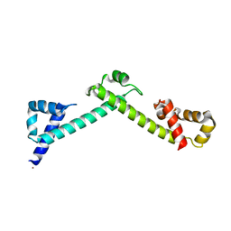
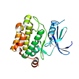
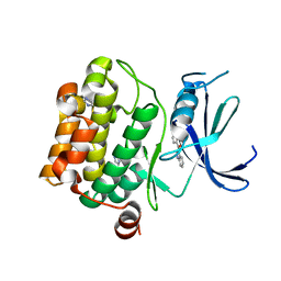
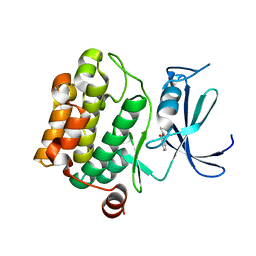
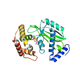
 | | Crystal structure of UBE2L3 bound to HOIP RING1 domain. | | 分子名称: | E3 ubiquitin-protein ligase RNF31, Ubiquitin-conjugating enzyme E2 L3, ZINC ION | | 著者 | Liu, J, Wang, Y, Pan, L. | | 登録日 | 2021-08-22 | | 公開日 | 2022-03-30 | | 最終更新日 | 2023-11-29 | | 実験手法 | X-RAY DIFFRACTION (1.66 Å) | | 主引用文献 | Mechanistic insights into the subversion of the linear ubiquitin chain assembly complex by the E3 ligase IpaH1.4 of Shigella flexneri.
Proc.Natl.Acad.Sci.USA, 119, 2022
|
|
7V8H
 | |
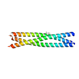
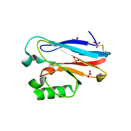
4FQG
 | | Crystal structure of the TCERG1 FF4-6 tandem repeat domain | | 分子名称: | CHLORIDE ION, NICKEL (II) ION, Transcription elongation regulator 1 | | 著者 | Liu, J, Fan, S, Lee, C.J, Greenleaf, A.L, Zhou, P. | | 登録日 | 2012-06-25 | | 公開日 | 2013-02-27 | | 最終更新日 | 2024-03-13 | | 実験手法 | X-RAY DIFFRACTION (2 Å) | | 主引用文献 | Specific Interaction of the Transcription Elongation Regulator TCERG1 with RNA Polymerase II Requires Simultaneous Phosphorylation at Ser2, Ser5, and Ser7 within the Carboxyl-terminal Domain Repeat.
J.Biol.Chem., 288, 2013
|
|
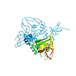
7V59
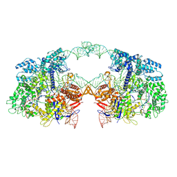 | | Cryo-EM structure of spyCas9-sgRNA-DNA dimer | | 分子名称: | CRISPR-associated endonuclease Cas9/Csn1, DNA (49-MER), RNA (115-MER) | | 著者 | Liu, J, Deng, P. | | 登録日 | 2021-08-16 | | 公開日 | 2022-08-17 | | 最終更新日 | 2024-06-12 | | 実験手法 | ELECTRON MICROSCOPY (5.26 Å) | | 主引用文献 | Nonspecific interactions between SpCas9 and dsDNA sites located downstream of the PAM mediate facilitated diffusion to accelerate target search.
Chem Sci, 12, 2021
|
|
4HLR
 | |
3U91
 | |
3UIA
 | |
3VBX
 | |
3VC4
 | |
3VBT
 | |
3VBV
 | |
3VBQ
 | |
5WEH
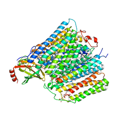 | | Cytochrome c oxidase from Rhodobacter sphaeroides in the reduced state | | 分子名称: | 1,2-Distearoyl-sn-glycerophosphoethanolamine, Aa3-type cytochrome c oxidase subunit IV, CALCIUM ION, ... | | 著者 | Liu, J, Ferguson-Miller, F, Ling, Q, Hiser, C. | | 登録日 | 2017-07-10 | | 公開日 | 2017-09-13 | | 最終更新日 | 2023-10-04 | | 実験手法 | X-RAY DIFFRACTION (3.45 Å) | | 主引用文献 | Role of conformational change and K-path ligands in controlling cytochrome c oxidase activity.
Biochem. Soc. Trans., 45, 2017
|
|
3VBY
 | |
3VBW
 | |
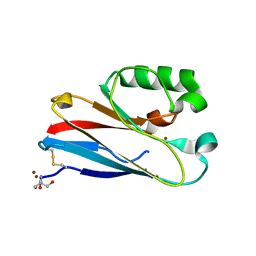
5X0W
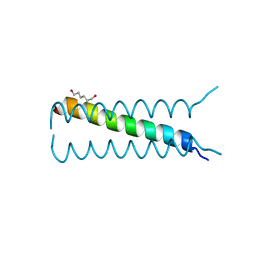
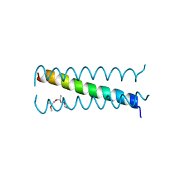
 | | Molecular mechanism for the binding between Sharpin and HOIP | | 分子名称: | E3 ubiquitin-protein ligase RNF31, Sharpin | | 著者 | Liu, J, Li, F, Cheng, X, Pan, L. | | 登録日 | 2017-01-23 | | 公開日 | 2017-10-18 | | 実験手法 | X-RAY DIFFRACTION (3 Å) | | 主引用文献 | Structural Insights into SHARPIN-Mediated Activation of HOIP for the Linear Ubiquitin Chain Assembly
Cell Rep, 21, 2017
|
|
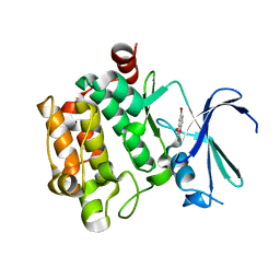
4QLW
 | | Azurin mutant M121E with iron | | 分子名称: | Azurin, FE (III) ION, NITRATE ION, ... | | 著者 | Liu, J, Robinson, H, Lu, Y. | | 登録日 | 2014-06-13 | | 公開日 | 2014-08-13 | | 最終更新日 | 2014-10-01 | | 実験手法 | X-RAY DIFFRACTION (2 Å) | | 主引用文献 | Redesigning the Blue Copper Azurin into a Redox-Active Mononuclear Nonheme Iron Protein: Preparation and Study of Fe(II)-M121E Azurin.
J.Am.Chem.Soc., 136, 2014
|
|
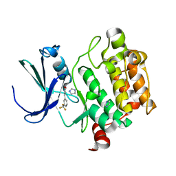
4QKT
 | | Azurin mutant M121EM44K with copper | | 分子名称: | 2-AMINO-2-HYDROXYMETHYL-PROPANE-1,3-DIOL, ACETATE ION, Azurin, ... | | 著者 | Liu, J, Robinson, H, Lu, Y. | | 登録日 | 2014-06-09 | | 公開日 | 2014-08-13 | | 最終更新日 | 2014-10-01 | | 実験手法 | X-RAY DIFFRACTION (1.641 Å) | | 主引用文献 | Redesigning the Blue Copper Azurin into a Redox-Active Mononuclear Nonheme Iron Protein: Preparation and Study of Fe(II)-M121E Azurin.
J.Am.Chem.Soc., 136, 2014
|
|
7D6H
 | |
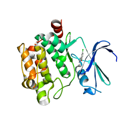
7E35
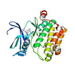
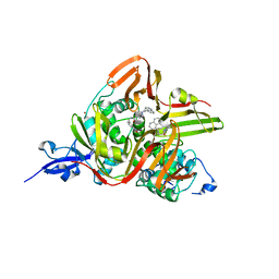 | | Crystal structure of the SARS-CoV-2 papain-like protease (PLPro) C112S mutant bound to compound S43 | | 分子名称: | N-[(3-acetamidophenyl)methyl]-1-[(1R)-1-naphthalen-1-ylethyl]piperidine-4-carboxamide, Non-structural protein 3, ZINC ION | | 著者 | Liu, J, Wang, Y, Xu, X, Pan, L. | | 登録日 | 2021-02-08 | | 公開日 | 2021-03-17 | | 最終更新日 | 2023-11-29 | | 実験手法 | X-RAY DIFFRACTION (2.4 Å) | | 主引用文献 | Development of potent and selective inhibitors targeting the papain-like protease of SARS-CoV-2.
Cell Chem Biol, 28, 2021
|
|
8XHR
 | |
7D65
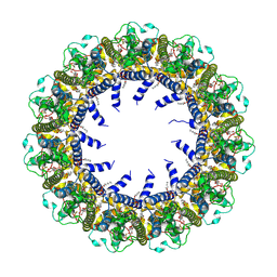 | | Cryo-EM Structure of human CALHM5 in the presence of Ca2+ | | 分子名称: | 1,2-DIOCTANOYL-SN-GLYCERO-3-PHOSPHATE, Calcium homeostasis modulator protein 5 | | 著者 | Liu, J, Guan, F.H, Wu, J, Wan, F.T, Lei, M, Ye, S. | | 登録日 | 2020-09-29 | | 公開日 | 2020-12-23 | | 実験手法 | ELECTRON MICROSCOPY (2.94 Å) | | 主引用文献 | Cryo-EM structures of human calcium homeostasis modulator 5.
Cell Discov, 6, 2020
|
|
7D61
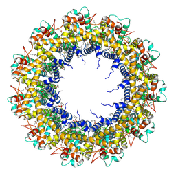 | | Cryo-EM Structure of human CALHM5 in the presence of EDTA | | 分子名称: | 1,2-DIOCTANOYL-SN-GLYCERO-3-PHOSPHATE, Calcium homeostasis modulator protein 5 | | 著者 | Liu, J, Guan, F.H, Wu, J, Wan, F.T, Lei, M, Ye, S. | | 登録日 | 2020-09-28 | | 公開日 | 2020-12-23 | | 実験手法 | ELECTRON MICROSCOPY (2.8 Å) | | 主引用文献 | Cryo-EM structures of human calcium homeostasis modulator 5.
Cell Discov, 6, 2020
|
|
7D60
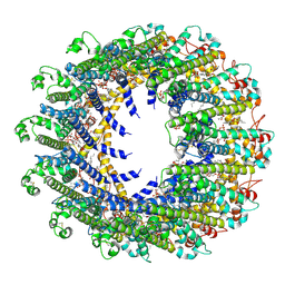 | | Cryo-EM Structure of human CALHM5 in the presence of rubidium red | | 分子名称: | 1,2-DIOCTANOYL-SN-GLYCERO-3-PHOSPHATE, Calcium homeostasis modulator protein 5 | | 著者 | Liu, J, Guan, F.H, Wu, J, Wan, F.T, Lei, M, Ye, S. | | 登録日 | 2020-09-28 | | 公開日 | 2020-12-23 | | 実験手法 | ELECTRON MICROSCOPY (2.61 Å) | | 主引用文献 | Cryo-EM structures of human calcium homeostasis modulator 5.
Cell Discov, 6, 2020
|
|


