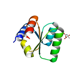
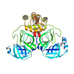
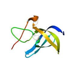
7YUL
 
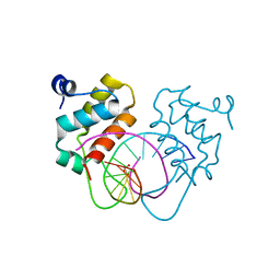 | | Crystal structure of human BEND6 BEN domain in complex with DNA | | 分子名称: | BEN domain-containing protein 6, DNA (5'-D(*CP*TP*CP*TP*CP*GP*CP*GP*AP*GP*AP*G)-3'), GLYCOLIC ACID | | 著者 | Liu, K, Xiao, Y.Q, Zhang, J, Min, J.R. | | 登録日 | 2022-08-17 | | 公開日 | 2023-04-26 | | 最終更新日 | 2023-11-29 | | 実験手法 | X-RAY DIFFRACTION (1.82 Å) | | 主引用文献 | Structural insights into DNA recognition by the BEN domain of the transcription factor BANP.
J.Biol.Chem., 299, 2023
|
|
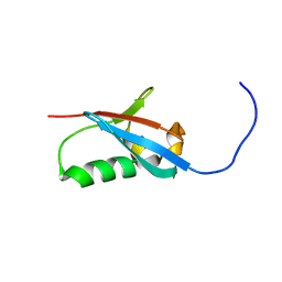
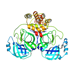
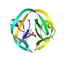
7YUN
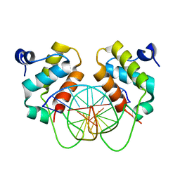 | | Crystal structure of human BEND6 BEN domain in complex with methylated DNA | | 分子名称: | BEN domain-containing protein 6, DNA (5'-D(*CP*TP*CP*TP*CP*GP*(5CM)P*GP*AP*GP*AP*G)-3') | | 著者 | Liu, K, Xiao, Y.Q, Zhang, J, Min, J.R. | | 登録日 | 2022-08-17 | | 公開日 | 2023-05-03 | | 最終更新日 | 2024-05-29 | | 実験手法 | X-RAY DIFFRACTION (2.13 Å) | | 主引用文献 | Structural insights into DNA recognition by the BEN domain of the transcription factor BANP.
J.Biol.Chem., 299, 2023
|
|
5YTE
 | | Large fragment of DNA Polymerase I from Thermus aquaticus in a closed ternary complex with with natural dT:dATP base pair | | 分子名称: | 2'-DEOXYADENOSINE 5'-TRIPHOSPHATE, DNA (5'-D(*AP*AP*AP*TP*GP*GP*CP*GP*CP*CP*GP*TP*GP*GP*TP*C)-3'), DNA (5'-D(*GP*AP*CP*CP*AP*CP*GP*GP*CP*GP*CP*(DOC))-3'), ... | | 著者 | Zeng, H, Mondal, M, Song, R.Y, Zhang, J, Xia, B, Gao, Y.Q, Yi, C.Q. | | 登録日 | 2017-11-17 | | 公開日 | 2018-11-21 | | 最終更新日 | 2023-11-22 | | 実験手法 | X-RAY DIFFRACTION (2.21 Å) | | 主引用文献 | Unnatural Cytosine Bases Recognized as Thymines by DNA Polymerases by the Formation of the Watson-Crick Geometry.
Angew. Chem. Int. Ed. Engl., 58, 2019
|
|
5YBB
 | | Structural basis underlying complex assembly andconformational transition of the type I R-M system | | 分子名称: | DNA, Restriction endonuclease S subunits, S-ADENOSYLMETHIONINE, ... | | 著者 | Liu, Y.P, Tang, Q, Zhang, J.Z, Tian, L.F, Gao, P, Yan, X.X. | | 登録日 | 2017-09-04 | | 公開日 | 2017-11-29 | | 最終更新日 | 2018-02-07 | | 実験手法 | X-RAY DIFFRACTION (3.2 Å) | | 主引用文献 | Structural basis underlying complex assembly and conformational transition of the type I R-M system.
Proc. Natl. Acad. Sci. U.S.A., 114, 2017
|
|
2KLI
 | | Structural Basis for the Photoconversion of A Phytochrome to the Activated FAR-RED LIGHT-ABSORBING Form | | 分子名称: | PHYCOCYANOBILIN, Sensor protein | | 著者 | Cornilescu, C.C, Cornilescu, G, Ulijasz, A.T, Zhang, J, Rivera, M, Vierstra, R.D, Markley, J.L, Center for Eukaryotic Structural Genomics (CESG) | | 登録日 | 2009-07-03 | | 公開日 | 2009-11-03 | | 最終更新日 | 2022-03-16 | | 実験手法 | SOLUTION NMR | | 主引用文献 | Structural basis for the photoconversion of a phytochrome to the activated Pfr form
Nature, 463, 2010
|
|
3V7Q
 | | Crystal structure of B. subtilis YlxQ at 1.55 A resolution | | 分子名称: | CITRIC ACID, POTASSIUM ION, Probable ribosomal protein ylxQ | | 著者 | Baird, N.J, Zhang, J, Hamma, T, Ferre-D'Amare, A.R. | | 登録日 | 2011-12-21 | | 公開日 | 2012-03-07 | | 最終更新日 | 2023-09-13 | | 実験手法 | X-RAY DIFFRACTION (1.55 Å) | | 主引用文献 | YbxF and YlxQ are bacterial homologs of L7Ae and bind K-turns but not K-loops.
Rna, 18, 2012
|
|
2KXJ
 | | Solution structure of UBX domain of human UBXD2 protein | | 分子名称: | UBX domain-containing protein 4 | | 著者 | Wu, Q, Huang, H, Zhang, J, Hu, Q, Wu, J, Shi, Y. | | 登録日 | 2010-05-06 | | 公開日 | 2011-05-18 | | 最終更新日 | 2024-05-01 | | 実験手法 | SOLUTION NMR | | 主引用文献 | Solution strcture of UBX domain of human UBXD2 protein
To be Published
|
|
4Q2W
 | | Crystal Structure of pneumococcal peptidoglycan hydrolase LytB | | 分子名称: | GLYCEROL, Putative endo-beta-N-acetylglucosaminidase | | 著者 | Bai, X.H, Chen, H.J, Jiang, Y.L, Wen, Z, Cheng, W, Li, Q, Zhang, J.R, Chen, Y, Zhou, C.Z. | | 登録日 | 2014-04-10 | | 公開日 | 2014-07-16 | | 最終更新日 | 2019-12-18 | | 実験手法 | X-RAY DIFFRACTION (1.65 Å) | | 主引用文献 | Structure of pneumococcal peptidoglycan hydrolase LytB reveals insights into the bacterial cell wall remodeling and pathogenesis.
J.Biol.Chem., 289, 2014
|
|
5HJG
 | | Crystal Structure of Human Transthyretin in Complex with Tetrabromobisphenol A (TBBPA) | | 分子名称: | 4,4'-propane-2,2-diylbis(2,6-dibromophenol), SODIUM ION, Transthyretin | | 著者 | Begum, A, Iakovleva, I, Brannstrom, K, Zhang, J, Andersson, P, Olofsson, A, Sauer-Eriksson, A.E. | | 登録日 | 2016-01-13 | | 公開日 | 2016-05-04 | | 最終更新日 | 2024-01-10 | | 実験手法 | X-RAY DIFFRACTION (1.4 Å) | | 主引用文献 | Tetrabromobisphenol A Is an Efficient Stabilizer of the Transthyretin Tetramer.
Plos One, 11, 2016
|
|
7X7H
 | | Crystal structure of Fructose regulator/Histidine phosphocarrier protein complex from Vibrio cholerae | | 分子名称: | CALCIUM ION, Catabolite repressor/activator, HPr family phosphocarrier protein | | 著者 | Kim, M.-K, Zhang, J, Yoon, C.-K, Seok, Y.-J. | | 登録日 | 2022-03-09 | | 公開日 | 2023-03-15 | | 最終更新日 | 2024-05-29 | | 実験手法 | X-RAY DIFFRACTION (2 Å) | | 主引用文献 | HPr prevents FruR-mediated facilitation of RNA polymerase binding to the fru promoter in Vibrio cholerae.
Nucleic Acids Res., 51, 2023
|
|
7XB3
 | |
7XAX
 | |
7WQI
 | | Crystal structure of SARS coronavirus main protease in complex with PF07304814 | | 分子名称: | 3C-like proteinase, [(3~{S})-3-[[(2~{S})-2-[(4-methoxy-1~{H}-indol-2-yl)carbonylamino]-4-methyl-pentanoyl]amino]-2-oxidanylidene-4-[(3~{R})-2-oxidanylidene-3,4-dihydropyrrol-3-yl]butyl] dihydrogen phosphate | | 著者 | Lin, C, Zhong, F.L, Zhou, X.L, Zeng, P, Zhang, J, Li, J. | | 登録日 | 2022-01-25 | | 公開日 | 2023-01-25 | | 最終更新日 | 2023-11-29 | | 実験手法 | X-RAY DIFFRACTION (1.93 Å) | | 主引用文献 | Crystal structure of SARS coronavirus main protease in complex with PF07304814
To Be Published
|
|
7XB4
 | | Crystal structure of SARS-Cov-2 main protease D48N mutant in complex with PF07321332 | | 分子名称: | (1R,2S,5S)-N-{(1E,2S)-1-imino-3-[(3S)-2-oxopyrrolidin-3-yl]propan-2-yl}-6,6-dimethyl-3-[3-methyl-N-(trifluoroacetyl)-L-valyl]-3-azabicyclo[3.1.0]hexane-2-carboxamide, Replicase polyprotein 1a | | 著者 | Hu, X.H, Li, J, Zhang, J. | | 登録日 | 2022-03-20 | | 公開日 | 2023-03-22 | | 最終更新日 | 2023-11-29 | | 実験手法 | X-RAY DIFFRACTION (2.07 Å) | | 主引用文献 | Crystal structure of SARS-Cov-2 main protease D48N mutant in complex with PF07321332
To Be Published
|
|
4NTJ
 | | Structure of the human P2Y12 receptor in complex with an antithrombotic drug | | 分子名称: | (2R)-2,3-dihydroxypropyl (9Z)-octadec-9-enoate, CHOLESTEROL, P2Y purinoceptor 12,Soluble cytochrome b562,P2Y purinoceptor 12, ... | | 著者 | Zhang, K, Zhang, J, Gao, Z.-G, Zhang, D, Zhu, L, Han, G.W, Moss, S.M, Paoletta, S, Kiselev, E, Lu, W, Fenalti, G, Zhang, W, Muller, C.E, Yang, H, Jiang, H, Cherezov, V, Katritch, V, Jacobson, K.A, Stevens, R.C, Wu, B, Zhao, Q, GPCR Network (GPCR) | | 登録日 | 2013-12-02 | | 公開日 | 2014-03-26 | | 最終更新日 | 2023-11-08 | | 実験手法 | X-RAY DIFFRACTION (2.62 Å) | | 主引用文献 | Structure of the human P2Y12 receptor in complex with an antithrombotic drug
Nature, 509, 2014
|
|
6LMS
 | |
5I0A
 | | RecA mini intein in complex with cisplatin | | 分子名称: | 3,3',3''-phosphanetriyltripropanoic acid, Cisplatin, Intein, ... | | 著者 | Li, Z, Zhang, J, Li, H.M. | | 登録日 | 2016-02-03 | | 公開日 | 2016-09-21 | | 最終更新日 | 2023-09-27 | | 実験手法 | X-RAY DIFFRACTION (1.5 Å) | | 主引用文献 | Structural insights into protein splicing inhibition by platinum therapeutics as potential anti-microbials
To be published
|
|
7N97
 | | State 2 of TcdB and FZD2 at pH5 | | 分子名称: | Frizzled-2, Toxin B | | 著者 | Jiang, M, Zhang, J. | | 登録日 | 2021-06-17 | | 公開日 | 2022-03-02 | | 最終更新日 | 2024-06-05 | | 実験手法 | ELECTRON MICROSCOPY (5.1 Å) | | 主引用文献 | Structural Basis for Receptor Recognition of the Clostridium difficile Toxin B and its Dissociation upon Acidification
To Be Published
|
|
7N8X
 | | Partial C. difficile TcdB and CSPG4 fragment | | 分子名称: | Chondroitin sulfate proteoglycan 4, Toxin B | | 著者 | Jiang, M, Zhang, J. | | 登録日 | 2021-06-16 | | 公開日 | 2022-03-02 | | 実験手法 | ELECTRON MICROSCOPY (3.4 Å) | | 主引用文献 | Structural Basis for Receptor Recognition of Clostridium difficile Toxin B and its Dissociation upon Acidification
To Be Published
|
|
7N9Q
 | | State 3 of TcdB and FZD2 at pH5 | | 分子名称: | Toxin B | | 著者 | Jiang, M, Zhang, J. | | 登録日 | 2021-06-18 | | 公開日 | 2022-03-02 | | 最終更新日 | 2024-06-05 | | 実験手法 | ELECTRON MICROSCOPY (4.6 Å) | | 主引用文献 | Structural Basis for Receptor Recognition of Clostridium difficile Toxin B and its Dissociation upon Acidification
To Be Published
|
|
7N9S
 | | TcdB and frizzled-2 CRD complex | | 分子名称: | Frizzled-2, Toxin B | | 著者 | Jiang, M, Zhang, J. | | 登録日 | 2021-06-18 | | 公開日 | 2022-03-02 | | 最終更新日 | 2024-06-05 | | 実験手法 | ELECTRON MICROSCOPY (5.1 Å) | | 主引用文献 | Structural Basis for Receptor Recognition of Clostridium difficile Toxin B and its Dissociation upon Acidification
To Be Published
|
|
7N9R
 | | state 4 of TcdB and FZD2 at pH5 | | 分子名称: | Toxin B | | 著者 | Jiang, M, Zhang, J. | | 登録日 | 2021-06-18 | | 公開日 | 2022-03-02 | | 最終更新日 | 2024-06-05 | | 実験手法 | ELECTRON MICROSCOPY (5.9 Å) | | 主引用文献 | Structural Basis for Receptor Recognition of Clostridium difficile Toxin B and its Dissociation upon Acidification
To Be Published
|
|
7N9Y
 | | Full-length TcdB and CSPG4 (401-560) complex | | 分子名称: | Chondroitin sulfate proteoglycan 4, Toxin B | | 著者 | Jiang, M, Zhang, J. | | 登録日 | 2021-06-18 | | 公開日 | 2022-03-02 | | 最終更新日 | 2024-06-05 | | 実験手法 | ELECTRON MICROSCOPY (4.8 Å) | | 主引用文献 | Structural Basis for Receptor Recognition of Clostridium difficile Toxin B and its Dissociation upon Acidification
To Be Published
|
|
7N95
 | | state 1 of TcdB and FZD2 at pH5 | | 分子名称: | Frizzled-2, Toxin B | | 著者 | Jiang, M, Zhang, J. | | 登録日 | 2021-06-16 | | 公開日 | 2022-03-02 | | 最終更新日 | 2024-06-05 | | 実験手法 | ELECTRON MICROSCOPY (4.1 Å) | | 主引用文献 | Structural Basis for Receptor Recognition of Clostridium difficile Toxin B and its Dissociation upon Acidification
To Be Published
|
|
3QYC
 | | Structure of a dimeric anti-HER2 single domain antibody | | 分子名称: | VH domain of IgG molecule | | 著者 | Baral, T.N, Chao, S, Li, S, Tanha, J, Arbabai, M, Wang, S, Zhang, J. | | 登録日 | 2011-03-03 | | 公開日 | 2012-02-08 | | 最終更新日 | 2014-02-05 | | 実験手法 | X-RAY DIFFRACTION (1.6 Å) | | 主引用文献 | Crystal Structure of a Human Single Domain Antibody Dimer Formed through V(H)-V(H) Non-Covalent Interactions.
Plos One, 7, 2012
|
|


