Loading
PDBj
MenuPDBj@FacebookPDBj@TwitterPDBj@YouTubewwPDB FoundationwwPDB
RCSB PDBPDBeBMRBAdv. SearchSearch help
PDB: 20 results

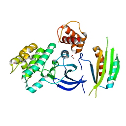
4YNZ
DownloadVisualize
BU of 4ynz by Molmil
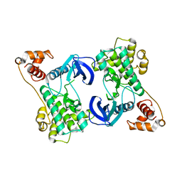
Structure of the N-terminal domain of SAD
Descriptor: Serine/threonine-protein kinase BRSK2
Authors:Wu, J.X, Wang, J, Chen, L, Wang, Z.X, Wu, J.W.
Deposit date:2015-03-11
Release date:2015-12-16
Last modified:2023-11-08
Method:X-RAY DIFFRACTION (2 Å)
Cite:Structural insight into the mechanism of synergistic autoinhibition of SAD kinases
Nat Commun, 6, 2015
4YOM
DownloadVisualize
BU of 4yom by Molmil
Structure of SAD kinase
Descriptor: 1,2-ETHANEDIOL, Serine/threonine-protein kinase BRSK2
Authors:Wu, J.X, Wang, J, Chen, L, Wang, Z.X, Wu, J.W.
Deposit date:2015-03-12
Release date:2015-12-16
Last modified:2023-11-08
Method:X-RAY DIFFRACTION (2.49 Å)
Cite:Structural insight into the mechanism of synergistic autoinhibition of SAD kinases
Nat Commun, 6, 2015
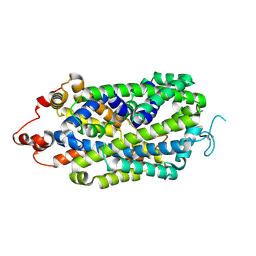
8XB4
DownloadVisualize
BU of 8xb4 by Molmil
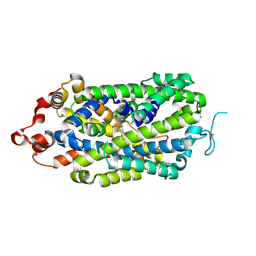
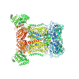
Structure of apo state of the human Norepinephrine Transporter
Descriptor: 2-acetamido-2-deoxy-beta-D-glucopyranose, GFP-MBP-solute carrier family 6 member 2
Authors:Wu, J.X, Ji, W.M.
Deposit date:2023-12-05
Release date:2024-06-26
Last modified:2024-09-25
Method:ELECTRON MICROSCOPY (2.92 Å)
Cite:Substrate binding and inhibition mechanism of norepinephrine transporter.
Nature, 633, 2024
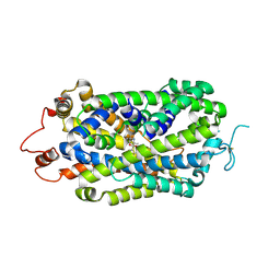
8XB2
DownloadVisualize
BU of 8xb2 by Molmil
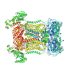
Structure of radafaxine-bound state of the human Norepinephrine Transporter
Descriptor: (2~{S},3~{S})-2-(3-chlorophenyl)-3,5,5-trimethyl-morpholin-2-ol, 2-acetamido-2-deoxy-beta-D-glucopyranose, GFP-MBP-solute carrier family 6 member 2,Maltose/maltodextrin-binding periplasmic protein,Sodium-dependent noradrenaline transporter,Maltose/maltodextrin-binding periplasmic protein,Sodium-dependent noradrenaline transporter
Authors:Wu, J.X, Ji, W.M.
Deposit date:2023-12-05
Release date:2024-07-10
Last modified:2024-10-16
Method:ELECTRON MICROSCOPY (3.04 Å)
Cite:Substrate binding and inhibition mechanism of norepinephrine transporter.
Nature, 633, 2024
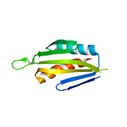
5IRI
DownloadVisualize
BU of 5iri by Molmil
Structure of the mouse SAD-B AIS-KA1 fragment
Descriptor: Serine/threonine-protein kinase BRSK1
Authors:Ma, H, Wu, J.X, Wang, J, Wu, J.W.
Deposit date:2016-03-13
Release date:2016-06-15
Last modified:2023-11-08
Method:X-RAY DIFFRACTION (2.8 Å)
Cite:Structure and inhibition analysis of the mouse SAD-B C-terminal fragment
Biosci.Biotechnol.Biochem., 2016
8XB3
DownloadVisualize
BU of 8xb3 by Molmil
Structural mechanism of substrate binding and inhibition of the human Norepinephrine Transporter
Descriptor: 1-[(3-iodanylphenyl)methyl]guanidine, 2-acetamido-2-deoxy-beta-D-glucopyranose, GFP-MBP-solute carrier family 6 member 2
Authors:Ji, W.M, Wu, J.X.
Deposit date:2023-12-05
Release date:2024-06-26
Last modified:2024-09-25
Method:ELECTRON MICROSCOPY (2.8 Å)
Cite:Substrate binding and inhibition mechanism of norepinephrine transporter.
Nature, 633, 2024
5YKF
DownloadVisualize
BU of 5ykf by Molmil
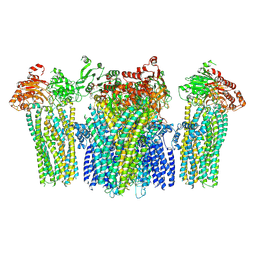
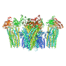
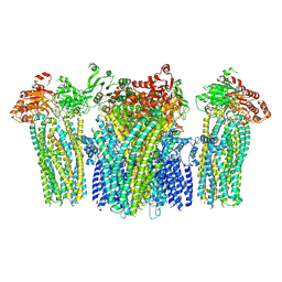
Structure of pancreatic ATP-sensitive potassium channel bound with glibenclamide and ATPgammaS (3D class1 at 4.33A)
Descriptor: 5-chloro-N-(2-{4-[(cyclohexylcarbamoyl)sulfamoyl]phenyl}ethyl)-2-methoxybenzamide, ATP-binding cassette sub-family C member 8 isoform X2, ATP-sensitive inward rectifier potassium channel 11, ...
Authors:Chen, L, Wu, J.X.
Deposit date:2017-10-14
Release date:2018-04-18
Last modified:2019-11-06
Method:ELECTRON MICROSCOPY (4.33 Å)
Cite:Ligand binding and conformational changes of SUR1 subunit in pancreatic ATP-sensitive potassium channels.
Protein Cell, 9, 2018
5YKG
DownloadVisualize
BU of 5ykg by Molmil
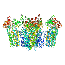
Structure of pancreatic ATP-sensitive potassium channel bound with glibenclamide and ATPgammaS (Class2 at 4.57A)
Descriptor: 5-chloro-N-(2-{4-[(cyclohexylcarbamoyl)sulfamoyl]phenyl}ethyl)-2-methoxybenzamide, ATP-binding cassette sub-family C member 8 isoform X2, ATP-sensitive inward rectifier potassium channel 11, ...
Authors:Chen, L, Wu, J.X.
Deposit date:2017-10-14
Release date:2018-04-18
Last modified:2018-06-13
Method:ELECTRON MICROSCOPY (4.57 Å)
Cite:Ligand binding and conformational changes of SUR1 subunit in pancreatic ATP-sensitive potassium channels.
Protein Cell, 9, 2018
6JPF
DownloadVisualize
BU of 6jpf by Molmil
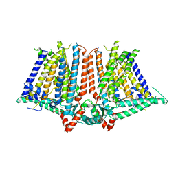
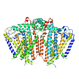
Structure of atOSCA1.1 channel at 3.52A
Descriptor: Protein OSCA1
Authors:Chen, L, Zhang, M, Kang, Y, Wu, J.X.
Deposit date:2019-03-26
Release date:2019-04-10
Last modified:2024-03-27
Method:ELECTRON MICROSCOPY (3.52 Å)
Cite:Structure of the mechanosensitive OSCA channels.
Nat. Struct. Mol. Biol., 25, 2018
5YKE
DownloadVisualize
BU of 5yke by Molmil
Structure of pancreatic ATP-sensitive potassium channel bound with glibenclamide and ATPgammaS (focused refinement on TM at 4.11A)
Descriptor: 5-chloro-N-(2-{4-[(cyclohexylcarbamoyl)sulfamoyl]phenyl}ethyl)-2-methoxybenzamide, ATP-binding cassette sub-family C member 8 isoform X2, ATP-sensitive inward rectifier potassium channel 11
Authors:Chen, L, Wu, J.X.
Deposit date:2017-10-14
Release date:2018-04-18
Last modified:2018-06-13
Method:ELECTRON MICROSCOPY (4.11 Å)
Cite:Ligand binding and conformational changes of SUR1 subunit in pancreatic ATP-sensitive potassium channels.
Protein Cell, 9, 2018
5YW9
DownloadVisualize
BU of 5yw9 by Molmil
Structure of pancreatic ATP-sensitive potassium channel bound with ATPgammaS (class1 5.0A)
Descriptor: ATP-binding cassette sub-family C member 8 isoform X2, ATP-sensitive inward rectifier potassium channel 11, PHOSPHOTHIOPHOSPHORIC ACID-ADENYLATE ESTER
Authors:Chen, L, Wu, J.X.
Deposit date:2017-11-29
Release date:2018-05-02
Last modified:2018-06-13
Method:ELECTRON MICROSCOPY (5 Å)
Cite:Ligand binding and conformational changes of SUR1 subunit in pancreatic ATP-sensitive potassium channels.
Protein Cell, 9, 2018
5YWB
DownloadVisualize
BU of 5ywb by Molmil
Structure of pancreatic ATP-sensitive potassium channel bound with Mg-ADP (CTD class2 at 5.2A)
Descriptor: ADENOSINE-5'-DIPHOSPHATE, ATP-binding cassette sub-family C member 8 isoform X2, ATP-sensitive inward rectifier potassium channel 11, ...
Authors:Chen, L, Wu, J.X.
Deposit date:2017-11-29
Release date:2018-05-30
Last modified:2024-10-23
Method:ELECTRON MICROSCOPY (5.2 Å)
Cite:Ligand binding and conformational changes of SUR1 subunit in pancreatic ATP-sensitive potassium channels.
Protein Cell, 9, 2018
5YWC
DownloadVisualize
BU of 5ywc by Molmil
Structure of pancreatic ATP-sensitive potassium channel bound with Mg-ADP (CTD class1 at 4.3A)
Descriptor: ADENOSINE-5'-DIPHOSPHATE, ATP-binding cassette sub-family C member 8 isoform X2, ATP-sensitive inward rectifier potassium channel 11, ...
Authors:Chen, L, Wu, J.X.
Deposit date:2017-11-29
Release date:2018-05-02
Last modified:2024-10-16
Method:ELECTRON MICROSCOPY (4.3 Å)
Cite:Ligand binding and conformational changes of SUR1 subunit in pancreatic ATP-sensitive potassium channels.
Protein Cell, 9, 2018
5YW8
DownloadVisualize
BU of 5yw8 by Molmil
Structure of pancreatic ATP-sensitive potassium channel bound with ATPgammaS (all particles at 4.4A)
Descriptor: ATP-binding cassette sub-family C member 8 isoform X2, ATP-sensitive inward rectifier potassium channel 11, PHOSPHOTHIOPHOSPHORIC ACID-ADENYLATE ESTER
Authors:Chen, L, Wu, J.X.
Deposit date:2017-11-29
Release date:2018-05-02
Last modified:2024-10-23
Method:ELECTRON MICROSCOPY (4.4 Å)
Cite:Ligand binding and conformational changes of SUR1 subunit in pancreatic ATP-sensitive potassium channels.
Protein Cell, 9, 2018
5Z1F
DownloadVisualize
BU of 5z1f by Molmil
Structure of atOSCA3.1 channel
Descriptor: CSC1-like protein ERD4
Authors:Chen, L, Zhang, M, Kang, Y, Wu, J.X.
Deposit date:2017-12-26
Release date:2018-09-12
Last modified:2024-03-27
Method:ELECTRON MICROSCOPY (4.8 Å)
Cite:Structure of the mechanosensitive OSCA channels.
Nat. Struct. Mol. Biol., 25, 2018
5YWD
DownloadVisualize
BU of 5ywd by Molmil
Structure of pancreatic ATP-sensitive potassium channel bound with Mg-ADP (focused refinement of SUR1 ABC transporter module at 4.22A)
Descriptor: ADENOSINE-5'-DIPHOSPHATE, ATP-binding cassette sub-family C member 8 isoform X2, MAGNESIUM ION
Authors:Chen, L, Wu, J.X.
Deposit date:2017-11-29
Release date:2018-05-02
Last modified:2024-03-27
Method:ELECTRON MICROSCOPY (4.2 Å)
Cite:Ligand binding and conformational changes of SUR1 subunit in pancreatic ATP-sensitive potassium channels.
Protein Cell, 9, 2018
5YW7
DownloadVisualize
BU of 5yw7 by Molmil
Structure of pancreatic ATP-sensitive potassium channel bound with glibenclamide and ATPgammaS (focused refinement on SUR1 ABC transporter module at 4.4A)
Descriptor: 5-chloro-N-(2-{4-[(cyclohexylcarbamoyl)sulfamoyl]phenyl}ethyl)-2-methoxybenzamide, ATP-binding cassette sub-family C member 8 isoform X2, PHOSPHOTHIOPHOSPHORIC ACID-ADENYLATE ESTER
Authors:Chen, L, Wu, J.X.
Deposit date:2017-11-29
Release date:2018-05-02
Last modified:2024-03-27
Method:ELECTRON MICROSCOPY (4.4 Å)
Cite:Ligand binding and conformational changes of SUR1 subunit in pancreatic ATP-sensitive potassium channels.
Protein Cell, 9, 2018
5YWA
DownloadVisualize
BU of 5ywa by Molmil
Structure of pancreatic ATP-sensitive potassium channel bound with ATPgammaS (CTD class 2 at 6.1A)
Descriptor: ATP-binding cassette sub-family C member 8 isoform X2, ATP-sensitive inward rectifier potassium channel 11, PHOSPHOTHIOPHOSPHORIC ACID-ADENYLATE ESTER
Authors:Chen, L, Wu, J.X.
Deposit date:2017-11-29
Release date:2018-05-02
Last modified:2024-10-16
Method:ELECTRON MICROSCOPY (6.1 Å)
Cite:Ligand binding and conformational changes of SUR1 subunit in pancreatic ATP-sensitive potassium channels.
Protein Cell, 9, 2018
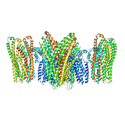
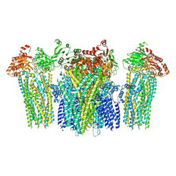
7D3E
DownloadVisualize
BU of 7d3e by Molmil
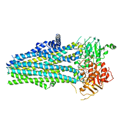
Cryo-EM structure of human DUOX1-DUOXA1 in low-calcium state
Descriptor: 2-acetamido-2-deoxy-beta-D-glucopyranose, Dual oxidase 1, FLAVIN-ADENINE DINUCLEOTIDE, ...
Authors:Chen, L, Wu, J.X.
Deposit date:2020-09-19
Release date:2020-12-09
Last modified:2021-06-23
Method:ELECTRON MICROSCOPY (2.8 Å)
Cite:Structures of human dual oxidase 1 complex in low-calcium and high-calcium states.
Nat Commun, 12, 2021
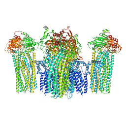
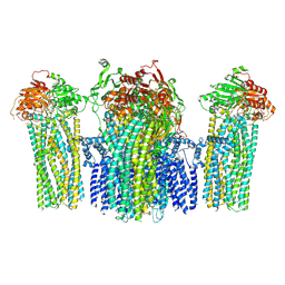
7D3F
DownloadVisualize
BU of 7d3f by Molmil
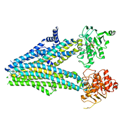
Cryo-EM structure of human DUOX1-DUOXA1 in high-calcium state
Descriptor: 2-acetamido-2-deoxy-beta-D-glucopyranose, CALCIUM ION, Dual oxidase 1, ...
Authors:Chen, L, Wu, J.X.
Deposit date:2020-09-19
Release date:2020-12-09
Last modified:2024-10-23
Method:ELECTRON MICROSCOPY (2.6 Å)
Cite:Structures of human dual oxidase 1 complex in low-calcium and high-calcium states.
Nat Commun, 12, 2021

226707

數據於2024-10-30公開中

PDB statisticsPDBj update infoContact PDBjnumon