3B54
 
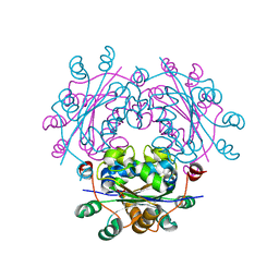 | |
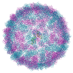
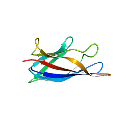
8R0F
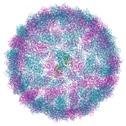 | | Capsid structure of Giardiavirus (GLV) HP strain | | 分子名称: | Capsid protein | | 著者 | Wang, H, Gianluca, M, Munke, A, Hassan, M.M, Lalle, M, Okamoto, K. | | 登録日 | 2023-10-31 | | 公開日 | 2024-04-03 | | 実験手法 | ELECTRON MICROSCOPY (2.14 Å) | | 主引用文献 | Capsid structure of Giardiavirus (GLV) HP strain
To Be Published
|
|
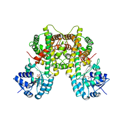
6N5C
 | | Crystal structure of the catalytic domain of PPIP5K2 in complex with AMPPNP and 5-PCF2Am-InsP5 | | 分子名称: | (1,1-difluoro-2-oxo-2-{[(1s,2R,3S,4s,5R,6S)-2,3,4,5,6-pentakis(phosphonooxy)cyclohexyl]amino}ethyl)phosphonic acid, 1,2-ETHANEDIOL, ACETATE ION, ... | | 著者 | Wang, H, Shears, S.B, Riley, A, Potter, B. | | 登録日 | 2018-11-21 | | 公開日 | 2019-08-21 | | 最終更新日 | 2023-10-11 | | 実験手法 | X-RAY DIFFRACTION (1.95 Å) | | 主引用文献 | Synthesis of an alpha-phosphono-alpha , alpha-difluoroacetamide analogue of the diphosphoinositol pentakisphosphate 5-InsP7.
Medchemcomm, 10, 2019
|
|
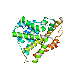
2NZ0
 | | Crystal structure of potassium channel Kv4.3 in complex with its regulatory subunit KChIP1 | | 分子名称: | CALCIUM ION, Kv channel-interacting protein 1, Potassium voltage-gated channel subfamily D member 3, ... | | 著者 | Wang, H, Yan, Y, Shen, Y, Chen, L, Wang, K. | | 登録日 | 2006-11-22 | | 公開日 | 2006-12-26 | | 最終更新日 | 2023-12-27 | | 実験手法 | X-RAY DIFFRACTION (3.2 Å) | | 主引用文献 | Structural basis for modulation of Kv4 K(+) channels by auxiliary KChIP subunits.
Nat.Neurosci., 10, 2007
|
|
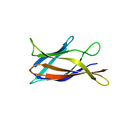
8R0G
 | | Capsid structure of Giardiavirus (GLV) CAT strain | | 分子名称: | Capsid protein | | 著者 | Wang, H, Gianluca, M, Munke, A, Hassan, M.M, Lalle, M, Okamoto, K. | | 登録日 | 2023-10-31 | | 公開日 | 2024-04-03 | | 実験手法 | ELECTRON MICROSCOPY (2.6 Å) | | 主引用文献 | Capsid structure of Giardiavirus (GLV) CAT strain
To Be Published
|
|
5YS5
 | | Crystal structure of Multicopper Oxidase CueO G304K mutant with seven copper ions | | 分子名称: | Blue copper oxidase CueO, COPPER (II) ION | | 著者 | Wang, H.Q, Liu, X.Q, Zhao, J.T, Yue, Q.X, Yan, Y.H, Dong, Y.H, Fan, Y.L, Tian, J, Wu, N.F, Gong, Y. | | 登録日 | 2017-11-13 | | 公開日 | 2018-10-17 | | 最終更新日 | 2023-11-22 | | 実験手法 | X-RAY DIFFRACTION (2.2 Å) | | 主引用文献 | Crystal structures of multicopper oxidase CueO G304K mutant: structural basis of the increased laccase activity
Sci Rep, 8, 2018
|
|
7U2L
 | | C5guano-uOR-Gi-scFv16 | | 分子名称: | Guanine nucleotide-binding protein G(I)/G(S)/G(O) subunit gamma-2, Guanine nucleotide-binding protein G(I)/G(S)/G(T) subunit beta-1, Guanine nucleotide-binding protein G(i) subunit alpha-1, ... | | 著者 | Wang, H, Qu, Q, Skiniotis, G, Kobilka, B. | | 登録日 | 2022-02-24 | | 公開日 | 2022-05-04 | | 最終更新日 | 2023-02-08 | | 実験手法 | ELECTRON MICROSCOPY (3.2 Å) | | 主引用文献 | Structure-based design of bitopic ligands for the μ-opioid receptor.
Nature, 613, 2023
|
|
6HBB
 | | Crystal Structure of the small subunit-like domain 1 of CcmM from Synechococcus elongatus (strain PCC 7942) | | 分子名称: | Carbon dioxide concentrating mechanism protein CcmM, SULFATE ION | | 著者 | Wang, H, Yan, X, Aigner, H, Bracher, A, Nguyen, N.D, Hee, W.Y, Long, B.M, Price, G.D, Hartl, F.U, Hayer-Hartl, M. | | 登録日 | 2018-08-10 | | 公開日 | 2018-12-12 | | 最終更新日 | 2024-01-17 | | 実験手法 | X-RAY DIFFRACTION (1.2 Å) | | 主引用文献 | Rubisco condensate formation by CcmM in beta-carboxysome biogenesis.
Nature, 566, 2019
|
|
7CGD
 | | Silver-bound E.coli malate dehydrogenase | | 分子名称: | Malate dehydrogenase, SILVER ION | | 著者 | Wang, H, Wang, M, Sun, H. | | 登録日 | 2020-07-01 | | 公開日 | 2020-09-23 | | 最終更新日 | 2023-11-29 | | 実験手法 | X-RAY DIFFRACTION (2.06 Å) | | 主引用文献 | Atomic differentiation of silver binding preference in protein targets: Escherichia coli malate dehydrogenase as a paradigm.
Chem Sci, 11, 2020
|
|
3Q3J
 | | Crystal structure of plexin A2 RBD in complex with Rnd1 | | 分子名称: | MAGNESIUM ION, PHOSPHOAMINOPHOSPHONIC ACID-GUANYLATE ESTER, Plexin-A2, ... | | 著者 | Wang, H, Tempel, W, Tong, Y, Guan, X, Shen, L, Buren, L, Zhang, N, Wernimont, A.K, Crombet, L, Arrowsmith, C.H, Edwards, A.M, Bountra, C, Weigelt, J, Park, H, Structural Genomics Consortium (SGC) | | 登録日 | 2010-12-21 | | 公開日 | 2011-01-12 | | 最終更新日 | 2023-09-13 | | 実験手法 | X-RAY DIFFRACTION (1.971 Å) | | 主引用文献 | Crystal structure of plexin A2 RBD in complex with Rnd1
to be published
|
|
2CVL
 | | Crystal structure of TTHA0137 from Thermus Thermophilus HB8 | | 分子名称: | protein translation initiation inhibitor | | 著者 | Wang, H, Murayama, K, Terada, T, Shirouzu, M, Kuramitsu, S, Yokoyama, S, RIKEN Structural Genomics/Proteomics Initiative (RSGI) | | 登録日 | 2005-06-08 | | 公開日 | 2005-12-08 | | 最終更新日 | 2011-07-13 | | 実験手法 | X-RAY DIFFRACTION (1.65 Å) | | 主引用文献 | Crystal structure of TTHA0137 from Thermus Thermophilus HB8
TO BE PUBLISHED
|
|
7CB0
 | |
7CB5
 | |
7CB6
 | |
3USI
 | |
4EGL
 | | Crystal structure of two tandem RNA recognition motifs of Human antigen R | | 分子名称: | ELAV-like protein 1, GLYCEROL, SULFATE ION | | 著者 | Wang, H, Zeng, F, Liu, H, Teng, M, Li, X. | | 登録日 | 2012-03-31 | | 公開日 | 2012-05-30 | | 最終更新日 | 2023-11-08 | | 実験手法 | X-RAY DIFFRACTION (2.9 Å) | | 主引用文献 | Crystal structure of two tandem RNA recognition motifs of Human antigen R
To be Published
|
|
4NND
 | |
2QYK
 | | Crystal structure of PDE4A10 in complex with inhibitor NPV | | 分子名称: | 4-[8-(3-nitrophenyl)-1,7-naphthyridin-6-yl]benzoic acid, Cyclic AMP-specific phosphodiesterase HSPDE4A10, MAGNESIUM ION, ... | | 著者 | Wang, H, Peng, M, Chen, Y, Geng, J, Robinson, H, Houslay, M. | | 登録日 | 2007-08-15 | | 公開日 | 2008-04-08 | | 最終更新日 | 2024-04-03 | | 実験手法 | X-RAY DIFFRACTION (2.1 Å) | | 主引用文献 | Structures of the four subfamilies of phosphodiesterase-4 provide insight into the selectivity of their inhibitors.
Biochem.J., 408, 2007
|
|
2YVR
 | | Crystal structure of MS1043 | | 分子名称: | Transcription intermediary factor 1-beta, ZINC ION | | 著者 | Wang, H, Kishishita, S, Murayama, K, Takemoto, C, Terada, T, Shirouzu, M, RIKEN Structural Genomics/Proteomics Initiative (RSGI) | | 登録日 | 2007-04-13 | | 公開日 | 2008-04-15 | | 最終更新日 | 2024-03-13 | | 実験手法 | X-RAY DIFFRACTION (1.8 Å) | | 主引用文献 | Crystal structure of MS1043
To be Published
|
|
7CB2
 | |
8H1L
 | | Crystal structure of glucose-2-epimerase in complex with D-Glucitol from Runella slithyformis Runsl_4512 | | 分子名称: | N-acylglucosamine 2-epimerase, sorbitol | | 著者 | Wang, H, Sun, X.M, Saburi, W, Yu, J, Yao, M. | | 登録日 | 2022-10-03 | | 公開日 | 2023-07-12 | | 最終更新日 | 2023-11-29 | | 実験手法 | X-RAY DIFFRACTION (2.33 Å) | | 主引用文献 | Structural insights into the substrate specificity and activity of a novel mannose 2-epimerase from Runella slithyformis.
Acta Crystallogr D Struct Biol, 79, 2023
|
|
5WH5
 | |
5YS1
 | | Crystal structure of Multicopper Oxidase CueO G304K mutant | | 分子名称: | Blue copper oxidase CueO, COPPER (II) ION | | 著者 | Wang, H.Q, Liu, X.Q, Zhao, J.T, Yue, Q.X, Yan, Y.H, Dong, Y.H, Fan, Y.L, Tian, J, Wu, N.F, Gong, Y. | | 登録日 | 2017-11-12 | | 公開日 | 2018-10-17 | | 最終更新日 | 2023-11-22 | | 実験手法 | X-RAY DIFFRACTION (1.49 Å) | | 主引用文献 | Crystal structures of multicopper oxidase CueO G304K mutant: structural basis of the increased laccase activity
Sci Rep, 8, 2018
|
|
5J07
 | | Monomeric Human Cu,Zn Superoxide dismutase, loops IV and VII deleted, apo form, circular permutant P1/2 | | 分子名称: | Superoxide dismutase [Cu-Zn],Superoxide dismutase [Cu-Zn],Superoxide dismutase [Cu-Zn],Superoxide dismutase [Cu-Zn] | | 著者 | Wang, H, Lang, L, Logan, D, Danielsson, J, Oliveberg, M. | | 登録日 | 2016-03-27 | | 公開日 | 2017-02-01 | | 最終更新日 | 2024-01-10 | | 実験手法 | X-RAY DIFFRACTION (2 Å) | | 主引用文献 | Tricking a Protein To Swap Strands.
J. Am. Chem. Soc., 138, 2016
|
|
5J0C
 | | Monomeric Human Cu,Zn Superoxide dismutase, loops IV and VII deleted, apo form, circular permutant P2/3 | | 分子名称: | Superoxide dismutase [Cu-Zn],Superoxide dismutase [Cu-Zn],OXIDOREDUCTASE,Superoxide dismutase [Cu-Zn] | | 著者 | Wang, H, Lang, L, Logan, D, Danielsson, J, Oliveberg, M. | | 登録日 | 2016-03-28 | | 公開日 | 2017-02-01 | | 最終更新日 | 2024-01-10 | | 実験手法 | X-RAY DIFFRACTION (1.6 Å) | | 主引用文献 | Tricking a Protein To Swap Strands.
J. Am. Chem. Soc., 138, 2016
|
|


