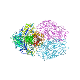
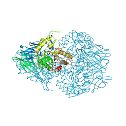
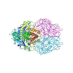
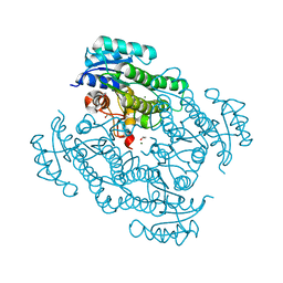
4EQR
 
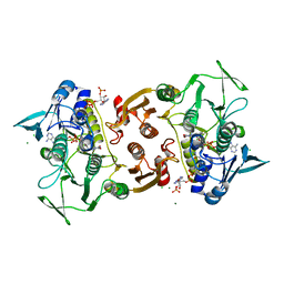 | | Crystal structure of the Y361F mutant of Staphylococcus aureus CoADR | | 分子名称: | CHLORIDE ION, COENZYME A, Coenzyme A disulfide reductase, ... | | 著者 | Wallace, B.D, Edwards, J.S, Wallen, J.R, Claiborne, A, Redinbo, M.R. | | 登録日 | 2012-04-19 | | 公開日 | 2012-10-17 | | 実験手法 | X-RAY DIFFRACTION (1.8 Å) | | 主引用文献 | Turnover-Dependent Covalent Inactivation of Staphylococcus aureus Coenzyme A-Disulfide Reductase by Coenzyme A-Mimetics: Mechanistic and Structural Insights.
Biochemistry, 51, 2012
|
|
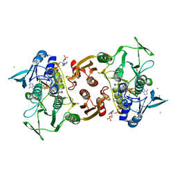
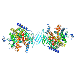
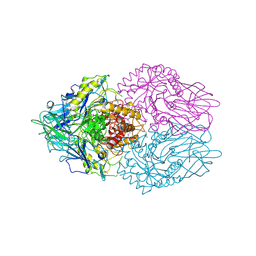
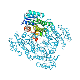
4EM3
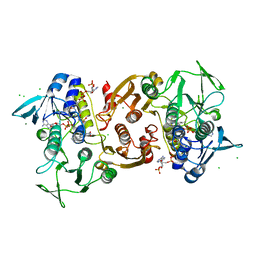 | | Crystal Structure of Staphylococcus aureus bound with the covalent inhibitor MeVS-CoA | | 分子名称: | CHLORIDE ION, Coenzyme A disulfide reductase, FLAVIN-ADENINE DINUCLEOTIDE, ... | | 著者 | Wallace, B.D, Edwards, J.S, Claiborne, A, Redinbo, M.R. | | 登録日 | 2012-04-11 | | 公開日 | 2012-10-17 | | 最終更新日 | 2017-11-15 | | 実験手法 | X-RAY DIFFRACTION (1.977 Å) | | 主引用文献 | Turnover-Dependent Covalent Inactivation of Staphylococcus aureus Coenzyme A-Disulfide Reductase by Coenzyme A-Mimetics: Mechanistic and Structural Insights.
Biochemistry, 51, 2012
|
|
6CXS
 | | Crystal Structure of Clostridium perfringens beta-glucuronidase bound with a novel, potent inhibitor 4-(8-(piperazin-1-yl)-1,2,3,4-tetrahydro-[1,2,3]triazino[4',5':4,5]thieno[2,3-c]isoquinolin-5-yl)morpholine | | 分子名称: | 4-(8-(piperazin-1-yl)-1,2,3,4-tetrahydro-[1,2,3]triazino[4',5':4,5]thieno[2,3-c]isoquinolin-5-yl)morpholine, Beta-glucuronidase, Maltose/maltodextrin-binding periplasmic protein | | 著者 | Wallace, B.D, Redinbo, M.R. | | 登録日 | 2018-04-04 | | 公開日 | 2019-04-17 | | 最終更新日 | 2023-10-04 | | 実験手法 | X-RAY DIFFRACTION (2.8 Å) | | 主引用文献 | Targeted inhibition of gut bacterial beta-glucuronidase activity enhances anticancer drug efficacy.
Proc.Natl.Acad.Sci.USA, 2020
|
|
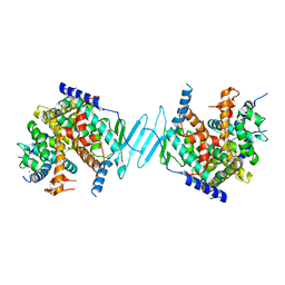
4EQS
 | | Crystal structure of the Y419F mutant of Staphylococcus aureus CoADR | | 分子名称: | CHLORIDE ION, COENZYME A, Coenzyme A disulfide reductase, ... | | 著者 | Wallace, B.D, Edwards, J.S, Wallen, J.R, Claiborne, A, Redinbo, M.R. | | 登録日 | 2012-04-19 | | 公開日 | 2012-10-17 | | 実験手法 | X-RAY DIFFRACTION (1.5 Å) | | 主引用文献 | Turnover-Dependent Covalent Inactivation of Staphylococcus aureus Coenzyme A-Disulfide Reductase by Coenzyme A-Mimetics: Mechanistic and Structural Insights.
Biochemistry, 51, 2012
|
|
3K4D
 | | Crystal structure of E. coli beta-glucuronidase with the glucaro-d-lactam inhibitor bound | | 分子名称: | (2S,3R,4S,5R)-3,4,5-trihydroxy-6-oxopiperidine-2-carboxylic acid, Beta-glucuronidase | | 著者 | Wallace, B.D, Redinbo, M.R. | | 登録日 | 2009-10-05 | | 公開日 | 2010-11-17 | | 最終更新日 | 2023-09-06 | | 実験手法 | X-RAY DIFFRACTION (2.393 Å) | | 主引用文献 | Alleviating cancer drug toxicity by inhibiting a bacterial enzyme.
Science, 330, 2010
|
|
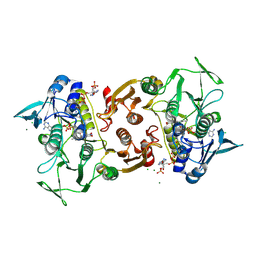
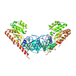
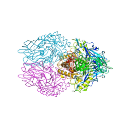
4EM4
 | | Crystal Structure of Staphylococcus aureus bound with the covalent inhibitor Pethyl-VS-CoA | | 分子名称: | CHLORIDE ION, Coenzyme A disulfide reductase, FLAVIN-ADENINE DINUCLEOTIDE, ... | | 著者 | Wallace, B.D, Edwards, J.S, Claiborne, A, Redinbo, M.R. | | 登録日 | 2012-04-11 | | 公開日 | 2012-10-17 | | 最終更新日 | 2017-11-15 | | 実験手法 | X-RAY DIFFRACTION (1.821 Å) | | 主引用文献 | Turnover-Dependent Covalent Inactivation of Staphylococcus aureus Coenzyme A-Disulfide Reductase by Coenzyme A-Mimetics: Mechanistic and Structural Insights.
Biochemistry, 51, 2012
|
|
5U6Z
 | |
1C4D
 | | GRAMICIDIN CSCL COMPLEX | | 分子名称: | CESIUM ION, CHLORIDE ION, GRAMICIDIN A, ... | | 著者 | Wallace, B.A. | | 登録日 | 1999-06-04 | | 公開日 | 2000-01-03 | | 最終更新日 | 2023-11-15 | | 実験手法 | X-RAY DIFFRACTION (2 Å) | | 主引用文献 | The Gramicidin Pore: Crystal Structure of a Cesium Complex.
Science, 241, 1988
|
|
1EPP
 | | ENDOTHIA ASPARTIC PROTEINASE (ENDOTHIAPEPSIN) COMPLEXED WITH PD-130,693 (MAS PHE LYS+MTF STA MBA) | | 分子名称: | ENDOTHIAPEPSIN, N-(dimethylsulfamoyl)-L-phenylalanyl-N-[(1S,2S)-2-hydroxy-4-{[(2S)-2-methylbutyl]amino}-1-(2-methylpropyl)-4-oxobutyl]-N~6~-(methylcarbamothioyl)-L-lysinamide, SULFATE ION | | 著者 | Wallace, B.A, Cooper, J.B, Blundell, T.L. | | 登録日 | 1994-07-27 | | 公開日 | 1994-12-20 | | 最終更新日 | 2017-11-29 | | 実験手法 | X-RAY DIFFRACTION (1.9 Å) | | 主引用文献 | Analyses of ligand binding in five endothiapepsin crystal complexes and their use in the design and evaluation of novel renin inhibitors.
J.Med.Chem., 36, 1993
|
|
4JKM
 | |
4JXG
 | | Crystal Structure of AmpC beta-lactamase from E. coli in Complex with Oxacillin | | 分子名称: | (2R,4S)-5,5-dimethyl-2-[(1R)-1-{[(5-methyl-3-phenyl-1,2-oxazol-4-yl)carbonyl]amino}-2-oxoethyl]-1,3-thiazolidine-4-carb oxylic acid, Beta-lactamase, PHOSPHATE ION, ... | | 著者 | Wallar, B.J, Powers, R.A, Docter, B.E. | | 登録日 | 2013-03-28 | | 公開日 | 2014-10-29 | | 最終更新日 | 2020-07-29 | | 実験手法 | X-RAY DIFFRACTION (1.65 Å) | | 主引用文献 | Complexed structures of AmpC beta-lactamase
To be Published
|
|
4JKL
 | |
4JKK
 | |
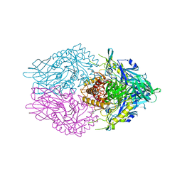
4J5X
 | | Crystal Structure of the SR12813-bound PXR/RXRalpha LBD Heterotetramer Complex | | 分子名称: | Nuclear receptor subfamily 1 group I member 2, Nuclear receptor coactivator 1, Retinoic acid receptor RXR-alpha, ... | | 著者 | Wallace, B.D, Betts, L, Redinbo, M.R. | | 登録日 | 2013-02-10 | | 公開日 | 2013-08-21 | | 最終更新日 | 2024-02-28 | | 実験手法 | X-RAY DIFFRACTION (2.8 Å) | | 主引用文献 | Structural and Functional Analysis of the Human Nuclear Xenobiotic Receptor PXR in Complex with RXRalpha.
J.Mol.Biol., 425, 2013
|
|
4J5W
 | | Crystal Structure of the apo-PXR/RXRalpha LBD Heterotetramer Complex | | 分子名称: | MAGNESIUM ION, Nuclear receptor subfamily 1 group I member 2, Nuclear receptor coactivator 1, ... | | 著者 | Wallace, B.D, Betts, L, Redinbo, M.R. | | 登録日 | 2013-02-10 | | 公開日 | 2013-08-21 | | 最終更新日 | 2024-02-28 | | 実験手法 | X-RAY DIFFRACTION (2.8 Å) | | 主引用文献 | Structural and Functional Analysis of the Human Nuclear Xenobiotic Receptor PXR in Complex with RXRalpha.
J.Mol.Biol., 425, 2013
|
|
3MBO
 | | Crystal Structure of the Glycosyltransferase BaBshA bound with UDP and L-malate | | 分子名称: | D-MALATE, GLYCEROL, Glycosyltransferase, ... | | 著者 | Wallace, B.D, Claiborne, A, Redinbo, M.R. | | 登録日 | 2010-03-25 | | 公開日 | 2010-09-29 | | 最終更新日 | 2023-09-06 | | 実験手法 | X-RAY DIFFRACTION (3.307 Å) | | 主引用文献 | Characterization of the N-Acetyl-alpha-d-glucosaminyl
l-Malate Synthase and Deacetylase Functions for Bacillithiol Biosynthesis
in Bacillus anthracis
Biochemistry, 49, 2010
|
|
3LPF
 | | Structure of E. coli beta-Glucuronidase bound with a novel, potent inhibitor 1-((6,7-dimethyl-2-oxo-1,2-dihydroquinolin-3-yl)methyl)-1-(2-hydroxyethyl)-3-(3-methoxyphenyl)thiourea | | 分子名称: | 1-[(6,7-dimethyl-2-oxo-1,2-dihydroquinolin-3-yl)methyl]-1-(2-hydroxyethyl)-3-(3-methoxyphenyl)thiourea, Beta-glucuronidase | | 著者 | Wallace, B.D, Redinbo, M.R. | | 登録日 | 2010-02-05 | | 公開日 | 2010-11-17 | | 最終更新日 | 2023-11-22 | | 実験手法 | X-RAY DIFFRACTION (2.26 Å) | | 主引用文献 | Alleviating cancer drug toxicity by inhibiting a bacterial enzyme.
Science, 330, 2010
|
|
3LPG
 | |
3K4A
 | |
3K46
 | |
1EDN
 | | HUMAN ENDOTHELIN-1 | | 分子名称: | ENDOTHELIN-1 | | 著者 | Wallace, B.A, Janes, R.W. | | 登録日 | 1994-09-19 | | 公開日 | 1995-10-15 | | 最終更新日 | 2011-07-13 | | 実験手法 | X-RAY DIFFRACTION (2.18 Å) | | 主引用文献 | The crystal structure of human endothelin.
Nat.Struct.Biol., 1, 1994
|
|
7P36
 | | X-ray structure of Lactobacillus kefir alcohol dehydrogenase (wild type) | | 分子名称: | 1,2-ETHANEDIOL, CHLORIDE ION, MAGNESIUM ION, ... | | 著者 | Bischoff, D, Walla, B, Janowski, R, Niessing, D, Weuster-Botz, D. | | 登録日 | 2021-07-07 | | 公開日 | 2021-07-21 | | 最終更新日 | 2024-01-31 | | 実験手法 | X-RAY DIFFRACTION (1.14 Å) | | 主引用文献 | Transfer of a Rational Crystal Contact Engineering Strategy between Diverse Alcohol Dehydrogenases
Crystals, 11, 2021
|
|
7P7Y
 | | X-ray structure of Lactobacillus kefir alcohol dehydrogenase mutant Q126K | | 分子名称: | 1,2-ETHANEDIOL, CHLORIDE ION, MAGNESIUM ION, ... | | 著者 | Bischoff, D, Walla, B, Janowski, R, Niessing, D, Weuster-Botz, D. | | 登録日 | 2021-07-21 | | 公開日 | 2021-07-28 | | 最終更新日 | 2024-01-31 | | 実験手法 | X-RAY DIFFRACTION (1.25 Å) | | 主引用文献 | Transfer of a Rational Crystal Contact Engineering Strategy between Diverse Alcohol Dehydrogenases
Crystals, 11, 2021
|
|
4NDB
 | | X-ray structure of a mutant (T61D) of calexcitin - a neuronal calcium-signalling protein | | 分子名称: | CALCIUM ION, Calexcitin | | 著者 | Erskine, P.T, Fokas, A, Muriithi, C, Razzall, E, Bowyer, A, Findlow, I.S, Werner, J.M, Wallace, B.A, Wood, S.P, Cooper, J.B. | | 登録日 | 2013-10-25 | | 公開日 | 2014-10-29 | | 最終更新日 | 2023-09-20 | | 実験手法 | X-RAY DIFFRACTION (2 Å) | | 主引用文献 | X-ray, spectroscopic and normal-mode dynamics of calexcitin: structure-function studies of a neuronal calcium-signalling protein.
Acta Crystallogr.,Sect.D, 71, 2015
|
|
5W13
 | |


