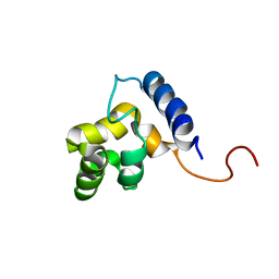
1KN5
 
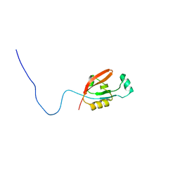
 | | SOLUTION STRUCTURE OF ARID DOMAIN OF ADR6 FROM SACCHAROMYCES CEREVISIAE | | 分子名称: | Transcription regulatory protein ADR6 | | 著者 | Tu, X, Wu, J, Xu, Y, Shi, Y. | | 登録日 | 2001-12-18 | | 公開日 | 2002-07-17 | | 最終更新日 | 2024-05-29 | | 実験手法 | SOLUTION NMR | | 主引用文献 | 1H, 13C and 15N resonance assignments and secondary structure of ADR6 DNA-binding domain.
J.Biomol.Nmr, 21, 2001
|
|
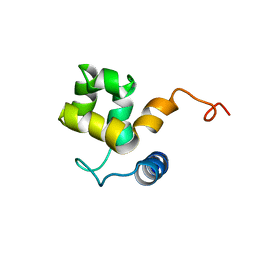
1KKX
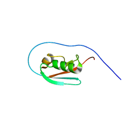
 | | Solution structure of the DNA-binding domain of ADR6 | | 分子名称: | Transcription regulatory protein ADR6 | | 著者 | Tu, X, Wu, J, Xu, Y, Shi, Y. | | 登録日 | 2001-12-10 | | 公開日 | 2002-07-17 | | 最終更新日 | 2024-05-29 | | 実験手法 | SOLUTION NMR | | 主引用文献 | 1H, 13C and 15N resonance assignments and secondary structure of ADR6 DNA-binding domain.
J.Biomol.Nmr, 21, 2001
|
|
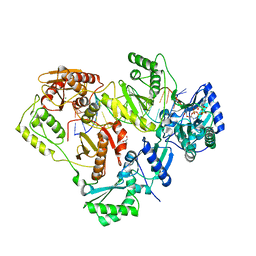
4DG1
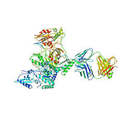
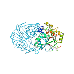
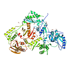 | | Crystal structure of HIV-1 reverse transcriptase (RT) with polymorphism mutation K172A and K173A | | 分子名称: | 1,2-ETHANEDIOL, GLYCEROL, MAGNESIUM ION, ... | | 著者 | Tu, X, Kirby, K.A, Marchand, B, Sarafianos, S.G. | | 登録日 | 2012-01-24 | | 公開日 | 2012-06-20 | | 最終更新日 | 2023-09-13 | | 実験手法 | X-RAY DIFFRACTION (2.15 Å) | | 主引用文献 | HIV-1 Reverse Transcriptase (RT) Polymorphism 172K Suppresses the Effect of Clinically Relevant Drug Resistance Mutations to Both Nucleoside and Non-nucleoside RT Inhibitors.
J.Biol.Chem., 287, 2012
|
|
4F3J
 | |
3KLF
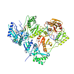
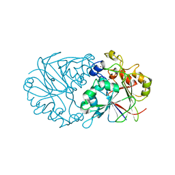
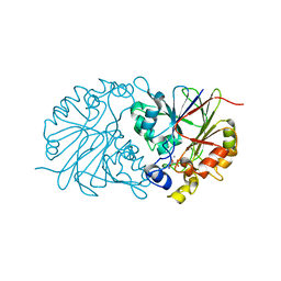
 | | Crystal structure of wild-type HIV-1 Reverse Transcriptase crosslinked to a DSDNA with a bound excision product, AZTPPPPA | | 分子名称: | DNA (5'-D(*A*CP*AP*GP*TP*CP*CP*CP*TP*GP*TP*TP*CP*GP*GP*(MRG)P*CP*GP*CP*CP*(2DA))-3'), DNA (5'-D(*AP*T*GP*CP*AP*TP*GP*GP*CP*GP*CP*CP*CP*GP*AP*AP*CP*AP*GP*GP*GP*AP*CP*TP*GP*TP*G)-3'), GLYCEROL, ... | | 著者 | Tu, X, Das, K, Sarafianos, S.G, Arnold, E. | | 登録日 | 2009-11-07 | | 公開日 | 2010-09-22 | | 最終更新日 | 2023-09-06 | | 実験手法 | X-RAY DIFFRACTION (3.15 Å) | | 主引用文献 | Structural basis of HIV-1 resistance to AZT by excision.
Nat.Struct.Mol.Biol., 17, 2010
|
|
3KLE
 | | Crystal structure of AZT-resistant HIV-1 Reverse Transcriptase crosslinked to a DSDNA with a bound excision product, AZTPPPPA | | 分子名称: | DNA (25-MER), DNA (5'-D(*AP*CP*AP*GP*TP*CP*CP*CP*TP*GP*TP*TP*CP*GP*GP*(MRG)P*CP*GP*CP*CP*(2DA))-3'), GLYCEROL, ... | | 著者 | Tu, X, Das, K, Sarafianos, S.G, Arnold, E. | | 登録日 | 2009-11-07 | | 公開日 | 2010-09-22 | | 最終更新日 | 2021-10-13 | | 実験手法 | X-RAY DIFFRACTION (3.2 Å) | | 主引用文献 | Structural basis of HIV-1 resistance to AZT by excision.
Nat.Struct.Mol.Biol., 17, 2010
|
|
3KLI
 | |
3KLG
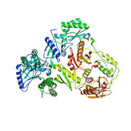
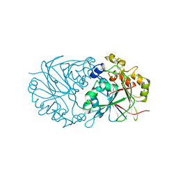
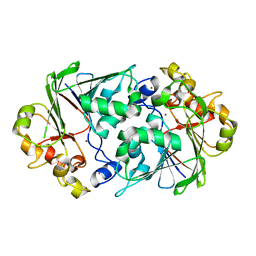
 | | Crystal structure of AZT-resistant HIV-1 Reverse Transcriptase crosslinked to pre-translocation AZTMP-Terminated DNA (COMPLEX N) | | 分子名称: | DNA (5'-D(*A*CP*AP*GP*TP*CP*CP*CP*TP*GP*TP*TP*CP*GP*GP*(MRG)P*CP*GP*CP*CP*AP*(ATM))-3'), DNA (5'-D(*AP*T*GP*CP*AP*TP*GP*GP*CP*GP*CP*CP*CP*GP*AP*AP*CP*AP*GP*GP*GP*AP*CP*TP*GP*TP*G)-3'), Reverse transcriptase/ribonuclease H, ... | | 著者 | Tu, X, Sarafianos, S.G, Arnold, E. | | 登録日 | 2009-11-07 | | 公開日 | 2010-09-22 | | 最終更新日 | 2023-09-06 | | 実験手法 | X-RAY DIFFRACTION (3.65 Å) | | 主引用文献 | Structural basis of HIV-1 resistance to AZT by excision.
Nat.Struct.Mol.Biol., 17, 2010
|
|
3KLH
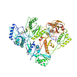
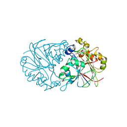
 | | Crystal structure of AZT-Resistant HIV-1 Reverse Transcriptase crosslinked to post-translocation AZTMP-Terminated DNA (COMPLEX P) | | 分子名称: | DNA (5'-D(*AP*CP*AP*GP*TP*CP*CP*CP*TP*GP*TP*TP*CP*GP*GP*(MRG)P*CP*GP*CP*CP*(ATM))-3'), DNA (5'-D(*AP*T*GP*CP*TP*AP*GP*GP*CP*GP*CP*CP*CP*GP*AP*AP*CP*AP*GP*GP*GP*AP*CP*TP*GP*TP*G)-3'), MAGNESIUM ION, ... | | 著者 | Tu, X, Sarafianos, S.G, Arnold, E. | | 登録日 | 2009-11-07 | | 公開日 | 2010-09-22 | | 最終更新日 | 2023-09-06 | | 実験手法 | X-RAY DIFFRACTION (2.9 Å) | | 主引用文献 | Structural basis of HIV-1 resistance to AZT by excision.
Nat.Struct.Mol.Biol., 17, 2010
|
|
4Z60
 | |
4Z6X
 | |
4Z5Z
 | |
4Z5Y
 | |
4Z67
 | |
4Z7R
 | |
4NN0
 | |
7DL8
 | |
1ONJ
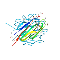
 | | Crystal structure of Atratoxin-b from Chinese cobra venom of Naja atra | | 分子名称: | Cobrotoxin b, SULFATE ION | | 著者 | Lou, X, Tu, X, Pan, G, Xu, C, Fan, R, Lu, W, Deng, W, Rao, P, Teng, M, Niu, L. | | 登録日 | 2003-02-28 | | 公開日 | 2004-02-28 | | 最終更新日 | 2017-10-11 | | 実験手法 | X-RAY DIFFRACTION (1.555 Å) | | 主引用文献 | Purification, N-terminal sequencing, crystallization and preliminary structural determination of atratoxin-b, a short-chain alpha-neurotoxin from Naja atra venom.
Acta Crystallogr.,Sect.D, 59, 2003
|
|
2L97
 | |
1V6P
 | | Crystal structure of Cobrotoxin | | 分子名称: | CHLORIDE ION, COPPER (II) ION, Cobrotoxin, ... | | 著者 | Lou, X, Tu, X, Wang, J, Teng, M, Niu, L, Liu, Q, Huang, Q, Hao, Q. | | 登録日 | 2003-12-03 | | 公開日 | 2004-12-21 | | 最終更新日 | 2023-12-27 | | 実験手法 | X-RAY DIFFRACTION (0.87 Å) | | 主引用文献 | The atomic resolution crystal structure of atratoxin determined by single wavelength anomalous diffraction phasing
J.Biol.Chem., 279, 2004
|
|
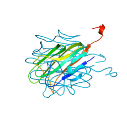
2K9X
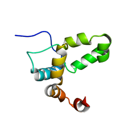
 | | Solution structure of Urm1 from Trypanosoma brucei | | 分子名称: | Uncharacterized protein | | 著者 | Zhang, W, Zhang, J, Xu, C, Wang, T, Zhang, X, Tu, X. | | 登録日 | 2008-10-27 | | 公開日 | 2009-03-10 | | 最終更新日 | 2024-05-29 | | 実験手法 | SOLUTION NMR | | 主引用文献 | Solution structure of Urm1 from Trypanosoma brucei
Proteins, 75, 2009
|
|
2L6K
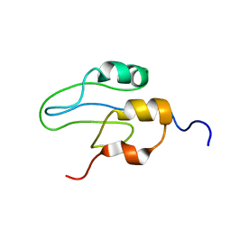
 | | Solution Structure of a Nonphosphorylated Peptide Recognizing Domain | | 分子名称: | Tensin-like C1 domain-containing phosphatase | | 著者 | Dai, K, Liao, S, Zhang, J, Zhang, X, Tu, X. | | 登録日 | 2010-11-22 | | 公開日 | 2011-10-12 | | 最終更新日 | 2024-05-15 | | 実験手法 | SOLUTION NMR | | 主引用文献 | Solution structure of Tensin2 SH2 domain and its phosphotyrosine-independent interaction with DLC-1
Plos One, 6, 2011
|
|
2K8H
 | |
2LI6
 | |
2L42
 | |


