6CO2
 
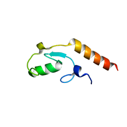
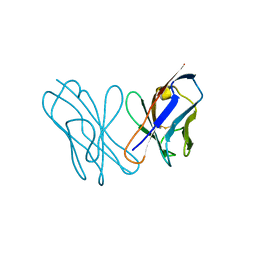
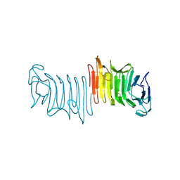
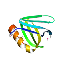
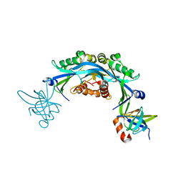 | | Structure of an engineered protein (NUDT16TI) in complex with 53BP1 Tudor domains | | 分子名称: | NUDT16-Tudor-interacting (NUDT16TI), TP53-binding protein 1 | | 著者 | Botuyan, M.V, Thompson, J.R, Cui, G, Mer, G. | | 登録日 | 2018-03-10 | | 公開日 | 2018-06-06 | | 最終更新日 | 2023-10-04 | | 実験手法 | X-RAY DIFFRACTION (2.49 Å) | | 主引用文献 | Mechanism of 53BP1 activity regulation by RNA-binding TIRR and a designer protein.
Nat. Struct. Mol. Biol., 25, 2018
|
|
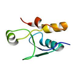
3KTF
 | |
3L1Z
 | |
3L1Y
 | |
3L1X
 | |
3Q66
 | |
3T1N
 | |
3SZM
 | |
3Q68
 | |
3SD4
 | |
3TW1
 | | Structure of Rtt106-AHN | | 分子名称: | GLYCEROL, Histone chaperone RTT106, N-[2-(1H-IMIDAZOL-4-YL)ETHYL]ACETAMIDE | | 著者 | Su, D, Thompson, J.R, Mer, G. | | 登録日 | 2011-09-21 | | 公開日 | 2012-02-01 | | 最終更新日 | 2023-12-06 | | 実験手法 | X-RAY DIFFRACTION (1.772 Å) | | 主引用文献 | Structural basis for recognition of H3K56-acetylated histone H3-H4 by the chaperone Rtt106.
Nature, 483, 2012
|
|
3TVV
 | |
3U7A
 | | AL-09 Y32F Y96F | | 分子名称: | Amyloidogenic immunoglobulin light chain protein AL-09 Y32F Y96F, variable domain | | 著者 | DiCostanzo, A.C, Thompson, J.R, Ramirez-Alvarado, M. | | 登録日 | 2011-10-13 | | 公開日 | 2012-07-04 | | 実験手法 | X-RAY DIFFRACTION (2 Å) | | 主引用文献 | Tyrosine Residues mediate crucial interactions in amyloid formation for immunoglobulin light chains
To be Published
|
|
3U79
 | | AL-103 Y32F Y96F | | 分子名称: | 2-(N-MORPHOLINO)-ETHANESULFONIC ACID, ACETATE ION, Amyloidogenic immunoglobulin light chain protein AL-103 Y32F Y96F, ... | | 著者 | DiCostanzo, A.C, Thompson, J.R, Ramirez-Alvarado, M. | | 登録日 | 2011-10-13 | | 公開日 | 2012-07-04 | | 実験手法 | X-RAY DIFFRACTION (1.62 Å) | | 主引用文献 | Tyrosine Residues mediate crucial interactions in amyloid formation for immunoglobulin light chains
To be Published
|
|
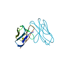
3U3Z
 | | Structure of human microcephalin (MCPH1) tandem BRCT domains in complex with an H2A.X peptide phosphorylated at Ser139 and Tyr142 | | 分子名称: | GLYCEROL, Histone H2A.X peptide, Microcephalin | | 著者 | Singh, N, Thompson, J.R, Heroux, A, Mer, G. | | 登録日 | 2011-10-06 | | 公開日 | 2012-07-25 | | 最終更新日 | 2023-12-06 | | 実験手法 | X-RAY DIFFRACTION (1.5 Å) | | 主引用文献 | Dual recognition of phosphoserine and phosphotyrosine in histone variant H2A.X by DNA damage response protein MCPH1.
Proc.Natl.Acad.Sci.USA, 109, 2012
|
|
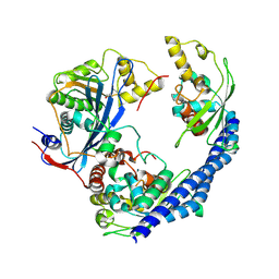
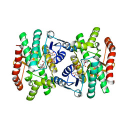
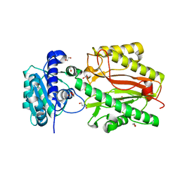
1IB6
 | | CRYSTAL STRUCTURE OF R153C E. COLI MALATE DEHYDROGENASE | | 分子名称: | MALATE DEHYDROGENASE, NICOTINAMIDE-ADENINE-DINUCLEOTIDE, SULFATE ION | | 著者 | Bell, J.K, Yennawar, H.P, Wright, S.K, Thompson, J.R, Viola, R.E, Banaszak, L.J. | | 登録日 | 2001-03-27 | | 公開日 | 2001-09-19 | | 最終更新日 | 2024-02-07 | | 実験手法 | X-RAY DIFFRACTION (2.1 Å) | | 主引用文献 | Structural Analyses of a Malate Dehydrogenase with a Variable Active Site
J.Biol.Chem., 276, 2001
|
|
1IE3
 | | CRYSTAL STRUCTURE OF R153C E. COLI MALATE DEHYDROGENASE | | 分子名称: | MALATE DEHYDROGENASE, NICOTINAMIDE-ADENINE-DINUCLEOTIDE, PYRUVIC ACID | | 著者 | Bell, J.K, Yennawar, H.P, Wright, S.K, Thompson, J.R, Viola, R.E, Banaszak, L.J. | | 登録日 | 2001-04-05 | | 公開日 | 2001-09-19 | | 最終更新日 | 2023-11-15 | | 実験手法 | X-RAY DIFFRACTION (2.5 Å) | | 主引用文献 | Structural Analyses of a Malate Dehydrogenase with a Variable Active Site
J.Biol.Chem., 276, 2001
|
|
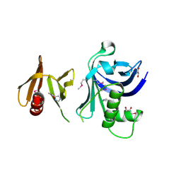
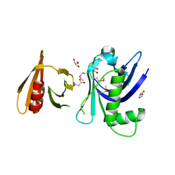
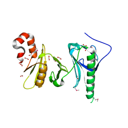
3FY3
 | | Crystal structure of truncated hemolysin A from P. mirabilis | | 分子名称: | Hemolysin | | 著者 | Weaver, T.M, Thompson, J.R, Bailey, L.J, Wawrzyn, G.T, Hocking, J.M, Howard, D.R. | | 登録日 | 2009-01-21 | | 公開日 | 2009-06-02 | | 最終更新日 | 2023-09-06 | | 実験手法 | X-RAY DIFFRACTION (1.8 Å) | | 主引用文献 | Structural and functional studies of truncated hemolysin A from Proteus mirabilis.
J.Biol.Chem., 284, 2009
|
|
3DVF
 | |
3FSS
 | |
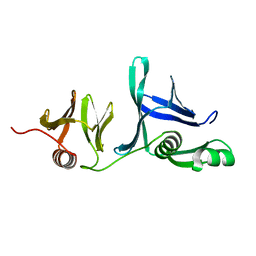
5UMR
 | | Crystal structure of N-terminal domain of human FACT complex subunit SSRP1 | | 分子名称: | FACT complex subunit SSRP1 | | 著者 | Su, D, Hu, Q, Thompson, J.R, Heroux, A, Botuyan, M.V, Mer, G. | | 登録日 | 2017-01-29 | | 公開日 | 2018-01-31 | | 最終更新日 | 2019-12-04 | | 実験手法 | X-RAY DIFFRACTION (1.501 Å) | | 主引用文献 | Crystal structure of N-terminal domain of human FACT complex subunit SSRP1
To Be Published
|
|
5UMT
 | | Crystal structure of N-terminal domain of human FACT complex subunit SPT16 | | 分子名称: | 1,2-ETHANEDIOL, FACT complex subunit SPT16, GLYCEROL | | 著者 | Su, D, Hu, Q, Thompson, J.R, Botuyan, M.V, Mer, G. | | 登録日 | 2017-01-29 | | 公開日 | 2018-01-31 | | 最終更新日 | 2023-10-04 | | 実験手法 | X-RAY DIFFRACTION (2.092 Å) | | 主引用文献 | Crystal structure of N-terminal domain of human FACT complex subunit SPT16
To Be Published
|
|
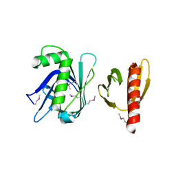
5UMU
 | | Crystal structure of the middle double PH domain of human FACT complex subunit SPT16 | | 分子名称: | ACETATE ION, FACT complex subunit SPT16, FORMIC ACID | | 著者 | Hu, Q, Thompson, J.R, Heroux, A, Su, D, Botuyan, M.V, Mer, G. | | 登録日 | 2017-01-29 | | 公開日 | 2018-01-31 | | 最終更新日 | 2019-12-04 | | 実験手法 | X-RAY DIFFRACTION (1.903 Å) | | 主引用文献 | Crystal structure of the middle double PH domain of human FACT complex subunit SPT16
To Be Published
|
|
5UMS
 | |
5UMV
 | |


