Loading
PDBj
MenuPDBj@FacebookPDBj@TwitterPDBj@YouTubewwPDB FoundationwwPDB
RCSB PDBPDBeBMRBAdv. SearchSearch help
PDB: 775 results

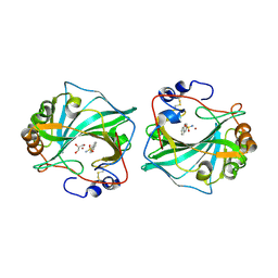
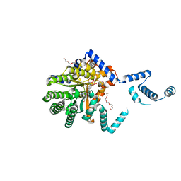
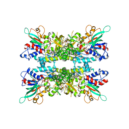
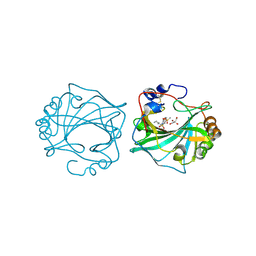
8Q1A
DownloadVisualize
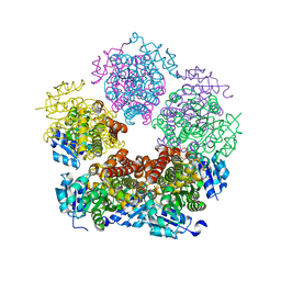
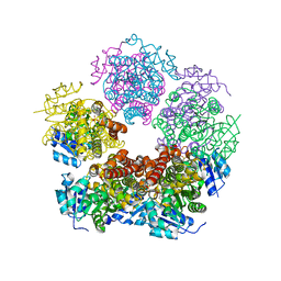
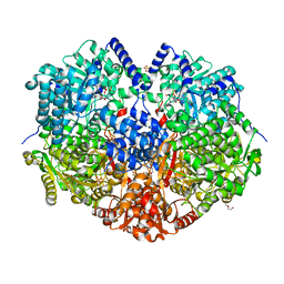
BU of 8q1a by Molmil
The Crystal Structure of Human Carbonic Anhydrase IX in Complex with inhibitor
Descriptor: 1,1,3-tris(oxidanylidene)-2-pentyl-1,2-benzothiazole-6-sulfonamide, Carbonic anhydrase 9, GLYCEROL, ...
Authors:Leitans, J, Tars, K.
Deposit date:2023-07-31
Release date:2023-11-08
Last modified:2023-12-06
Method:X-RAY DIFFRACTION (2.35 Å)
Cite:Structural Basis of Saccharin Derivative Inhibition of Carbonic Anhydrase IX.
Chemmedchem, 18, 2023
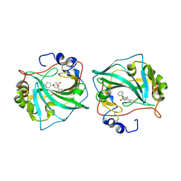
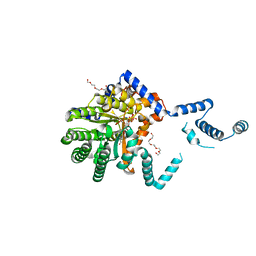
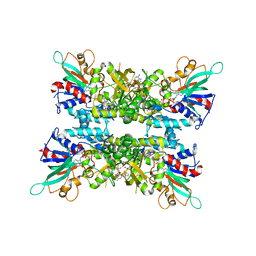
6TL6
DownloadVisualize
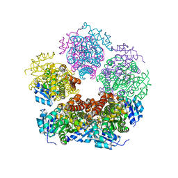
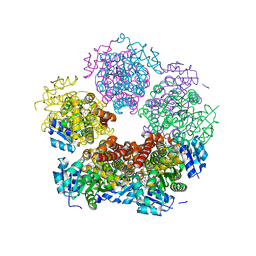
BU of 6tl6 by Molmil
Three dimensional structure of human carbonic anhydrase IX in complex with sulfonamide
Descriptor: Carbonic anhydrase 9, GLYCEROL, ZINC ION, ...
Authors:Leitans, J, Tars, K.
Deposit date:2019-12-01
Release date:2020-12-16
Last modified:2024-01-24
Method:X-RAY DIFFRACTION (2.15 Å)
Cite:Isoform-Selective Enzyme Inhibitors by Exploring Pocket Size According to the Lock-and-Key Principle.
Biophys.J., 119, 2020
6TL5
DownloadVisualize
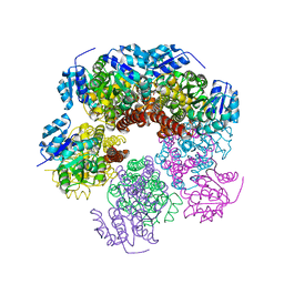
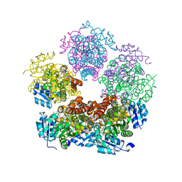
BU of 6tl5 by Molmil
Three dimensional structure of human carbonic anhydrase IX in complex with sulfonamide
Descriptor: 3,5-diphenylbenzenesulfonamide, Carbonic anhydrase 9, ZINC ION
Authors:Leitans, J, Tars, K.
Deposit date:2019-12-01
Release date:2020-12-16
Last modified:2024-01-24
Method:X-RAY DIFFRACTION (2.21 Å)
Cite:Isoform-Selective Enzyme Inhibitors by Exploring Pocket Size According to the Lock-and-Key Principle.
Biophys.J., 119, 2020
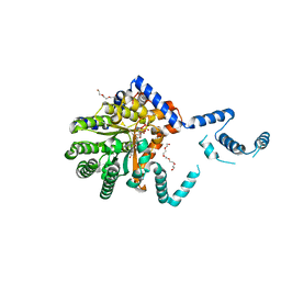
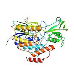
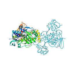
7SQN
DownloadVisualize
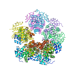
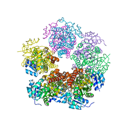
BU of 7sqn by Molmil
Structure of the E. coli PutA proline dehydrogenase domain (residues 86-630) complexed with (2S)-oxetane-2-carboxylic acid
Descriptor: (2S)-oxetane-2-carboxylic acid, Bifunctional protein PutA, FLAVIN-ADENINE DINUCLEOTIDE, ...
Authors:Tanner, J.J, Bogner, A.N.
Deposit date:2021-11-05
Release date:2022-01-19
Last modified:2023-10-18
Method:X-RAY DIFFRACTION (2.25 Å)
Cite:Structure-affinity relationships of reversible proline analog inhibitors targeting proline dehydrogenase.
Org.Biomol.Chem., 20, 2022
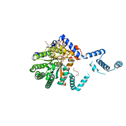
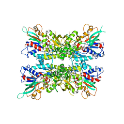
7MWT
DownloadVisualize
BU of 7mwt by Molmil
Structure of the E. coli PutA proline dehydrogenase domain (residues 86-630) complexed with 1,1-Cyclobutanedicarboxylate
Descriptor: Bifunctional protein PutA, FLAVIN-ADENINE DINUCLEOTIDE, SULFATE ION, ...
Authors:Tanner, J.J, Bogner, A.N.
Deposit date:2021-05-17
Release date:2022-01-19
Last modified:2023-10-18
Method:X-RAY DIFFRACTION (2.19 Å)
Cite:Structure-affinity relationships of reversible proline analog inhibitors targeting proline dehydrogenase.
Org.Biomol.Chem., 20, 2022
7MWU
DownloadVisualize
BU of 7mwu by Molmil
Structure of the E. coli PutA proline dehydrogenase domain (residues 86-630) complexed with cyclobutanecarboxylic acid
Descriptor: Bifunctional protein PutA, FLAVIN-ADENINE DINUCLEOTIDE, PENTAETHYLENE GLYCOL, ...
Authors:Tanner, J.J, Bogner, A.N.
Deposit date:2021-05-17
Release date:2022-01-19
Last modified:2024-05-22
Method:X-RAY DIFFRACTION (1.69 Å)
Cite:Structure-affinity relationships of reversible proline analog inhibitors targeting proline dehydrogenase.
Org.Biomol.Chem., 20, 2022
7MWV
DownloadVisualize
BU of 7mwv by Molmil
Structure of the E. coli PutA proline dehydrogenase domain (residues 86-630) complexed with cyclopropanecarboxylic acid
Descriptor: Bifunctional protein PutA, FLAVIN-ADENINE DINUCLEOTIDE, PENTAETHYLENE GLYCOL, ...
Authors:Tanner, J.J, Bogner, A.N.
Deposit date:2021-05-17
Release date:2022-01-19
Last modified:2023-10-18
Method:X-RAY DIFFRACTION (1.69 Å)
Cite:Structure-affinity relationships of reversible proline analog inhibitors targeting proline dehydrogenase.
Org.Biomol.Chem., 20, 2022
6WPU
DownloadVisualize
BU of 6wpu by Molmil
Structure of S-allyl-L-cysteine S-oxygenase from Allium sativum
Descriptor: FLAVIN-ADENINE DINUCLEOTIDE, Flavin-containing monooxygenase, SULFATE ION
Authors:Tanner, J.J, Campbell, A.C, Schuermann, J.P.
Deposit date:2020-04-27
Release date:2020-06-17
Last modified:2024-03-06
Method:X-RAY DIFFRACTION (2.084 Å)
Cite:Structure and function of a flavin-dependent S-monooxygenase from garlic (Allium sativum).
J.Biol.Chem., 295, 2020
6X0H
DownloadVisualize
BU of 6x0h by Molmil
Structure of oxidized SidA ornithine hydroxylase with the FAD in the "out" conformation
Descriptor: ACETATE ION, CALCIUM ION, FLAVIN-ADENINE DINUCLEOTIDE, ...
Authors:Tanner, J.J, Campbell, A.C.
Deposit date:2020-05-15
Release date:2020-08-05
Last modified:2023-10-18
Method:X-RAY DIFFRACTION (2.087 Å)
Cite:Trapping conformational states of a flavin-dependent N -monooxygenase in crystallo reveals protein and flavin dynamics.
J.Biol.Chem., 295, 2020
6X0J
DownloadVisualize
BU of 6x0j by Molmil
Structure of reduced SidA ornithine hydroxylase with the FAD "in" and complexed with NADP and L-ornithine
Descriptor: DIHYDROFLAVINE-ADENINE DINUCLEOTIDE, L-ornithine, L-ornithine N(5)-monooxygenase, ...
Authors:Tanner, J.J, Campbell, A.C.
Deposit date:2020-05-15
Release date:2020-08-05
Last modified:2023-10-18
Method:X-RAY DIFFRACTION (2.335 Å)
Cite:Trapping conformational states of a flavin-dependent N -monooxygenase in crystallo reveals protein and flavin dynamics.
J.Biol.Chem., 295, 2020
6X0I
DownloadVisualize
BU of 6x0i by Molmil
Structure of oxidized SidA ornithine hydroxylase with the FAD "in" and complexed with NADP
Descriptor: ACETATE ION, CALCIUM ION, FLAVIN-ADENINE DINUCLEOTIDE, ...
Authors:Tanner, J.J, Campbell, A.C.
Deposit date:2020-05-15
Release date:2020-08-05
Last modified:2023-10-18
Method:X-RAY DIFFRACTION (1.95 Å)
Cite:Trapping conformational states of a flavin-dependent N -monooxygenase in crystallo reveals protein and flavin dynamics.
J.Biol.Chem., 295, 2020
5KOX
DownloadVisualize
BU of 5kox by Molmil
Structure of rifampicin monooxygenase complexed with rifampicin
Descriptor: FLAVIN-ADENINE DINUCLEOTIDE, Pentachlorophenol 4-monooxygenase, RIFAMPICIN
Authors:Tanner, J.J, Liu, L.-K.
Deposit date:2016-07-01
Release date:2016-09-07
Last modified:2023-10-04
Method:X-RAY DIFFRACTION (1.8 Å)
Cite:The Structure of the Antibiotic Deactivating, N-hydroxylating Rifampicin Monooxygenase.
J.Biol.Chem., 291, 2016
8TCV
DownloadVisualize
BU of 8tcv by Molmil
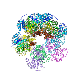
Structure of PYCR1 complexed with 4-bromobenzene-1,3-dicarboxylic acid
Descriptor: 4-bromobenzene-1,3-dicarboxylic acid, Pyrroline-5-carboxylate reductase 1, mitochondrial, ...
Authors:Tanner, J.J, Meeks, K.R.
Deposit date:2023-07-02
Release date:2024-03-06
Last modified:2024-03-20
Method:X-RAY DIFFRACTION (1.74 Å)
Cite:Novel Fragment Inhibitors of PYCR1 from Docking-Guided X-ray Crystallography.
J.Chem.Inf.Model., 64, 2024
8TCZ
DownloadVisualize
BU of 8tcz by Molmil
Structure of PYCR1 complexed with 2-(pyridin-2-yl)cyclopropane-1-carboxylic acid
Descriptor: (1S,2S)-2-(pyridin-2-yl)cyclopropane-1-carboxylic acid, Pyrroline-5-carboxylate reductase 1, mitochondrial, ...
Authors:Tanner, J.J, Meeks, K.R.
Deposit date:2023-07-02
Release date:2024-03-06
Last modified:2024-03-20
Method:X-RAY DIFFRACTION (2.1 Å)
Cite:Novel Fragment Inhibitors of PYCR1 from Docking-Guided X-ray Crystallography.
J.Chem.Inf.Model., 64, 2024
8TCW
DownloadVisualize
BU of 8tcw by Molmil
Structure of PYCR1 complexed with 2-methyl-3-(2-oxoimidazolidin-1-yl)benzoic acid
Descriptor: 2-methyl-3-(2-oxoimidazolidin-1-yl)benzoic acid, Pyrroline-5-carboxylate reductase 1, mitochondrial, ...
Authors:Tanner, J.J, Meeks, K.R.
Deposit date:2023-07-02
Release date:2024-03-06
Last modified:2024-03-20
Method:X-RAY DIFFRACTION (1.94 Å)
Cite:Novel Fragment Inhibitors of PYCR1 from Docking-Guided X-ray Crystallography.
J.Chem.Inf.Model., 64, 2024
6XP1
DownloadVisualize
BU of 6xp1 by Molmil
Structure of human PYCR1 complexed with L-thiazolidine-2-carboxylate
Descriptor: (2S)-1,3-thiazolidine-2-carboxylic acid, Pyrroline-5-carboxylate reductase 1, mitochondrial, ...
Authors:Tanner, J.J, Christensen, E.M.
Deposit date:2020-07-07
Release date:2020-11-04
Last modified:2023-10-18
Method:X-RAY DIFFRACTION (1.75 Å)
Cite:In crystallo screening for proline analog inhibitors of the proline cycle enzyme PYCR1.
J.Biol.Chem., 295, 2020
8TCX
DownloadVisualize
BU of 8tcx by Molmil
Structure of PYCR1 complexed with 2,4-dioxo-1,2,3,4-tetrahydroquinazoline-6-carboxylic acid
Descriptor: 2,4-dioxo-1,2,3,4-tetrahydroquinazoline-6-carboxylic acid, Pyrroline-5-carboxylate reductase 1, mitochondrial, ...
Authors:Tanner, J.J, Meeks, K.R.
Deposit date:2023-07-02
Release date:2024-03-06
Last modified:2024-03-20
Method:X-RAY DIFFRACTION (1.72 Å)
Cite:Novel Fragment Inhibitors of PYCR1 from Docking-Guided X-ray Crystallography.
J.Chem.Inf.Model., 64, 2024
8TD0
DownloadVisualize
BU of 8td0 by Molmil
Structure of PYCR1 complexed with 5-oxo-7a-phenyl-hexahydropyrrolo[2,1-b][1,3]thiazole-3-carboxylic acid
Descriptor: (3R,4S,7aR)-5-oxo-7a-phenylhexahydropyrrolo[2,1-b][1,3]thiazole-3-carboxylic acid, Pyrroline-5-carboxylate reductase 1, mitochondrial, ...
Authors:Tanner, J.J, Meeks, K.R.
Deposit date:2023-07-02
Release date:2024-03-06
Last modified:2024-03-20
Method:X-RAY DIFFRACTION (2.2 Å)
Cite:Novel Fragment Inhibitors of PYCR1 from Docking-Guided X-ray Crystallography.
J.Chem.Inf.Model., 64, 2024
8TCU
DownloadVisualize
BU of 8tcu by Molmil
Structure of PYCR1 complexed with 2-chloro-5-(2-oxoimidazolidin-1-yl)benzoic acid
Descriptor: 2-chloro-5-(2-oxoimidazolidin-1-yl)benzoic acid, Pyrroline-5-carboxylate reductase 1, mitochondrial, ...
Authors:Tanner, J.J, Meeks, K.R.
Deposit date:2023-07-02
Release date:2024-03-06
Last modified:2024-03-20
Method:X-RAY DIFFRACTION (2 Å)
Cite:Novel Fragment Inhibitors of PYCR1 from Docking-Guided X-ray Crystallography.
J.Chem.Inf.Model., 64, 2024
8TD1
DownloadVisualize
BU of 8td1 by Molmil
Structure of PYCR1 complexed with 3-(6-Oxa-9-azaspiro(4.5)decane-9-carbonyl)benzoic acid
Descriptor: 3-(6-oxa-9-azaspiro[4.5]decane-9-carbonyl)benzoic acid, DI(HYDROXYETHYL)ETHER, Pyrroline-5-carboxylate reductase 1, ...
Authors:Tanner, J.J, Meeks, K.R.
Deposit date:2023-07-02
Release date:2024-03-06
Last modified:2024-03-20
Method:X-RAY DIFFRACTION (1.88 Å)
Cite:Novel Fragment Inhibitors of PYCR1 from Docking-Guided X-ray Crystallography.
J.Chem.Inf.Model., 64, 2024
8TCY
DownloadVisualize
BU of 8tcy by Molmil
Structure of PYCR1 complexed with 7-fluoro-2-oxo-1,2,3,4-tetrahydroquinoline-6-carboxylic acid
Descriptor: 7-fluoro-2-oxo-1,2,3,4-tetrahydroquinoline-6-carboxylic acid, DI(HYDROXYETHYL)ETHER, Pyrroline-5-carboxylate reductase 1, ...
Authors:Tanner, J.J, Meeks, K.R.
Deposit date:2023-07-02
Release date:2024-03-06
Last modified:2024-03-20
Method:X-RAY DIFFRACTION (1.95 Å)
Cite:Novel Fragment Inhibitors of PYCR1 from Docking-Guided X-ray Crystallography.
J.Chem.Inf.Model., 64, 2024
6X9D
DownloadVisualize
BU of 6x9d by Molmil
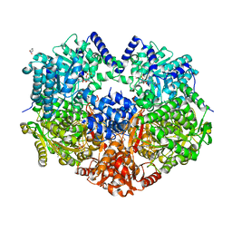
Structure of proline utilization A with trans-4-hydroxy-L-proline bound in the L-glutamate-gamma-semialdehyde dehydrogenase active site
Descriptor: 4-HYDROXYPROLINE, Bifunctional protein PutA, DI(HYDROXYETHYL)ETHER, ...
Authors:Tanner, J.J, Campbell, A.C.
Deposit date:2020-06-02
Release date:2020-12-30
Last modified:2023-10-18
Method:X-RAY DIFFRACTION (1.54 Å)
Cite:Structural analysis of prolines and hydroxyprolines binding to the l-glutamate-gamma-semialdehyde dehydrogenase active site of bifunctional proline utilization A.
Arch.Biochem.Biophys., 698, 2020
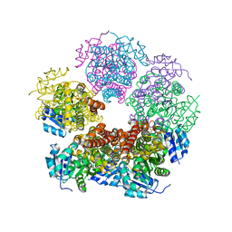
7STE
DownloadVisualize
BU of 7ste by Molmil
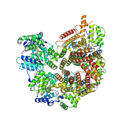
Rad24-RFC ADP state
Descriptor: ADENOSINE-5'-DIPHOSPHATE, ADENOSINE-5'-TRIPHOSPHATE, Checkpoint protein RAD24, ...
Authors:Castaneda, J.C, Schrecker, M, Remus, D, Hite, R.K.
Deposit date:2021-11-12
Release date:2022-04-06
Last modified:2024-06-05
Method:ELECTRON MICROSCOPY (2.73 Å)
Cite:Mechanisms of loading and release of the 9-1-1 checkpoint clamp.
Nat.Struct.Mol.Biol., 29, 2022
6XP2
DownloadVisualize
BU of 6xp2 by Molmil
Structure of human PYCR1 complexed with L-thiazolidine-4-carboxylate
Descriptor: Pyrroline-5-carboxylate reductase 1, mitochondrial, SULFATE ION, ...
Authors:Tanner, J.J, Christensen, E.M.
Deposit date:2020-07-07
Release date:2020-11-04
Last modified:2023-10-18
Method:X-RAY DIFFRACTION (2.3 Å)
Cite:In crystallo screening for proline analog inhibitors of the proline cycle enzyme PYCR1.
J.Biol.Chem., 295, 2020
6X9C
DownloadVisualize
BU of 6x9c by Molmil
Structure of proline utilization A with L-proline bound in the L-glutamate-gamma-semialdehyde dehydrogenase active site
Descriptor: 1,4-DIHYDRONICOTINAMIDE ADENINE DINUCLEOTIDE, Bifunctional protein PutA, DI(HYDROXYETHYL)ETHER, ...
Authors:Tanner, J.J, Campbell, A.C.
Deposit date:2020-06-02
Release date:2020-12-30
Last modified:2023-10-18
Method:X-RAY DIFFRACTION (1.44 Å)
Cite:Structural analysis of prolines and hydroxyprolines binding to the l-glutamate-gamma-semialdehyde dehydrogenase active site of bifunctional proline utilization A.
Arch.Biochem.Biophys., 698, 2020

222415

PDB entries from 2024-07-10

PDB statisticsPDBj update infoContact PDBjnumon